Comprehensive Description
provided by Smithsonian Contributions to Zoology
Eucidaris tribuloides (Lamarck)
Cidarites tribuloides Lamarck, 1816:56. [For a synonymy, see Mortensen, 1928:400. The biology of this species has been described by McPherson, 1968.]
This echinoid lives both on the reef and in the lagoon. It lives in crevices in the coral in the following zones: reef crest, sand and rubble, patch reef, rubble and pavement, buttress, and spur and groove. It is not common in any of these environments. It occurs in great numbers in the lagoon, particularly east of Water Cay Range in Thalassia beds where the grass is especially luxurious and the water depth is between 5 and 7 meters. Here the echinoid feeds on sponges and Thalassia and has a density of approximately one specimen per square meter. This distribution is not regular—commonly several specimens occur close together eating the same sponge.
- bibliographic citation
- Kier, Porter M. 1975. "The echinoids of Carrie Bow Cay, Belize." Smithsonian Contributions to Zoology. 1-45. https://doi.org/10.5479/si.00810282.206
Comprehensive Description
provided by Smithsonian Contributions to Zoology
Eucidaris tribuloides (Lamarck)
Eucidaris tribuloides.—For a complete synonymy and additional description see Mortensen, 1928, p. 400.
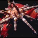
The test of Eucidaris tribuloides is moderately flattened above and below (Plate 17: figure 5). There is a moderate incurving of the basicoronal plates at the edge of the peristome. The test at the ambitus is commonly circular, but numerous specimens are subpentagonal and a small percentage pentagonal. The radius in the pentagonal specimens is greatest in the interambulacral midzone and shortest in the ambulacra. The flattening of the test above and below is relatively more prominent on small specimens than on large ones of 50 mm or more. The apical system is commonly slightly larger than one-third of the horizontal diameter of the test and approximately the same size as the peristome. On small to medium-size specimens the apical system may be noticeably smaller than the peristome. The madreporite is commonly enlarged and prominent on denuded specimens.
The ambulacra are only slightly sinuate (Plate 17: figures 4, 5). Marginal tubercles are prominent, the bosses on some specimens contiguous and transversely oval, arranged in uniform series. Inner tubercles crowd the interporiferous zone and the crowding exists regardless of the presence of one, two, or three inner tubercles per plate. Two inner tubercles per plate are the most common, but there is only a very slight tendency for the inner tubercles to form in series.
Interambulacral plates in the region of the ambitus are approximately twice as wide as high and have shallow small areoles. The tubercles of the scrobicular ring are noticeably more prominent than the extrascrobicular tubercles of the interambulacral midzone (Plate 17: figure 5). There is less difference in size between the scrobicular tubercles and the extrascrobicular tubercles bordering the ambulacra (Plate 17: figures 4, 5). In the interambulacral midzone the extrascrobicular tubercles are in somewhat horizontal rows separated by furrows. The large number of primary tubercles in vertical series is an important diagnostic feature (Plate 17: figure 5). A specimen 35 mm in diameter commonly has 9 to 10 primary tubercles in a vertical series.
The primary spines are short, from about half the horizontal diameter of the test to only slightly larger than the horizontal diameter (Plate 16: figures 2–4). They are fairly stout, either cylindrical or noticeably tapered. The tip of the spine (Plate 17: figure 1) has a crown formed by lamellae, each of which is at the tip end of a row of warts (Plate 17: figures 1, 2). At the center of the crown is a small prominence. Except when very small, specimens lack pointed spinules. The shaft of each spine is covered by a dense spongy covering of anastomosing hairs. The extent of development of this covering varies between individuals. Under water those with a very dense covering appear to have a coat of fur (Plate 17: figure 3). The neck of the primary spines is very short and very difficult to observe on nondenuded specimens. Oral primaries are oval in transverse outline, only slightly different from transitional and ambital primaries. The scrobicular spines are broad, straight sided. The tips are very blunt and concave, almost shovellike on some specimens (Plate 16: figure 1).
The large globiferous pedicellariae lack an end tooth and are similar to the kind shown in Figure 4.
The specimens are brown, commonly darkest at the collar and tips of the marginal and scrobicular spines. The brown pigment of the primary spines is commonly obscured by the gray or white covering of anastomosing hairs. The denuded test is olive; the areoles white or almost so.
Eucidaris tribuloides is usually found in fairly shallow water, but its depth range extends into the upper limits of the distribution of Stylocidaris affinis. The two species are commonly collected at the same station, especially off the west coast of Florida. Very small specimens of both species are strikingly similar but can be differentiated by the following features.
Both species have light and dark banding of the primary spines with well-developed thorns. Close examination of the dark bands of E. tribuloides will reveal that some of the thorns have already developed into typical warts (Plate 18: figure 3). Observe the dark bands nearest the base of the spines as this is where the first warts appear. As the individual grows, wart development progresses more rapidly in the dark bands than the light ones, and a stage is reached where the thoms remain only in the light bands (Plate 18: figures 1–3). These also change to warts and the color banding usually leaves. The crownlike tip develops on at least one primary spine even on extremely small individuals of E. tribuloides (Plate 18: figures 1–3). One or two very broad tipped large scrobicular spines will be present even on the very small specimens (Plate 18: figure 2).
The thoms on the primary spines of S. affinis remain and, as the spines grow, simply become the spinules. The scrobicular spines of even the smallest specimens of S. affinis have the prominent reddish midline stripe (Plate 18: figure 5).
DISTRIBUTION.—West Indies, Gulf of Mexico, and Bermuda. Littoral to 450 meters.
- bibliographic citation
- Phelan, Thomas Francis. 1970. "A field guide to the Cidaroid echinoids of the northwestern Atlantic Ocean, Gulf of Mexico, and the Caribbean Sea." Smithsonian Contributions to Zoology. 1-67. https://doi.org/10.5479/si.00810282.40