-
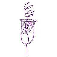
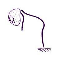
Bicosoeca pulchra Hilliard, 1971. Bicosoeca cells with a lorica that is 19.3-20.2 microns long, 9.7-10.6 microns wide. The lorica is delicately urn-shaped, posteriorly pointed, with a short strip appended to the base. There is a slight constriction just below a moderately flaring opening, while the lip at the oral rim may converge slightly. The lorica wall is thin, hyaline, and appears serrate in optical profile when observed in aqueous methylene blue. Staining reveals the presence of transverse bands, which number 10-12 in 10 microns The spherical protoplast (6.5-7 microns long, 4.5-6 microns wide) is colourless, granular, and has a central nucleus and a vacuole. A single flagellum bearing two rows of submicroscopic hairs extends dorsally from the cell and is about three times its length.
-
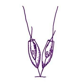
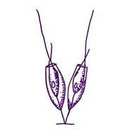
Bicosoeca tenuis Kent, 1880. Bicosoeca cells with a lorica that is elongate-ovate or subfusiforme, nearly three times as long as broad, tapering equally at each extremity, pedicel scarcely one-half the height of the lorica, protoplast with a lip-like projection, slightly exsert anteriorly. Length of lorica 8.5 to 10.2 microns The assignment of this species to Bicosoeca is incorrect, because in Bicosoeca, one flagellum attaches the cell to the lower end of the lorica, whereas here both flagella project from the opening of the lorica.
-
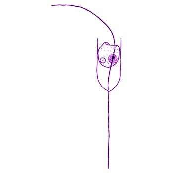
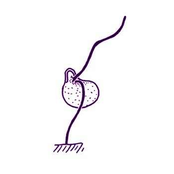
Bicosoeca vacillans Stole, 1888. Bicosoeca cells located in a stalked lorica, the chamber of which measured 17-25 microns long. The lorica chamber was approximately cylindrical with a slightly pointed posterior and a pedicel of 1-1.5 times the lorica length. Loricas may differ slightly in shape. The lorica wall was most frequently curved slightly outwards at the aperture to a greater or lesser extent and in some cases the lorica was waisted below the aperture. The lorica had fine horizontal bands spaced about 0.4 microns apart, with numerous fine perpendicular fibres in each band. Cells were sub-spherical and had a flattened indistinct peristome. The anterior flagellum was about 3 times the cell length and held at an angle to the longitudinal axis of the cell. The posterior flagellum was attached to the base of the lorica via a fine thread.
-
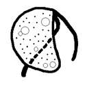
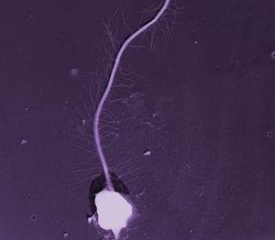
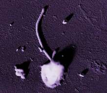
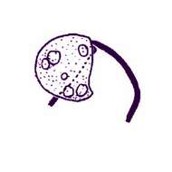
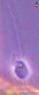
This image was of material in samples taken during a scientific cruise in the Pacific. Water was filtered to concentrate the organisms that were present, then dried onto a thin sheet of plastic and then shadowed with a fine layer of metal to provide contrast. The preparation was then observed with an electron-microscope. This technique has been used to document the diversity of marine microbes, especially, protists in the oceans.
-
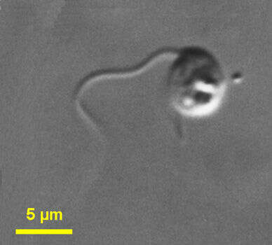
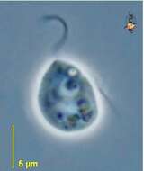
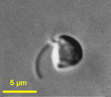
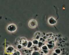
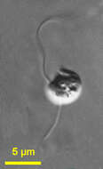
Cafeteria (cafeteria) is probably the most abundant heterotrophic flagellate in marine ecosystems. It is a voracious bacterivore. Sessile feeding cells are D-shaped, 1.5 to 10 microns long, and laterally compressed. There is a ventral groove, and the flagella insert at the head of the groove. The anterior flagellum draws a current of water towards the cell, or pulls the cell forward when it is swimming around. Feeding cells are usually attached to the substrate by the tip of the second flagellum. This cell is a little swollen. Animations by Rosemary Arbur of flagellar beat patterns are available
here.Phase contrast.
-
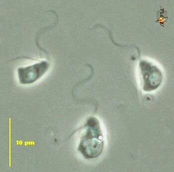
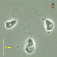
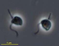
Cafeteria (cafeteria) is probably the most abundant heterotrophic flagellate in marine ecosystems. It is a voracious bacterivore. Sessile feeding cells are D-shaped, 1.5 to 10 microns long, and laterally compressed. There is a ventral groove, and the flagella insert at the head of the groove. These cells have been disturbed, and are not feeding. The anterior flagella have the sine-wave beat pattern that is characteristic of stramenopiles. The posterior flagella are shorter. Phase contrast.
-
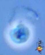
Cafeteria (cafeteria) is probably the most abundant heterotrophic flagellate in marine ecosystems. It is a voracious bacterivore. Sessile feeding cells are D-shaped, 1.5 to 10 microns long, and laterally compressed. There is a ventral groove, and the flagella insert at the head of the groove. The anterior flagellum draws a current of water towards the cell, or pulls the cell forward when it is swimming around. Feeding cells are usually attached to the substrate by the tip of the second flagellum. Phase contrast. Rotten picture.
-
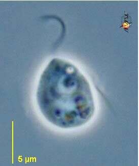
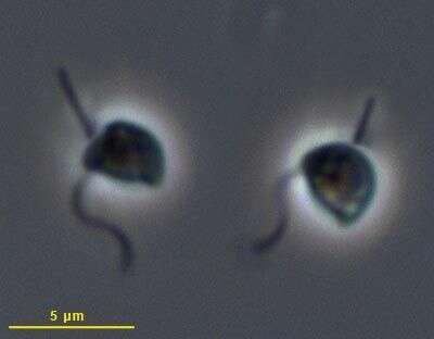
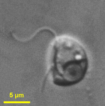
Cafeteria (cafeteria) roenbergensis Fenchel and Patterson, 1988. Cells are D-shaped, 2 to 5 microns long, and laterally compressed. There is a shallow groove on the left side of the cell. Two flagella of similar length emerge subapically and are slightly longer than the cell. The anterior flagellum is directed perpendicular to the ventral face of the cell of attached cells. The posterior flagellum is reflexed, passing over one face of the cell and then attaching to the substrate by the tip. In swimming cells, the anterior flagellum is directed forwards and beats with a sine-wave, and the posterior flagellum is directed backwards and trails. Usually the cells move fast following a spiral path, but sometimes move slowly. Bacteria may be ingested near the anterior part or posterior part of the ventral groove. Not common in sediments, widespread and abundant in the water column.
-
Cafeteria roenbergensis Fenchel and Patterson, 1988. Cells are D-shaped, 2 to 5 microns long, and laterally compressed. There is a shallow groove on the left side of the cell. Two flagella of similar length emerge subapically and are slightly longer than the cell. The anterior (= feeding = hairy - the hairs are not visible by light microscopy) flagellum is directed perpendicular to the ventral face of the cell of attached cells. The posterior flagellum is reflexed, passing over one face of the cell and then attaching to the substrate by the tip. In swimming cells, the anterior flagellum is directed forwards and beats with a sine-wave, and the posterior flagellum is directed backwards and trails. Usually the cells move quickly following a spiral path, but sometimes they move slowly. Bacteria may be ingested near the anterior part or posterior part of the ventral groove.
-
-
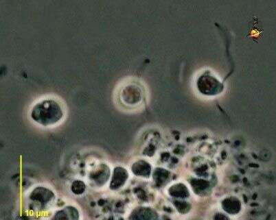
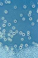
Cafeteria is a weed. When plenty of bacterial food is available, species of this genus will grow in great profusion. Dozens of cells are seen here near a piece of detritus. Animations by Rosemary Arbur of flagellar beat patterns are available
here. Phase contrast microscopy.
-
Acronema sippewissettensis Teal et al., 1998. Reniform slightly dorsoventrally flattened flagellates range in size from 3-7 microns in length by 3-5 microns in width. They are spherical in two to three day old cultures, and more oblong in six to seven day-old cultures. A slight peristome originates from the anterior dorsal edge. Both acronematic (tapered) flagella emerge from a mid-ventral depression in young mastigotes or from a subapical ventral depression in older ones. A ventral groove originates below the peristome in a depression where the undulipodium emerges, and runs posteriorly on the right-hand side of the cell to a midventral level. The short posterior flagellum is 4-6 microns long, while the anterior one is 5-7 microns long. The tapered region of the posterior flagellum, 1.5-2.25 microns, is approximately three times that of the anterior flagellum, 0.5-0.75 microns Probably this is Cafeteria roenbergensis.
-
Cafeteria (cafeteria) marsupialis Larsen and Patterson, 1990. Cells are D-shaped and somewhat variable in shape. When attached the cells are 5 to 12 microns long (mostly about 7 to 8 microns) and 5 to 10 microns wide, When swimming the cells are up to 7 to 10 microns long and 3 to 5 microns wide. The cells attach to the substrate by the tip of the posterior flagellum, which lies in a deep ventral groove. The anterior flagellum is directed normal to the groove. The organism may feed on suspended bacteria, which are drawn towards the body by the activity of the anterior flagellum, entering along a curved channel at the posterior end of the ventral groove. The anterior flagellum of the attached cell is about 1.5 to 2 times the cell length and the posterior flagellum is slightly longer than the cell. A single nucleus with a rounded nucleolus lies just below the insertion of the flagella. The cell body may include many - sometimes large - food vacuoles. Undigested residues of food are egested by the fusion of old food vacuoles with the plasma membrane. Swimming cells are more rounded, with anterior flagellum directed to the anterior, recurrent flagellum trailing. It often occurs with Carpediemonas membranifera and C. bialata. More frequent in slightly anaerobic preparations.
-
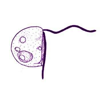
Cafeteria marsupialis Larsen and Patterson, 1990. Cells are normally D-shaped but may be somewhat variable in shape. When attached the cells are 5 to 12 microns long (mostly about 7 to 8 microns) and 5 to 10 microns wide, When swimming the cells are up to 7 to 10 microns long and 3 to 5 microns wide. The cells attach to the substrate by the tip of the posterior flagellum, which lies in a deep ventral groove. The anterior flagellum is directed at right-angles to the groove. The organism may feed on suspended bacteria, which are drawn towards the body by the activity of the anterior flagellum, entering along a curved channel at the posterior end of the ventral groove. The anterior flagellum of the attached cell is about 1.5 to 2 times the cell length and the posterior flagellum is slightly longer than the cell. A single nucleus with a rounded nucleolus lies just below the insertion of the flagella. The cell body may include many - sometimes large - food vacuoles. Undigested residues of food are egested by the fusion of old food vacuoles with the plasma membrane. Swimming cells are more rounded, with anterior flagellum directed to the anterior, recurrent flagellum trailing. Common and widespread.
-
Cafeteria (cafeteria) minuta (Ruinen, 1938) Larsen and Patterson, 1990. Cells are about 4 - 6 microns long with a small ventral groove. Two flagella emerge from the anterior end of the groove. The long anterior flagellum is about 2.5 - 4 times the cell length and beats with a sine-wave, and the posterior flagellum is about 1 - 1.5 times the cell length. When the cells swim, the anterior flagellum is directed forward and the posterior one trails. The cells may attach to the substrate by the tip of the posterior flagellum. Not common.
-
Cafeteria minuta (Ruinen, 1938) Larsen and Patteron, 1990. Cells are almost globular, 3.5-4 microns but with a subapical ventral depression from which the flagella emerge. The hairy or feeding flagellum (the hairs are not visible with the light microscope), which projects in front of the swimming cell, measures 10-12 microns, and may beat with more than one complete sine wave along its length. Second flagellum about 5 microns, curves over the side of the body and may attach by its tip to the substratum.
-
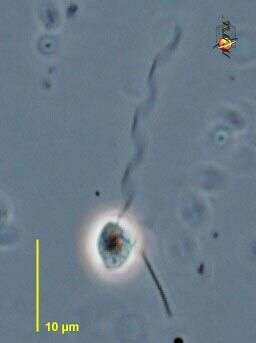
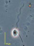
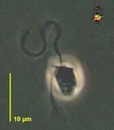
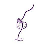
Pseudobodo (sue-doe-bow-dough) is a naked bicosoecid stramenopile. As with other bicosoecids, it attaches to the substrate by the tip of the recurrent flagellum. The anterior flagellum is directed away from the substrate, beats with an undulating pattern, and draws a current of water with suspended bacteria (its food) towards the cell. The cell has a ridge to one side of the flagellum and this marks the margin of the ingestion region. Phase contrast.
-
Pseudobodo (sue-doe-bow-dough) is a naked bicosoecid stramenopile. As with other bicosoecids, it attaches to the substrate by the tip of the recurrent flagellum. The anterior flagellum is directed away from the substrate, beats with an undulating pattern, and draws a current of water with suspended bacteria (its food) towards the cell. Phase contrast.
-
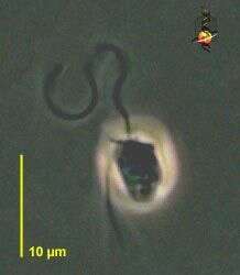
Pseudobodo (sue-doe-bow-dough) is a naked bicosoecid stramenopile. As with other bicosoecids, it attaches to the substrate by the tip of the recurrent flagellum, although this cell has detached. The anterior flagellum is directed away from the substrate, beats with an undulating pattern, and draws a current of water with suspended bacteria (its food) towards the cell. The cell has a ridge to one side of the flagellum and this marks the margin of the ingestion region. Phase contrast.
-
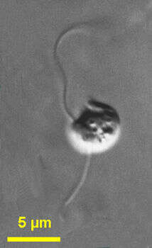
Pseudobodo (sue-doe-bow-dough) is a naked bicosoecid stramenopile. As with other bicosoecids, it attaches to the substrate by the tip of the recurrent flagellum. The anterior flagellum is directed away from the substrate, beats with an undulating pattern, and draws a current of water with suspended bacteria (its food) towards the cell. The cell has a ridge to one side of the flagellum and this marks the margin of the ingestion region. Phase contrast.
-
-
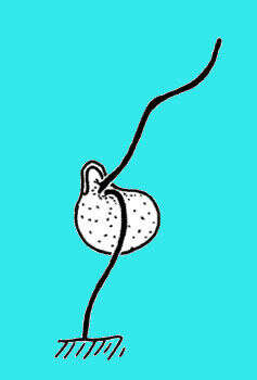
Pseudobodo (sued-oh-boe-dough) tremulans Griessmann, 1913. Cells are about 4.5 - 6 microns long with an anterior collar around the anterior part of the cell in unstressed feeding cells. The insertion sites of the two flagella are separated by a protrusion at the anterior of the cell. The anterior flagellum has a sine-wave beating pattern and is about 3.5 times the length of the cell, and the posterior flagellum is about twice the length of the cell and may attach to the substrate by its tip. When the cells move, the anterior collar may be hard to see. The cells move by swimming with the anterior flagellum directed forwards. Not common.
-
Pseudobodo tremulans Griessmann, 1913. Cells are about 4.5 - 6 microns long with an anterior collar around the anterior part of the cell in unstressed feeding cells. The insertion sites of the two flagella are separated by a protrusion at the anterior of the cell. The anterior flagellum has a sine-wave beating pattern and is about 3.5 times the length of the cell, and the posterior flagellum is about twice the length of the cell and may attach to the substrate by its tip. When the cells move, the anterior collar may be hard to see. The cells move by swimming with the anterior flagellum directed forwards.
-
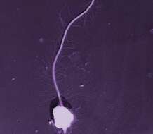
This image was made from samples taken during a scientific cruise in the Pacific. Water was filtered to concentrate the organisms that were present, then dried onto a thin sheet of plastic and then shadowed with a fine layer of metal to provide contrast. The preparation was then observed with an electron-microscope. This technique has been used to document the diversity of marine microbes, especially, protists in the oceans.