-
-
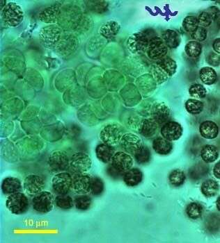
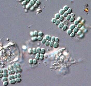
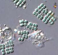
Microcystis flos-aquae (Cyanobacteria, Chroococcales) is a together with Microcystis aeruginosa are common in Lake Kinneret throughout the year, occasionally forming blooms with surface scums in winter or spring. The individual cells in a M. flos-aquae colony are 3-4 um in diameter, the colony is spherical or lens-shaped, with varying degree of spacing between cells within a colony. The dark spots clearly showing in the individual cells in this picture are due to the reflection of light from the gas vesicles. A bunch of choanoflagellates are attached at the upper left side of the colony.
-
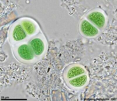
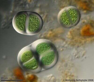
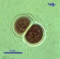
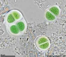
Chroococcus turgidus, (Cyanobacteria, Chroococales), from Lake Kinneret pelagic waters, April 2006, showing 2 daughter cells after division by simple binary fission â as characteristic for most Chroococcales species. This species is common in the plankton of Lake Kinneret throughout the year. Usually there are 2 â 8 cells in a colony. Clearly delimited colorless mucilaginous envelopes surround the individual cells following their contours, and the entire colony. Cell diameter: 8 â 11 µm.
-
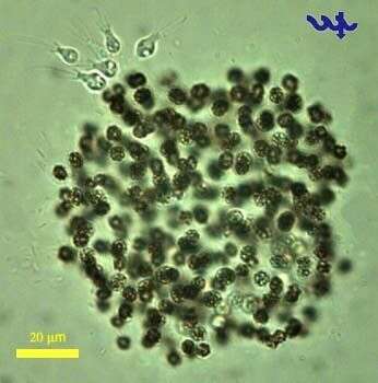
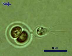
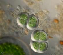
Chroococcus turgidus (Cyanobacteria, Chroococcales) is common in Lake Kinneret in recent years. In this unusual photograph a choanoflagellate is attached to its outer shell.
-
High resolution photo of Chroococcus turgidus using Planapo 63/1.4. Sample from sphagnum pond situated in the northern alpine region of Austria near Salzburg. Images were taken using Zeiss Universal with Olympus C7070 CCD camera.
-
High resolution photo of Chroococcus turgidus using Planapo 63/1.4 with DIC. Sample from sphagnum pond situated in the northern alpine region of Austria near Salzburg. Images were taken using Zeiss Universal with Olympus C7070 CCD camera.
-
High resolution photo of Chroococcus turgidus using Planapo 63/1.4 with DIC. Sample from sphagnum pond situated in the northern alpine region of Austria near Salzburg. Images were taken using Zeiss Universal with Olympus C7070 CCD camera.
-
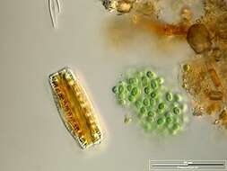
Colony accompanied by Epithemia adnata. Scale bar indicates 50 µm. Sample from a wetland at the Pillersee (Tyrol, Austria). The image was built up using several photomicrographic frames with manual stacking technique. Images were taken using Zeiss Universal with Olympus C7070 CCD camera.Image under Creative Commons License V 3.0 (CC BY-NC-SA).
-
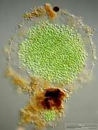
Scale bar indicates 100 µm. Sample from the pond Hegne Moor situated in the vicinity of Lake Constance. Images were taken using Zeiss Universal with Olympus C7070 CCD camera.Image under Creative Commons License V 3.0 (CC BY-NC-SA).
-
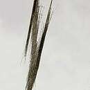
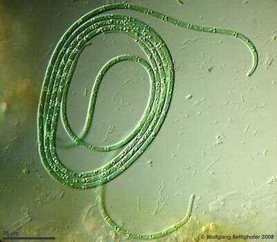
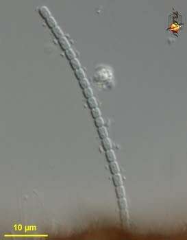
Filamentous cyanobacteria which can move like Oscillatoria, aka Phormidium splendidum. Sample from sphagnum pond situated in the northern alpine region of Austria near Salzburg. Images were taken using Zeiss Universal with Olympus C7070 CCD camera.
-
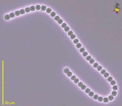
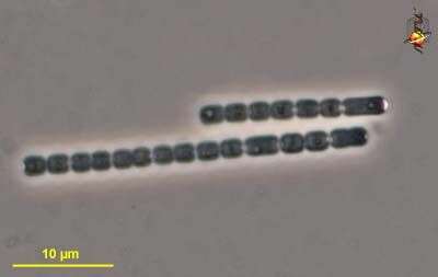
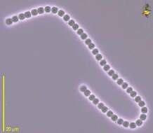
Unidentified cyanobacterium. Cells in pairs form a filament. Phase contrast.
-
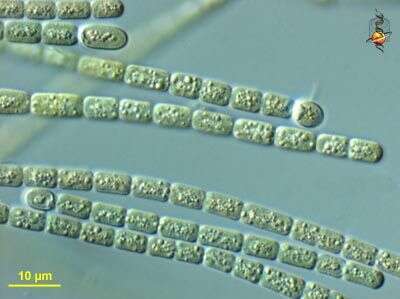
Filamentous blue green algae with several morphologies, all lack a mucus sheath, glide and had no evident heterocysts (differentiated cells), and are probably (though not certainly) assignable to Oscillatoria. Phase contrast.
-
Filamentous cyanobacterium, unidentified, found as one of several cyanobacterial epibionts on the leaves of the moss Hygrohypnum, a site which seems to be a focus for nitrogen fixation. Differential interference contrast.
-
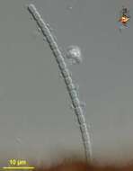
Filamentous cyanobacterium, unidentified. Phase contrast.
-
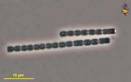
Unidentified cyanobacterium, filamentous, blue-green or olive green, often with a differentiated cell at one end of a filament. Differential interference contrast. Material from cooler waters at LaDuke spring, a thermal site within Gallatin National Forest, photograph by Kathy Sheehan and David Patterson.
-
Blue green algae forming gelatinous aggregates, this image forming tight clump with filaments emerging. Phase contrast microscopy.
data on this strain.
-
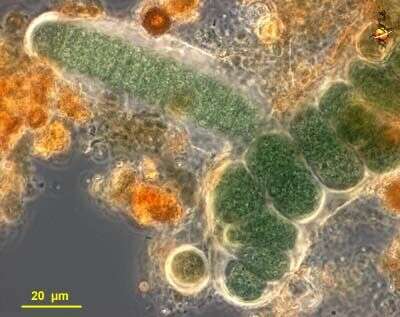
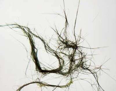
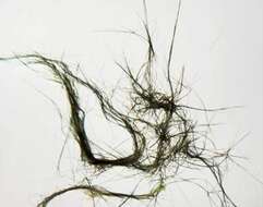
Intertwined filaments of cyanobacteria grow together to form a microbial mat.
-
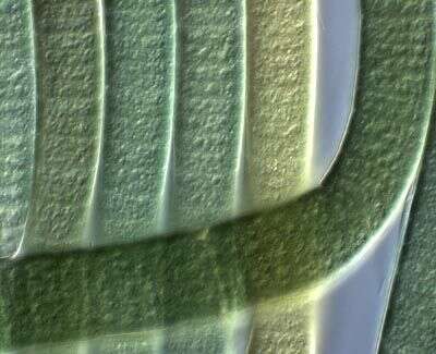
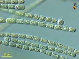
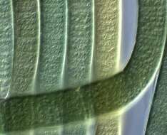
This image of cyanobacteria shows how the filaments are made up of many disc-shaped cells joined end to end. Although all are the same species, the colors of the filaments vary. This can cause mats to have regions of different colors, even when they are formed by the same species
-
Cyanobacteria. Group of cells observed in sandy and muddy marine sediments in the vicinity of Broome, Western Australia in September 2003. This image was taken using differential interference contrast optics. This work was supported by the Australian Biological Resources Study.
-
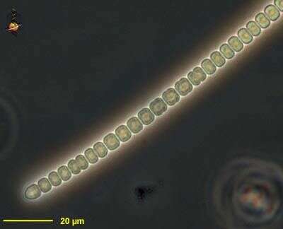
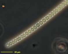
Cyanobacteria. Filament observed in sandy and muddy marine sediments in the vicinity of Broome, Western Australia in September 2003. This image was taken using phase contrast optics. This work was supported by the Australian Biological Resources Study.
-
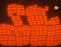
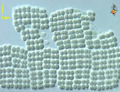
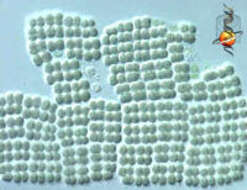
Merismopedia (mer-is-mo-pea-dee-a) is a blue-green alga or cyanobacterium (blue-green alga). The genus is distinguished by the square-packed pattern of the coccoid cells. It is common in sediments, but the number of cells which occur in a colony may vary from only a few to thousands. As a cyanobacterium, the photosynthetic pigments are located throughout the cytoplasm. Differential interference contrast
-
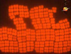
Merismopedia (mer-is-mo-pea-dee-a) is a blue-green alga or cyanobacterium. The genus is distinguished by the square-packed pattern of the coccoid cells. It is common in sediments, but the number of cells which occur in a colony may vary from only a few to thousands. As a cyanobacterium, the photosynthetic pigments are located throughout the cytoplasm. These can be seen if the cells are illuminated with ultra-violet light and then the distribution of the pigment detected by the resulting fluorescence. Fluorescence microscopy.
-
Merismopedia (mer-is-mo-pea-dee-a) is a blue-green alga or cyanobacterium. The genus is distinguished by the square-packed pattern of the coccoid cells. It is common in sediments, but the number of cells which occur in a colony may vary from only a few to thousands. As a cyanobacterium, the photosynthetic pigments are located throughout the cytoplasm. Differential interference contrast.
-
Merismopedia (mer-is-mo-pea-dee-a) is a blue-green alga or cyanobacterium. The genus is distinguished by the square-packed pattern of the coccoid cells. It is common in sediments, but the number of cells which occur in a colony may vary from only a few to thousands. As a cyanobacterium, the photosynthetic pigments are located throughout the cytoplasm. Differential interference contrast