-
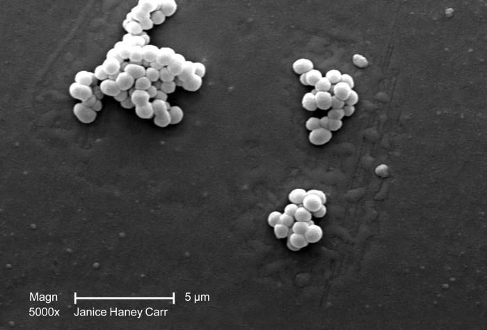
Under a moderately-high magnification of 5,000X, this scanning electron micrograph (SEM) revealed several small clusters of Gram-positive, beta-hemolytic Group C Streptococcus sp. bacteria.Created: 2008
-
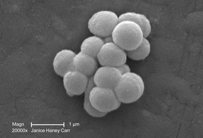
Under a high magnification of 20,000X, this scanning electron micrograph (SEM) revealed a small clustered group of Gram-positive, beta-hemolytic Group C Streptococcus sp. bacteria. See PHIL 10586 for a colorized version of this image.Created: 2008
-
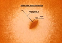
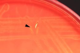
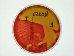
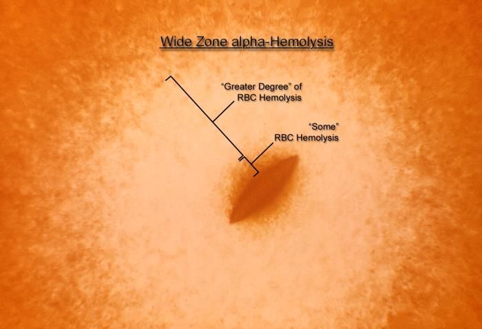
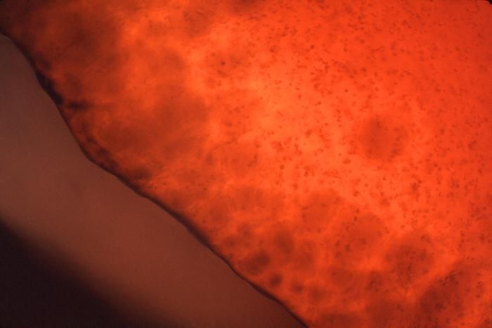
Magnified 100x, this 1977 photograph depicted a Petri dish filled with heart infusion agar medium containing 5% defibrinated rabbit blood, i.e., blood agar plate (BAP). After having been inoculated, using the "stab" technique using a culture of Streptococcus anginosus bacteria, of the Gram-positive viridans group of streptococci (VGS), the BAP was incubated in a carbon dioxide enriched atmosphere at 35oC for 24 hours. The culture grew bacterial colonies around the stab site, surrounded by what is known as "wide zone alpha hemolytic" (WZα) color changes. Characteristics of WZα reactivity are described as, "the area immediately adjacent to the colony has some red blood cells (RBCs), but an area outside of that may be completely, or nearly completely, cleared of RBCs. Therefore, there are no reactive zones where "complete" RBC hemolysis has occurred, as in the case in beta-hemolytic reactions, hence the Wide Zone "alpha" terminology.Created: 1977
-
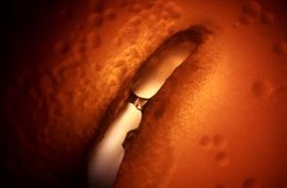
Magnified 100x, this 1977 photograph depicted a Petri dish filled with heart infusion agar medium containing 5% defibrinated rabbit blood, i.e., blood agar plate (BAP). A loop of diluted culture of Streptococcus anginosus was put into the melted agar (50oC) just before the blood was added to the melted agar. The agar was allowed to solidify, and then incubated at 35oC for 24 hours in a normal atmosphere. The culture grew subsurface bacterial colonies, one of which is seen here, surrounded by what is known as "wide zone alpha hemolytic" (WZα) color changes. Characteristics of WZα reactivity are described as, "the area immediately adjacent to the colony has some red blood cells (RBCs), but an area outside of that may be completely, or nearly completely, cleared of RBCs. Therefore, there are no reactive zones where "complete" RBC hemolysis has occurred, as in the case in beta-hemolytic reactions, hence the Wide Zone "alpha" terminology.Created: 1977
-
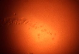
Magnified 100x, this 1977 photograph depicted a Petri dish filled with heart infusion agar medium containing 5% defibrinated rabbit blood, i.e., blood agar plate (BAP). After having been inoculated with a culture of Streptococcus anginosus bacteria, of the Gram-positive viridans group of streptococci (VGS), the BAP was incubated in a carbon dioxide enriched atmosphere at 35oC for 24 hours. In this view, one can see numbers of growing "surface" colonies surrounded by what is known as "wide zone alpha hemolytic" (WZα) color changes. Characteristics of WZα reactivity are described as, "the area immediately adjacent to the colony has some red blood cells (RBCs), but an area outside of that may be completely, or nearly completely, cleared of RBCs. Therefore, there is no reactive zones where "complete" RBC hemolysis has occurred, as is the case in beta-hemolytic reactions, hence the Wide Zone "alpha" terminology.Created: 1977
-
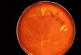
Magnified 10x, this image depicted a Petri dish filled with heart infusion agar medium containing 5% defibrinated rabbit blood, i.e., blood agar plate (BAP). After having been inoculated using both a streak and stab technique with a culture of Streptococcus anginosus bacteria, of the Gram-positive viridans group of streptococci (VGS), the BAP was incubated in a carbon dioxide enriched atmosphere at 35oC for 24 hours. In this view, one can see numbers of growing "surface" and "stab" colonies surrounded by what is known as "wide zone alpha hemolytic" (WZα) color changes. Characteristics of WZα reactivity are described as, "the area immediately adjacent to the colony has some red blood cells (RBCs), but an area outside of that may be completely, or nearly completely, cleared of RBCs. Therefore, there is no reactive zones where "complete" RBC hemolysis has occurred, as is the case in beta-hemolytic reactions, hence the Wide Zone "alpha" terminology.Created: 1977
-
This image depicted a Petri dish filled with heart infusion agar medium containing 5% defibrinated rabbit blood, i.e., blood agar plate (BAP). After having been inoculated, using both a streak and stab technique with culture of Streptococcus anginosus bacteria, a member of the Gram-positive viridans group of streptococci (VGS), the BAP was incubated in a carbon dioxide enriched atmosphere at 35oC for 24 hours. The culture grew bacterial colonies. In this view, one can see numbers of growing colonies surrounded by what is known as "wide zone alpha hemolytic" (WZα) color changes. Characteristics of WZα reactivity are described as, "the area immediately adjacent to the colony has some red blood cells (RBCs), but an area outside of that may be completely, or nearly completely, cleared of RBCs. Therefore, there is no reactive zones where "complete" RBC hemolysis had occurred, as is the case in beta-hemolytic reactions, hence the Wide Zone "alpha" terminology.Created: 1977
-
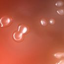
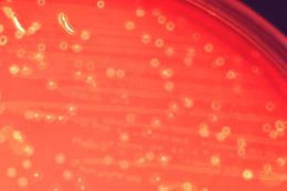
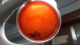
Magnified 100x, this 1977 photograph depicted a Petri dish filled with trypticase soy agar medium containing 5% defibrinated sheep's blood, i.e., blood agar plate (BAP). After having been inoculated, using a stab technique, with alpha-hemolytic Streptococcus anginosus bacteria, i.e., a member of the Gram-positive viridans group of streptococci (VGS), the BAP was incubated in a carbon dioxide enriched atmosphere at 35oC for 24 hours. The culture grew bacterial colonies. In this view, one can see numbers of colonies that were growing at the edge of the stab, surrounded by the characteristic color changes, i.e., a hazy, faded, and indistinct region in which some of the red blood cells (RBCs) were destroyed in the blood agar medium, or "hemolyzed", indicating that these bacteria were indeed alpha-hemolytic in nature.Created: 1977
-
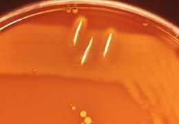
Magnified 100x, this image depicted a Petri dish filled with trypticase soy agar medium containing 5% defibrinated sheep's blood, i.e., blood agar plate (BAP). After having been inoculated with alpha-hemolytic Streptococcus anginosus bacteria, i.e., members of the Gram-positive viridans group of streptococci (VGS), just before the blood was added to the agar, a loop of diluted culture was put into the melted agar (50oC). The melted agar with blood was allowed to solidify and then incubated at 35oC for 24 hours in a normal atmosphere. The culture grew subsurface bacterial colonies, one of which was seen here. Surrounded by a characteristic hazy, faded, and indistinct region (arrow) in which some of the red blood cells were destroyed in the agar medium, or "hemolyzed", indicating that these bacteria were indeed alpha-hemolytic in nature.Created: 1977
-
Magnified 100x, this 1977 photograph depicted a Petri dish filled with trypticase soy agar medium containing 5% defibrinated sheep's blood, i.e., blood agar plate (BAP). After having been inoculated with alpha-hemolytic Streptococcus anginosus bacteria, i.e., a member of the Gram-positive viridans group of streptococci (VGS), the BAP was incubated in a carbon dioxide enriched atmosphere at 35oC for 24 hours. The culture grew bacterial colonies, which were seen here. The characteristic color changes, i.e., a hazy, faded, indistinct region surrounding each colony in which some of the red blood cells (RBCs) were destroyed in the blood agar medium, or "hemolyzed", indicated that these bacteria were indeed alpha-hemolytic in nature.It is the incomplete nature of the hemolytic reaction adjacent to the colonies, which spares numbers of RBCs in the blood agar medium, that is of qualitative importance when distinguishing alpha from beta-hemolysisCreated: 1977
-
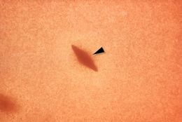
Magnified 100x, this 1977 photograph depicted a Petri dish filled with trypticase soy agar medium containing 5% defibrinated sheep's blood, i.e., blood agar plate (BAP). After having been inoculated with alpha-hemolytic Streptococcus anginosus bacteria, i.e., a member of the Gram-positive viridans group of streptococci (VGS), using a streak/stab technique, the BAP was incubated in a carbon dioxide enriched atmosphere at 35oC for 24 hours. The culture grew bacterial colonies. Typical alpha-hemolytic colonies were seen adjacent to the stab sites (arrow). The hazy, faded, indistinct region surrounding each colony indicated that red blood cells (RBCs) were destroyed, or "hemolyzed", in the BAP, and that these bacteria were indeed alpha-hemolytic in nature.It is the incomplete nature of the hemolytic reaction adjacent to the colonies, which spares numbers of RBCs in the blood agar medium, that is of qualitative importance when distinguishing alpha from beta-hemolysis.Created: 1977
-
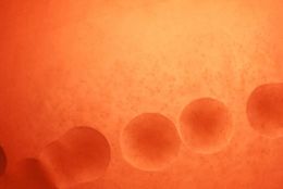
This 1977 image depicts a Petri dish filled with trypticase soy agar medium containing 5% defibrinated sheep's blood, i.e., blood agar plate (BAP). After having been inoculated with alpha-hemolytic Streptococcus anginosus bacteria, i.e., a member of the Gram-positive viridans group of streptococci (VGS), the BAP was incubated in a carbon dioxide enriched atmosphere at 35oC for 24 hours. The culture grew numbers of surface bacterial colonies. The characteristic color changes, i.e., a hazy, faded, indistinct region surrounding each colony in which some of the red blood cells (RBCs) were destroyed in the blood agar medium, or "hemolyzed", indicated that these bacteria were indeed alpha-hemolytic in nature.It is the incomplete nature of the hemolytic reaction adjacent to the colonies, which spares numbers of RBCs in the blood agar medium, that is of qualitative importance when distinguishing alpha from beta-hemolysis. No magnification was used here.Created: 1977
-
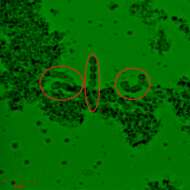
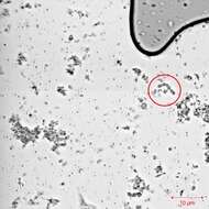
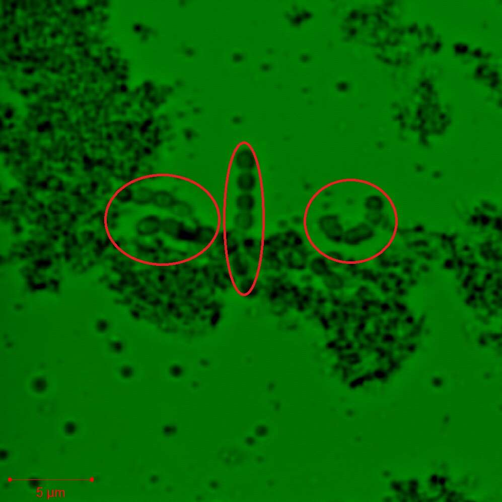
Description: Français : Image (colorée pour améliorer le contraste et donc la visibilité des bactéries) représentant une agglomération de chaînes de Streptococcus thermophilus prise à l'aide d'un microscope confocal ZEISS LSM 780 (avec échelle). Date: 24 May 2013, 20:11:47. Source: Own work. Author:
Adam Benyoussef.
-
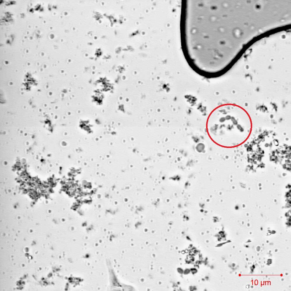
Description: Français : Image représentant une agglomération de chaînes de Streptococcus thermophilus prise à l'aide d'un microscope confocal ZEISS LSM 780 (avec echelle). Date: 24 May 2013, 20:11:48. Source: Own work. Author:
Adam Benyoussef.
-
-
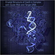
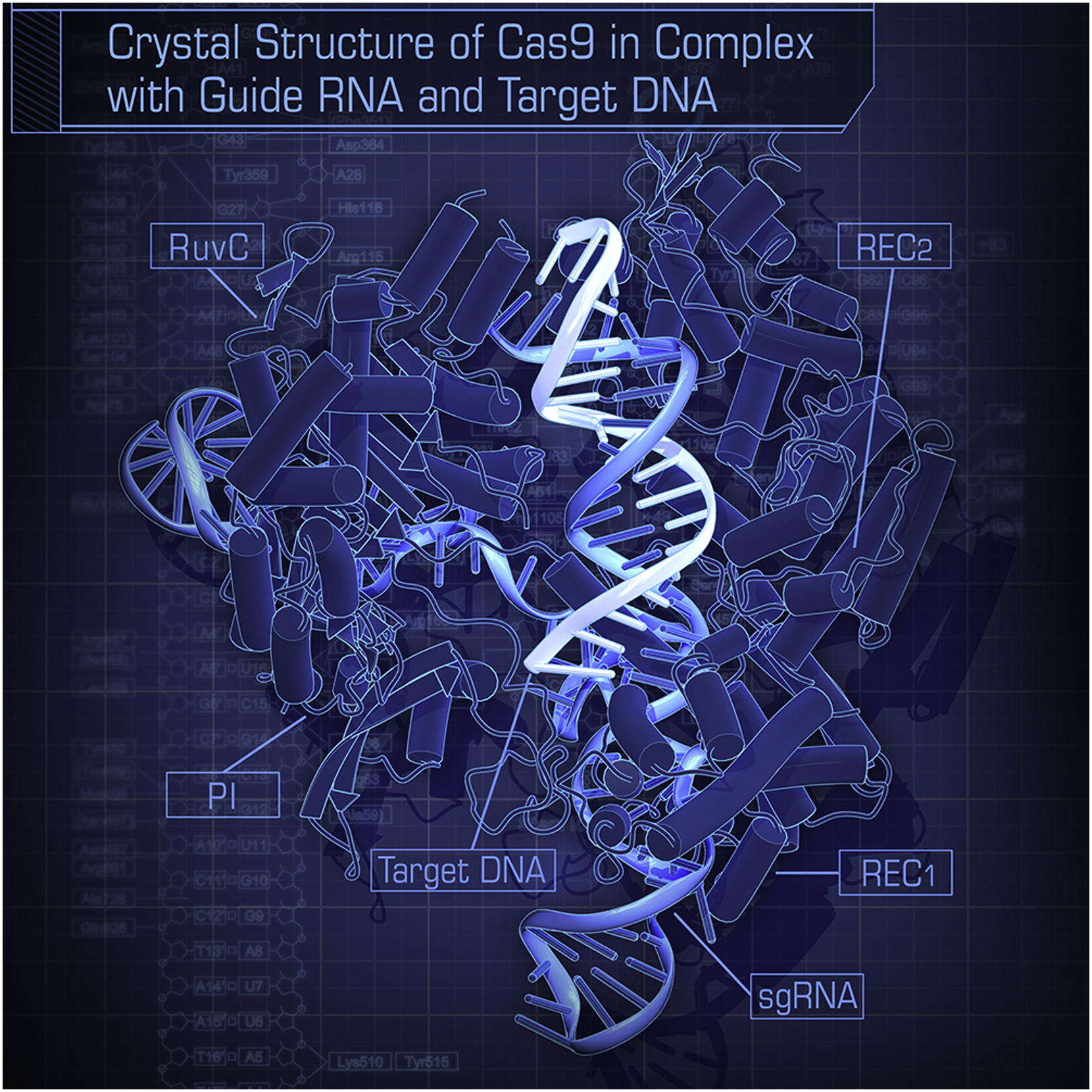
Hiroshi Nishimasu, F. Ann Ran, Patrick D. Hsu, Silvana Konermann, Soraya I. Shehata,
Wikimedia Commons
Description: English: the crystal structure of Streptococcus pyogenes Cas9 in complex with sgRNA and its target DNA at 2.5 A ˚ resolution. Date: 1 May 2014, 11:09:15. Source: Crystal Structure of Cas9 in Complex with Guide RNA and Target DNA
https://dx.doi.org/10.1016/j.cell.2014.02.001. Author: Hiroshi Nishimasu, F. Ann Ran, Patrick D. Hsu, Silvana Konermann, Soraya I. Shehata, Naoshi Dohmae, Ryuichiro Ishitani, Feng Zhang, and Osamu Nureki.
-
-
NIAID|sourceurl=https://flickr.com/photos/54591706@N02/52602062457%7Carchive=%7Creviewdate=2023-02-13 03:45:00|reviewlicense=cc-by-2.0|reviewer=FlickreviewR 2
Wikimedia Commons
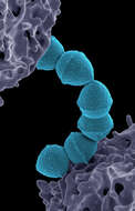
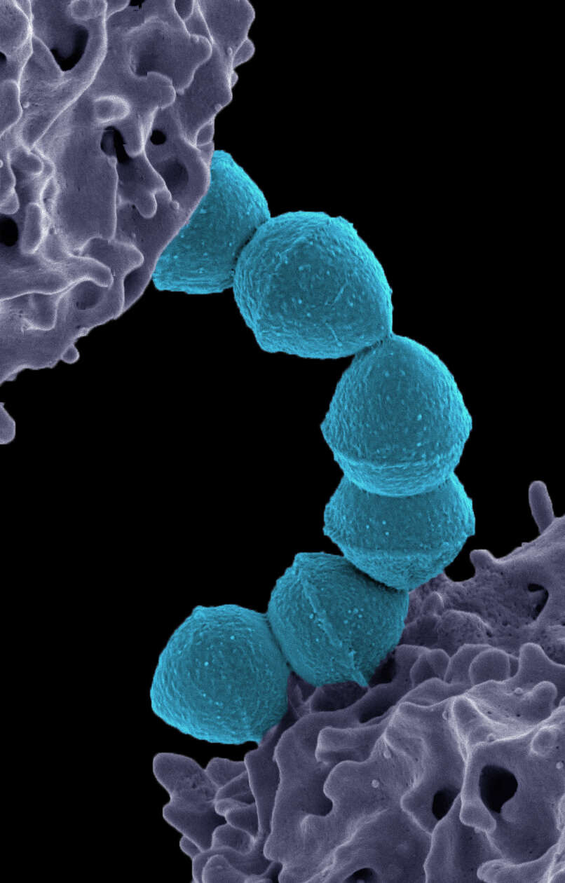
Summary.mw-parser-output table.commons-file-information-table,.mw-parser-output.fileinfotpl-type-information{border:1px solid #a2a9b1;background-color:#f8f9fa;padding:5px;font-size:95%;border-spacing:2px;box-sizing:border-box;margin:0;width:100%}.mw-parser-output table.commons-file-information-table>tbody>tr,.mw-parser-output.fileinfotpl-type-information>tbody>tr{vertical-align:top}.mw-parser-output table.commons-file-information-table>tbody>tr>td,.mw-parser-output table.commons-file-information-table>tbody>tr>th,.mw-parser-output.fileinfotpl-type-information>tbody>tr>td,.mw-parser-output.fileinfotpl-type-information>tbody>tr>th{padding:4px}.mw-parser-output.fileinfo-paramfield{background:#ccf;text-align:right;padding-right:0.4em;width:15%;font-weight:bold}.mw-parser-output.commons-file-information-table+table.commons-file-information-table,.mw-parser-output.commons-file-information-table+div.commons-file-information-table>table{border-top:0;padding-top:0;margin-top:-8px}@media only screen and (max-width:719px){.mw-parser-output table.commons-file-information-table,.mw-parser-output.commons-file-information-table.fileinfotpl-type-information{border-spacing:0;padding:0;word-break:break-word;width:100%!important}.mw-parser-output.commons-file-information-table>tbody,.mw-parser-output.fileinfotpl-type-information>tbody{display:block}.mw-parser-output.commons-file-information-table>tbody>tr>td,.mw-parser-output.commons-file-information-table>tbody>tr>th,.mw-parser-output.fileinfotpl-type-information>tbody>tr>td,.mw-parser-output.fileinfotpl-type-information>tbody>tr>th{padding:0.2em 0.4em;text-align:left;text-align:start}.mw-parser-output.commons-file-information-table>tbody>tr,.mw-parser-output.fileinfotpl-type-information>tbody>tr{display:flex;flex-direction:column}.mw-parser-output.commons-file-information-table+table.commons-file-information-table,.mw-parser-output.commons-file-information-table+div.commons-file-information-table>table{margin-top:-1px}.mw-parser-output.fileinfo-paramfield{box-sizing:border-box;flex:1 0 100%;width:100%}} Description: Colorized scanning electron micrograph of Group A Streptococcus (Streptococcus pyogenes) bacteria (teal) and a human neutrophil (purple). Credit: NIAID. Date: 29 December 2022, 18:22. Source:
Streptococcus Pyogenes (Group A Strep). Author:
NIAID.
-
-
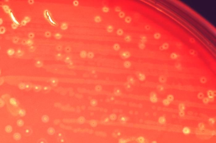
Description: English: Beta-haemolytic colonies of Streptococcus pyogenes on 5% shhep blood agar after 48 hours of incubation. Date: 26 September 2021, 14:02:35. Source: Own work. Author:
Ajay Kumar Chaurasiya.
-
-
-
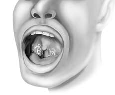
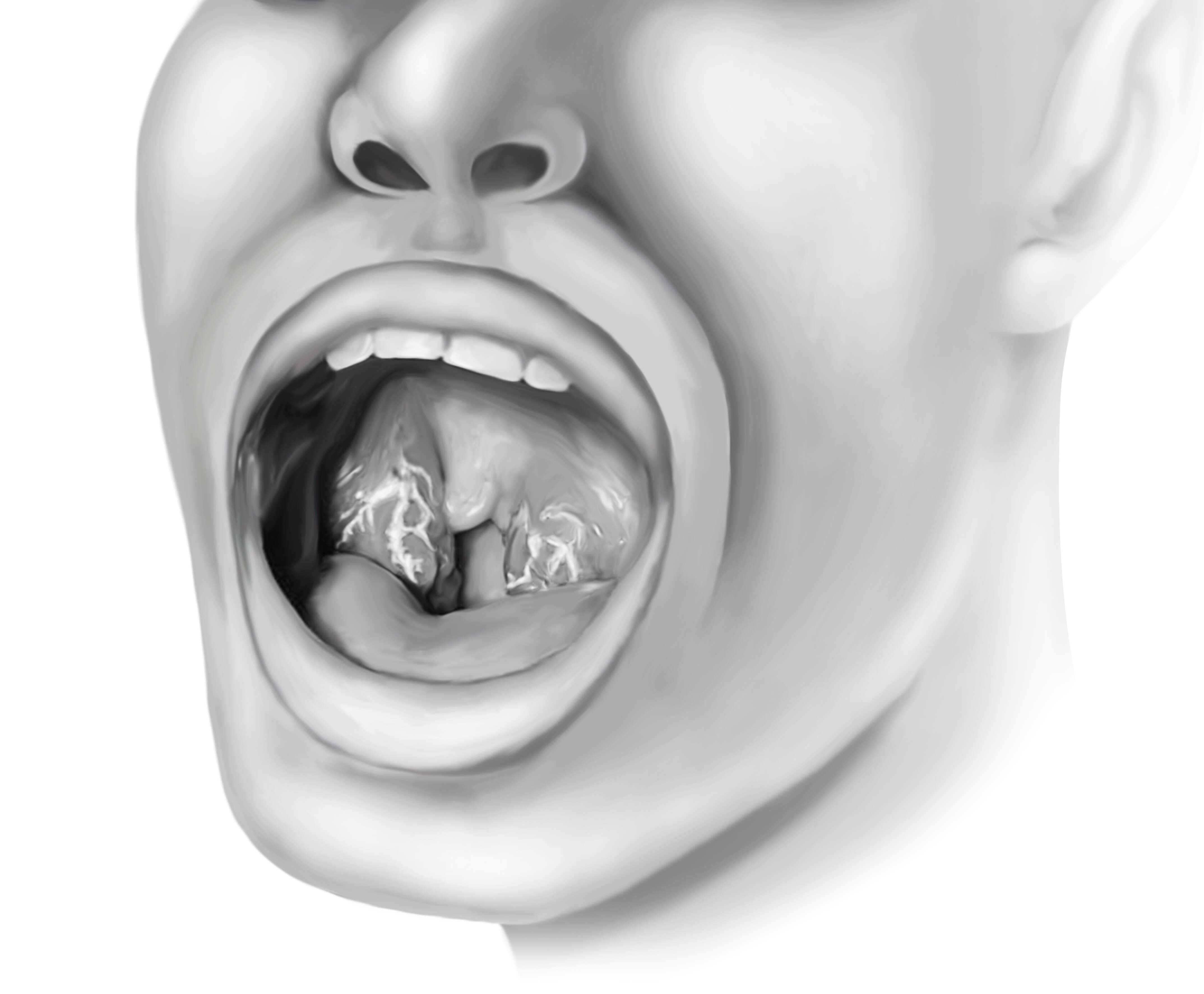
Description: English: A sketch of an oropharynx with Streptococcus Pyogenes induced pharyngitis. Date: 24 January 2019, 04:35:39. Source: Own work. Author:
Sam.bulloo.
-


