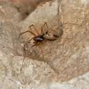
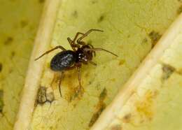
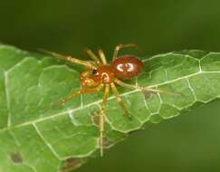
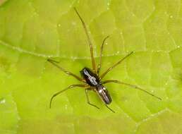
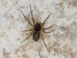
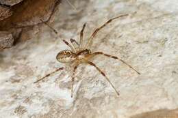
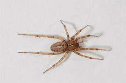
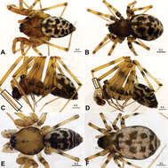
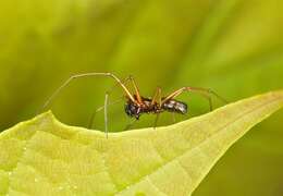
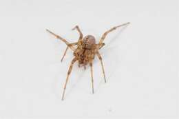
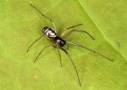
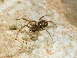
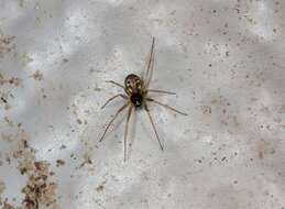
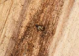
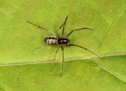
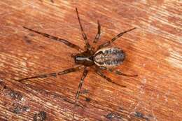
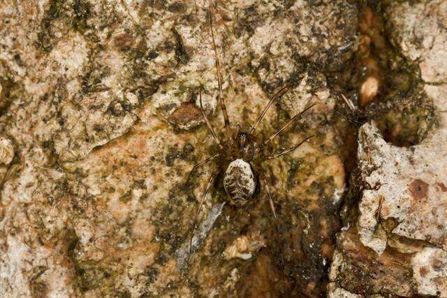
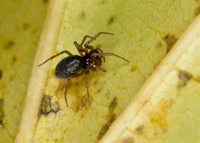
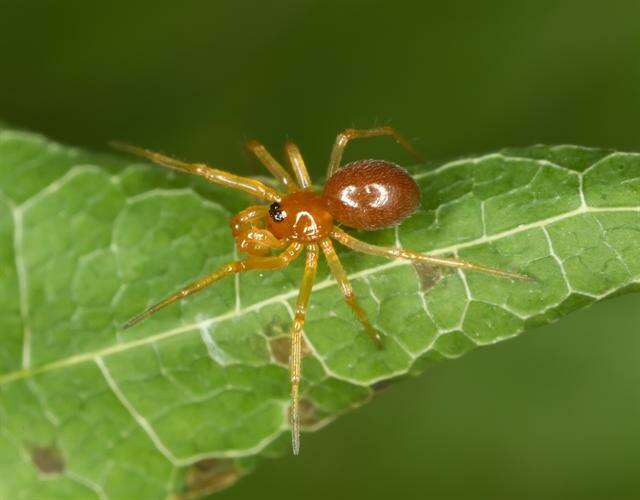
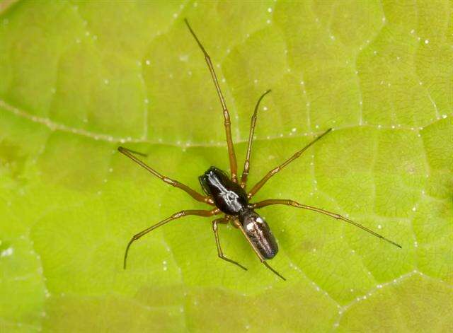
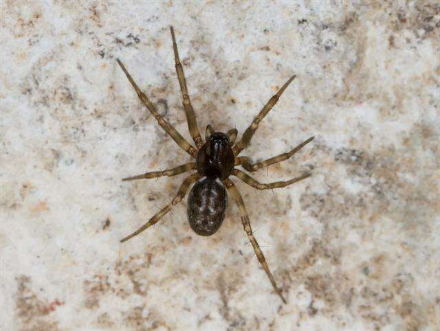
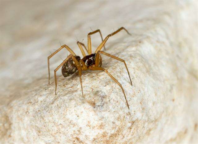
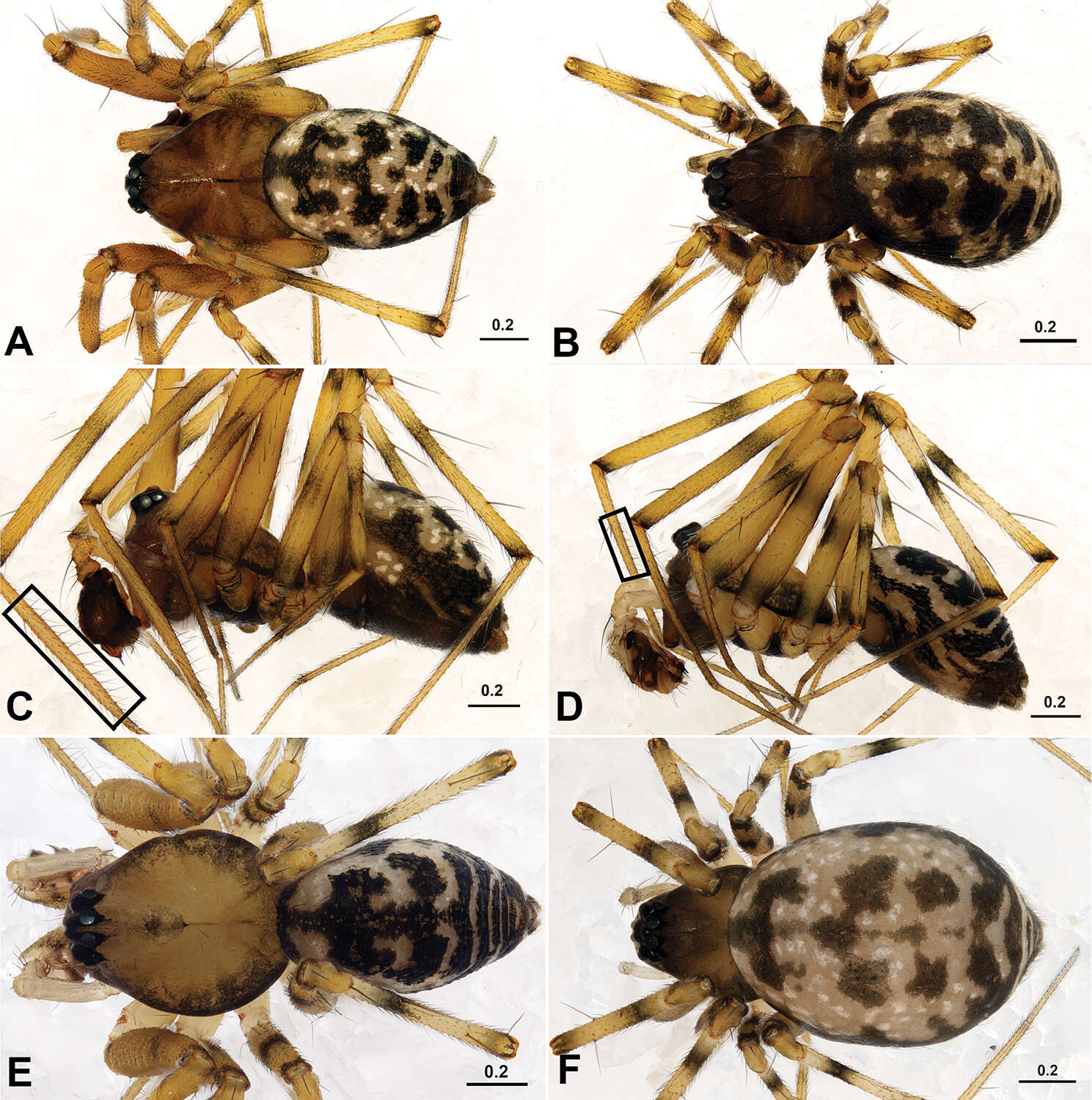
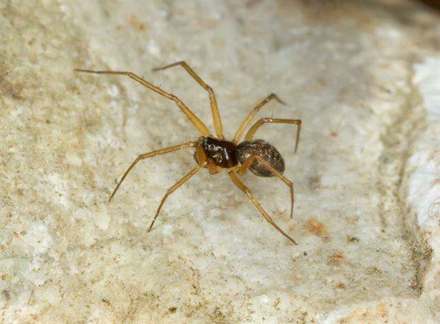
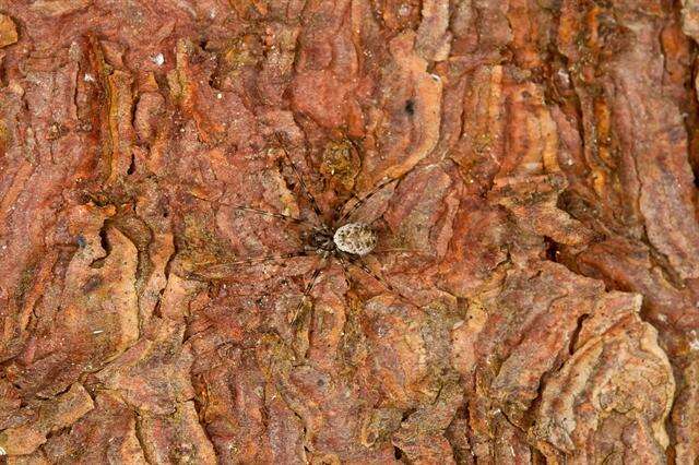
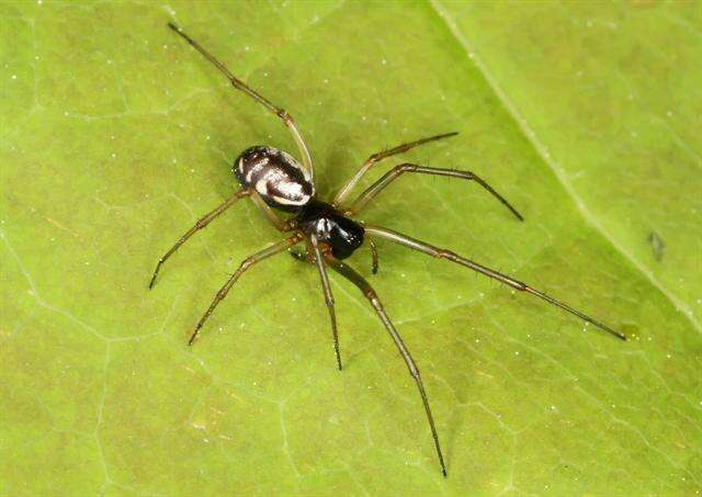
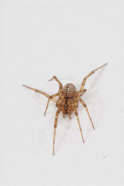
Figure 1.Acanoides beijingensis sp. n. (A–C) and Acanoides hengshanensis (D–F). A male, dorsal B female, dorsal C male, lateral, rectangle indicates ventrolateral rows of bristles on Mt I D male, lateral, rectangle indicates ventrolateral rows of bristles on Mt I E male, dorsal F female, dorsal. [Scale bars: mm].
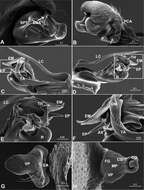
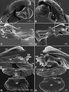
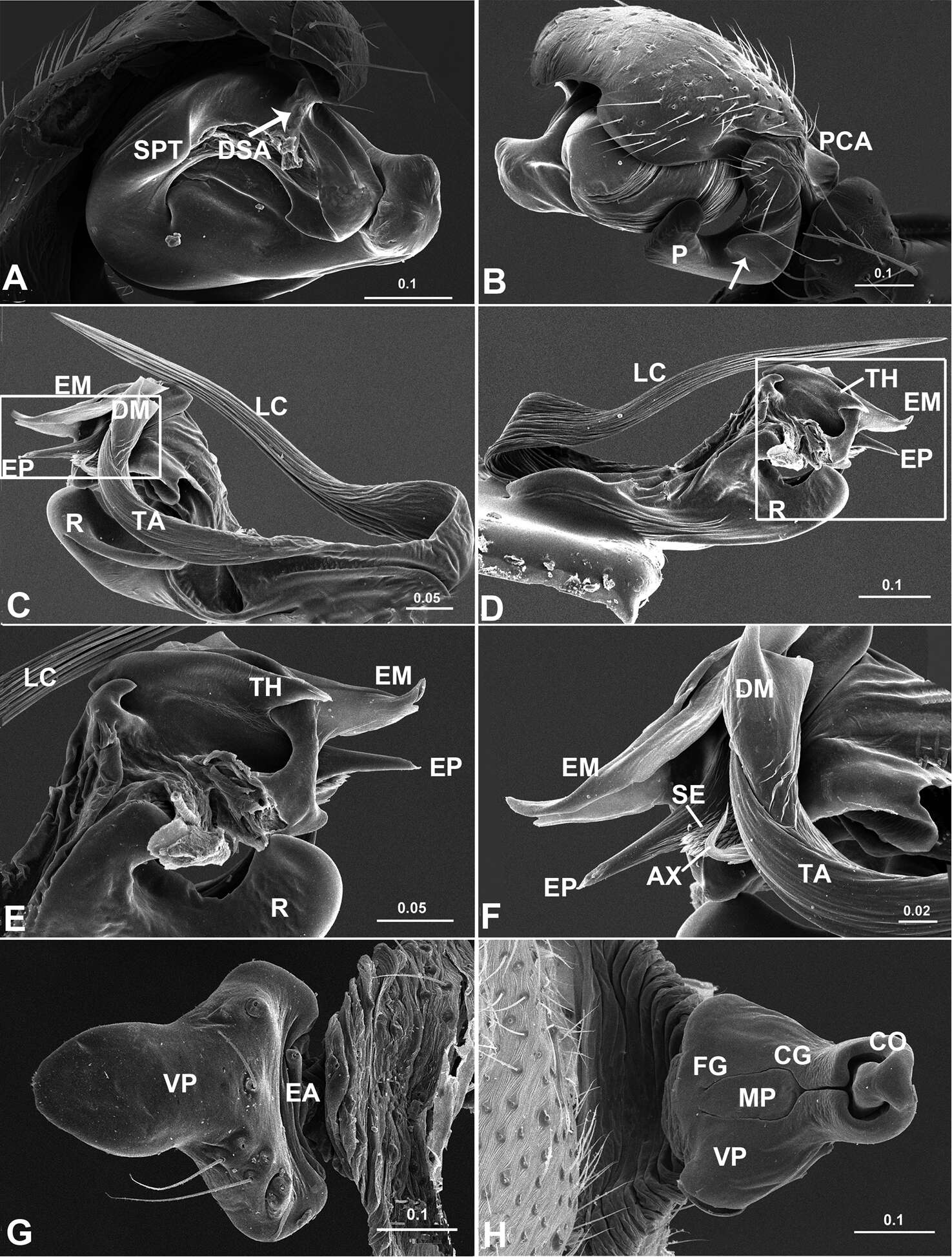
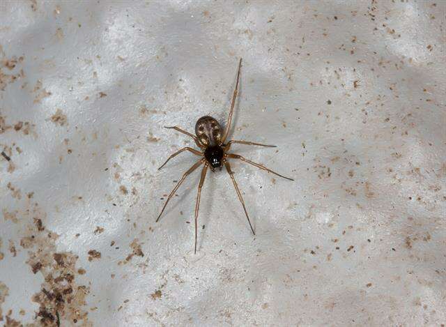
Figure 4.Acanoides beijingensis sp. n. A palp (embolic division removed), prolateral B palp, retrolateral, arrow indicates half rounded lateral tooth on paracymbium C embolic division, ventral D embolic division, dorsal E detail of D F detail of C G epigynum, ventral H epigynum, dorsal. AX apex of embolus; CG copulatory groove; CO copulatory opening; DM distal membrane of terminal apophysis; DSA distal suprategular apophysis; EA extensible area of epigynal basal part; EM embolic membrane; EP embolus proper; FG fertilization groove; LC lamella characteristica; MP median plate; P paracymbium; PCA proximal cymbial apophysis; R radix; S spermatheca; SE serrated area on embolus; SPT suprategulum; TA terminal apophysis; TH thumb of embolus; VP ventral plate. [Scale bars: mm].
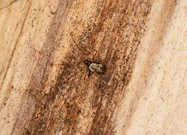
Figure 5.Acanoides hengshanensis. A palp (embolic division removed), prolateral B palp, retrolateral, arrow indicates pointed tooth on posterolateral margin C embolic division, ventral D embolic division, dorsal E detail of D F detail of C G epigynum, ventral H epigynum, dorsal. AX apex of embolus; CG copulatory groove; CO copulatory opening; DM distal membrane of terminal apophysis; EA extensible area of epigynal basal part; EM embolic membrane; EP embolus proper; FG fertilization groove; LC lamella characteristica; P paracymbium; PCA proximal cymbial apophysis; R radix; S spermatheca; SPT suprategulum; TA terminal apophysis; TH thumb of embolus; VP ventral plate. [Scale bars: mm].