-
Michael G. Rix, Mark S. Harvey
Zookeys
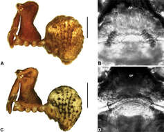
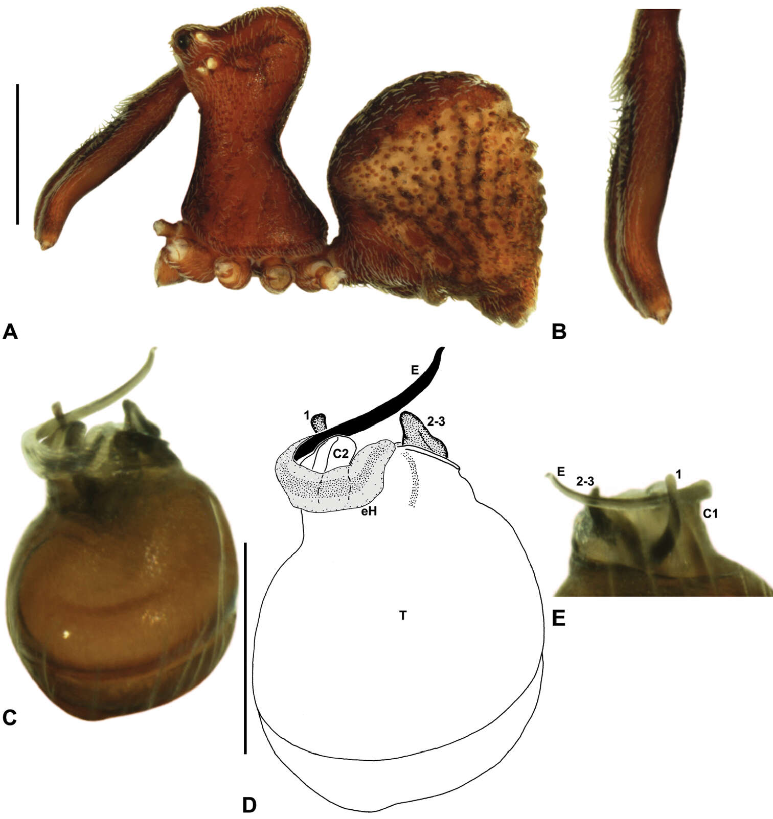
Figure 18.Zephyrarchaea porchi sp. n. A–E, Holotype male (MV K11581) from Bimbi Park, Otway Range, Victoria: A, cephalothorax and abdomen, lateral view; B, chelicerae, lateral view, showing accessory setae; C–D, pedipalpal bulb (partially expanded), retrolateral view; E, detail of distal tegular sclerites, prolateral view. C1–2 = conductor sclerites 1–2; E = embolus; eH = embolic (distal) haematodocha; T = tegulum; (TS)1–3 = tegular sclerites 1–3. Scale bars: A = 1.0 mm; D = 0.2 mm.
-
Michael G. Rix, Mark S. Harvey
Zookeys
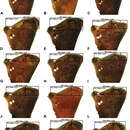
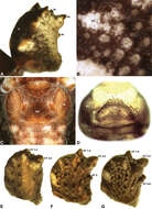
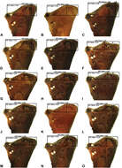
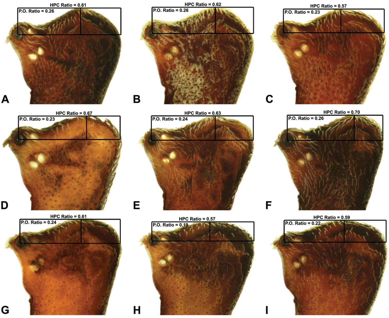
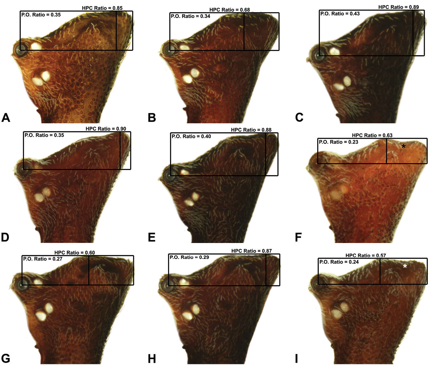
Figure 8.Lateral ‘head’ profiles of males of species of Austrarchaea from south-eastern Queensland and extreme north-eastern New South Wales (including the Border Ranges), showing variation in carapace shape as quantified by the post-ocular ratio (P.O. Ratio) and ratio of highest point of carapace relative to post-ocular length (HPC Ratio): A, holotype A. alani sp. n.; B, holotype A. aleenae sp. n.; C, holotype A. judyae sp. n.; D, holotype A. raveni sp. n.; E, holotype A. harmsi sp. n.; F, holotype A. clyneae sp. n.; G, holotype A. cunninghami sp. n.; H, holotype A. dianneae sp. n.; I, A. nodosa (Forster, 1956) (QMB S75416). Asterisks (*) denote concave depressions.
-
Michael G. Rix, Mark S. Harvey
Zookeys
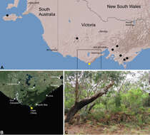
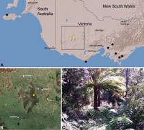
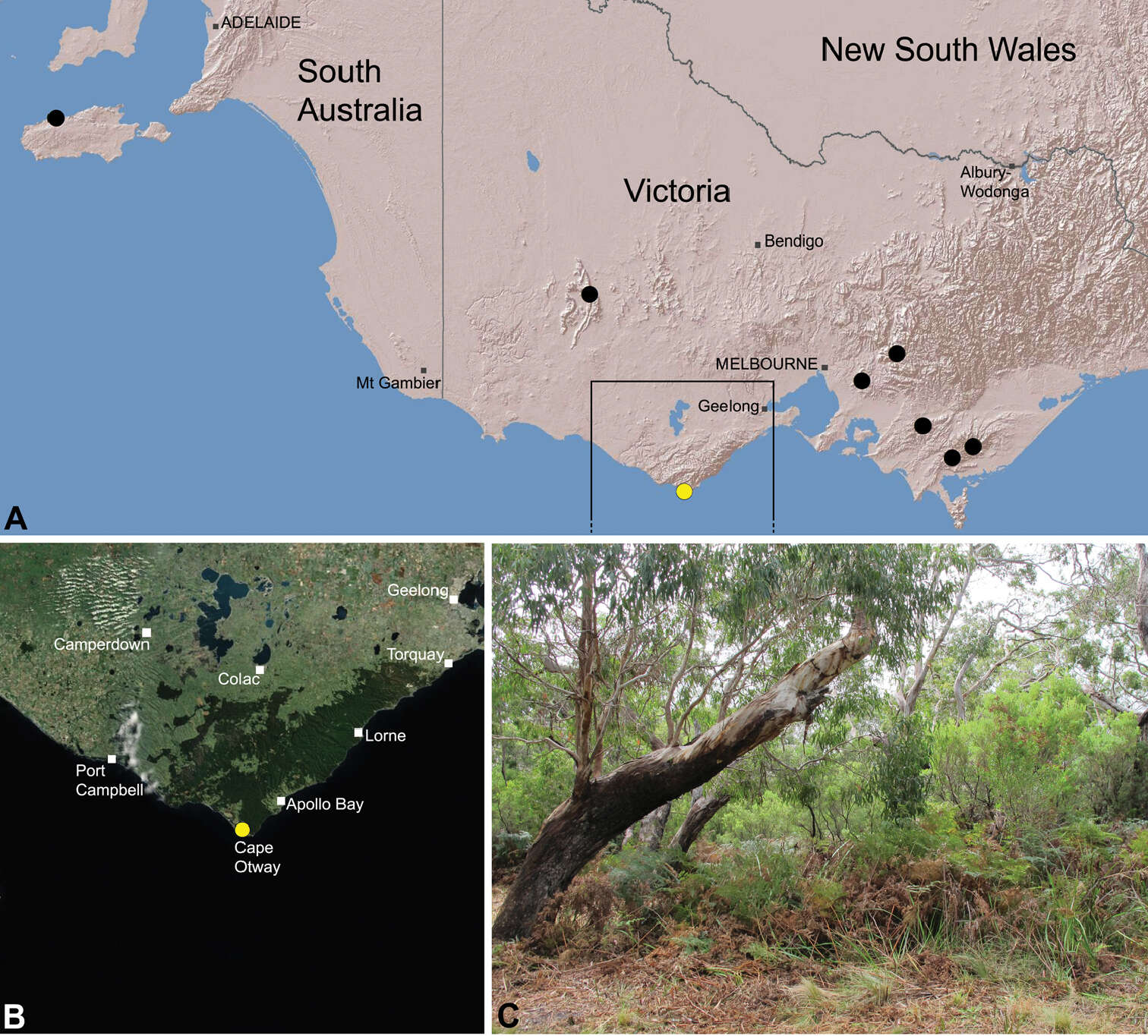
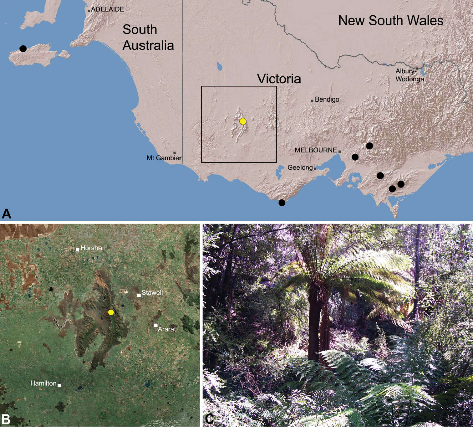
Figure 28.Zephyrarchaea porchi sp. n., distribution and habitat: A, topographic map showing the known distribution of Archaeidae in Victoria and South Australia, with collection localities for Zephyrarchaea porchi highlighted in yellow; B, satellite image showing detail of inset (A); C, bracken-rich eucalypt forest at the type locality – Bimbi Park, Otway Range, Victoria (March 2012). Image (C) by N. Porch, used with permission.
-
Michael G. Rix, Mark S. Harvey
Zookeys
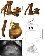
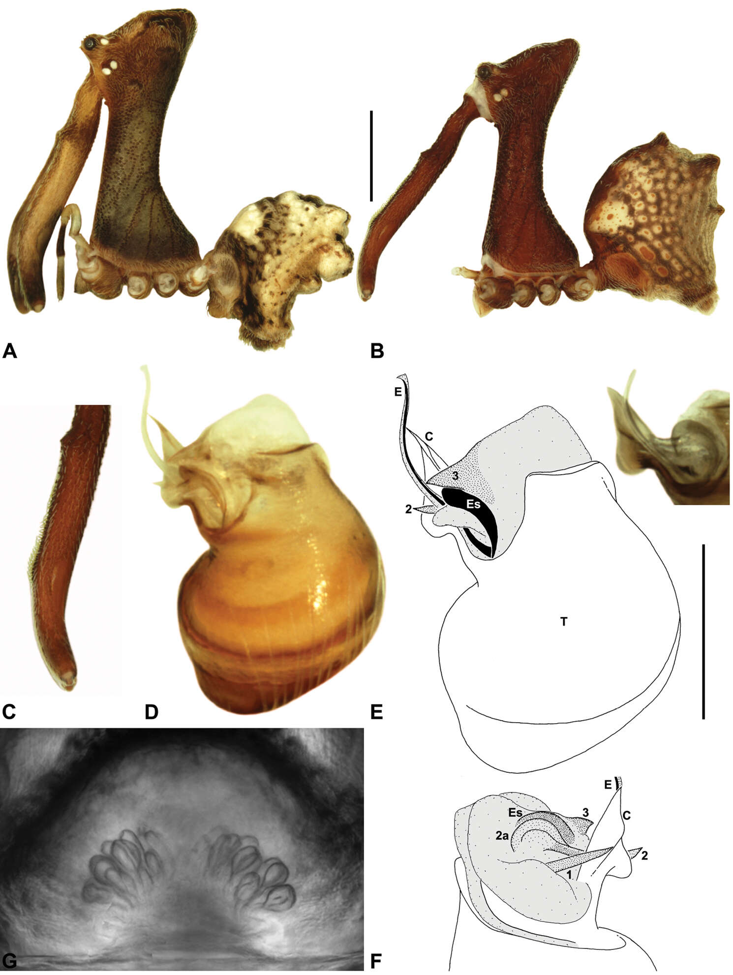
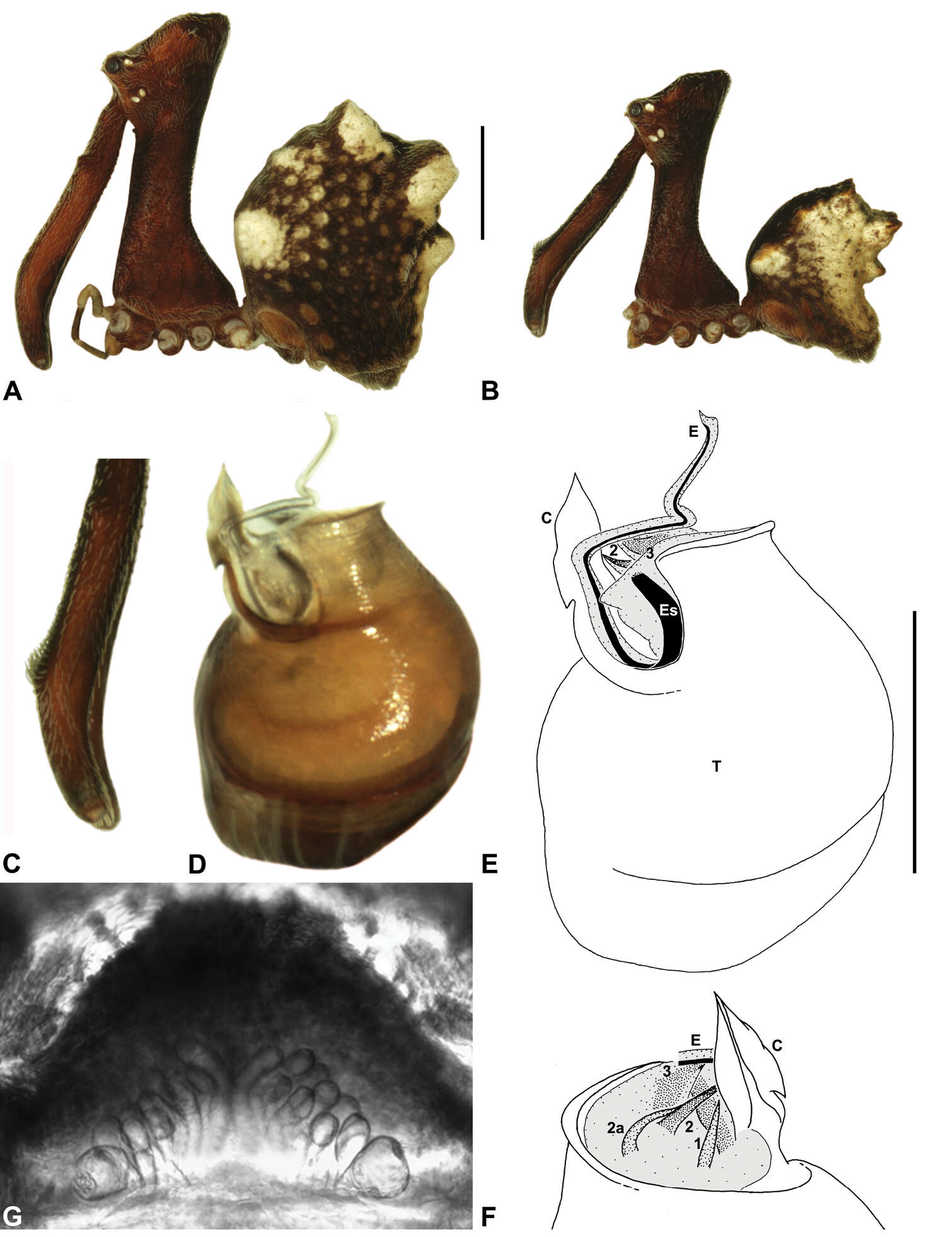
Figure 12.Austrarchaea cunninghami sp. n. A–B, Cephalothorax and abdomen, lateral view: A, allotype female (QMB S90183) from Main Range National Park, Queensland; B, holotype male (QMB S90184) from Main Range National Park, Queensland. C, Holotype male chelicerae, lateral view, showing accessory setae. D–F, Holotype male right pedipalp (flipped horizontal for inter-specific comparison): D–E, bulb, retrolateral view; F, detail of distal tegular sclerites, prodistal view. G, Allotype female internal genitalia, dorsal view. C = conductor; E = embolus; Es = embolic sclerite; T = tegulum; (TS)1–3 = tegular sclerites 1–3. Scale bars: A–B = 1.0 mm; E = 0.2 mm.
-
Michael G. Rix, Mark S. Harvey
Zookeys
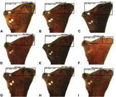
Figure 9.Lateral ‘head’ profiles of females of species of Zephyrarchaea, showing variation in carapace shape as quantified by the post-ocular ratio (P.O. Ratio) and ratio of highest point of carapace relative to post-ocular length (HPC Ratio) (see Fig. 8): A, allotype Zephyrarchaea vichickmani sp. n.; B, allotype Zephyrarchaea marae sp. n.; C, holotype Zephyrarchaea grayi sp. n.; D, holotype Zephyrarchaea austini sp. n.; E, Zephyrarchaea mainae (Platnick, 1991b); F, allotype Zephyrarchaea janineae sp. n.; G, holotype Zephyrarchaea robinsi (Harvey, 2002a); H, allotype Zephyrarchaea melindae sp. n.; I, allotype Zephyrarchaea barrettae sp. n.
-
Michael G. Rix, Mark S. Harvey
Zookeys
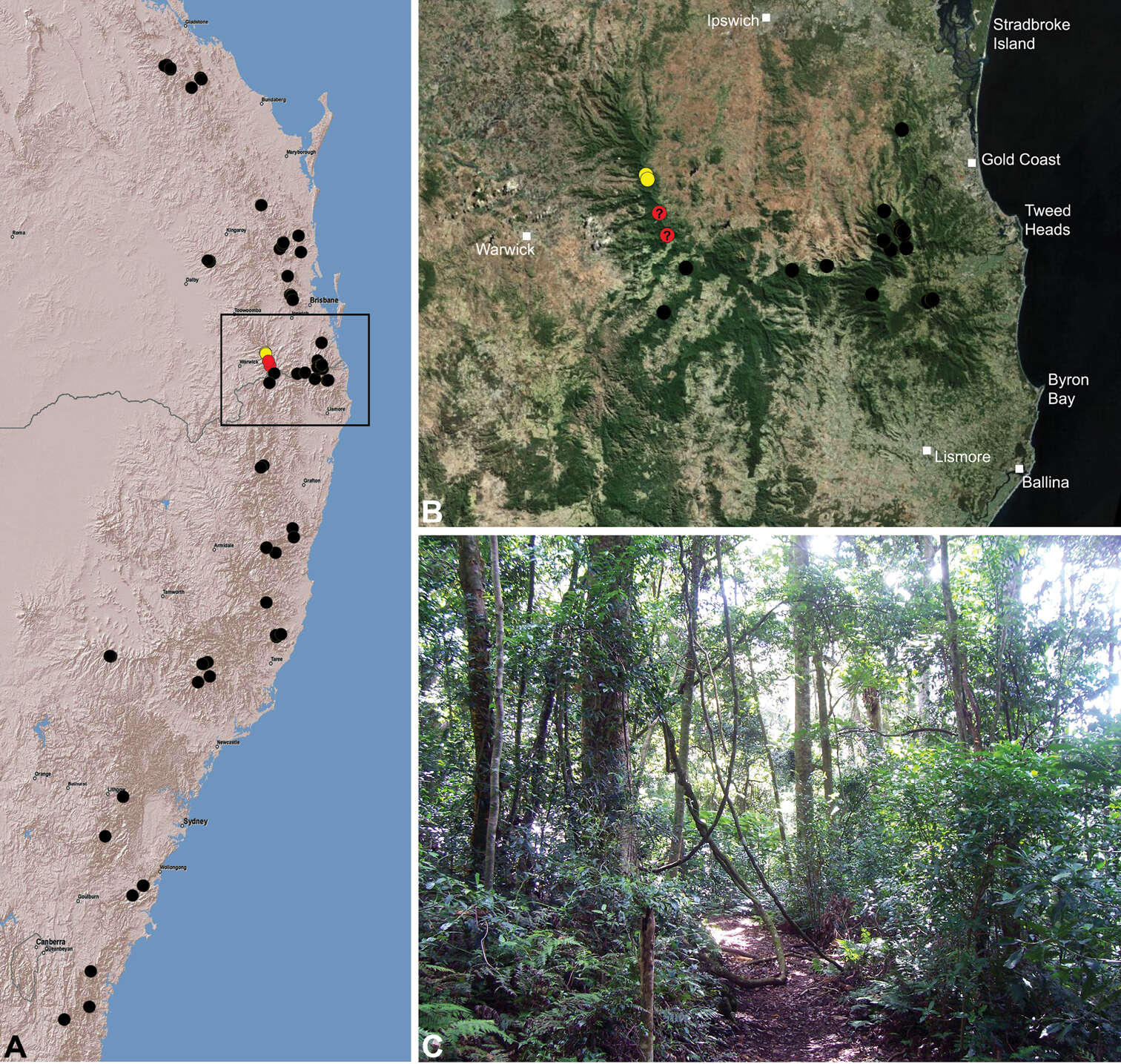
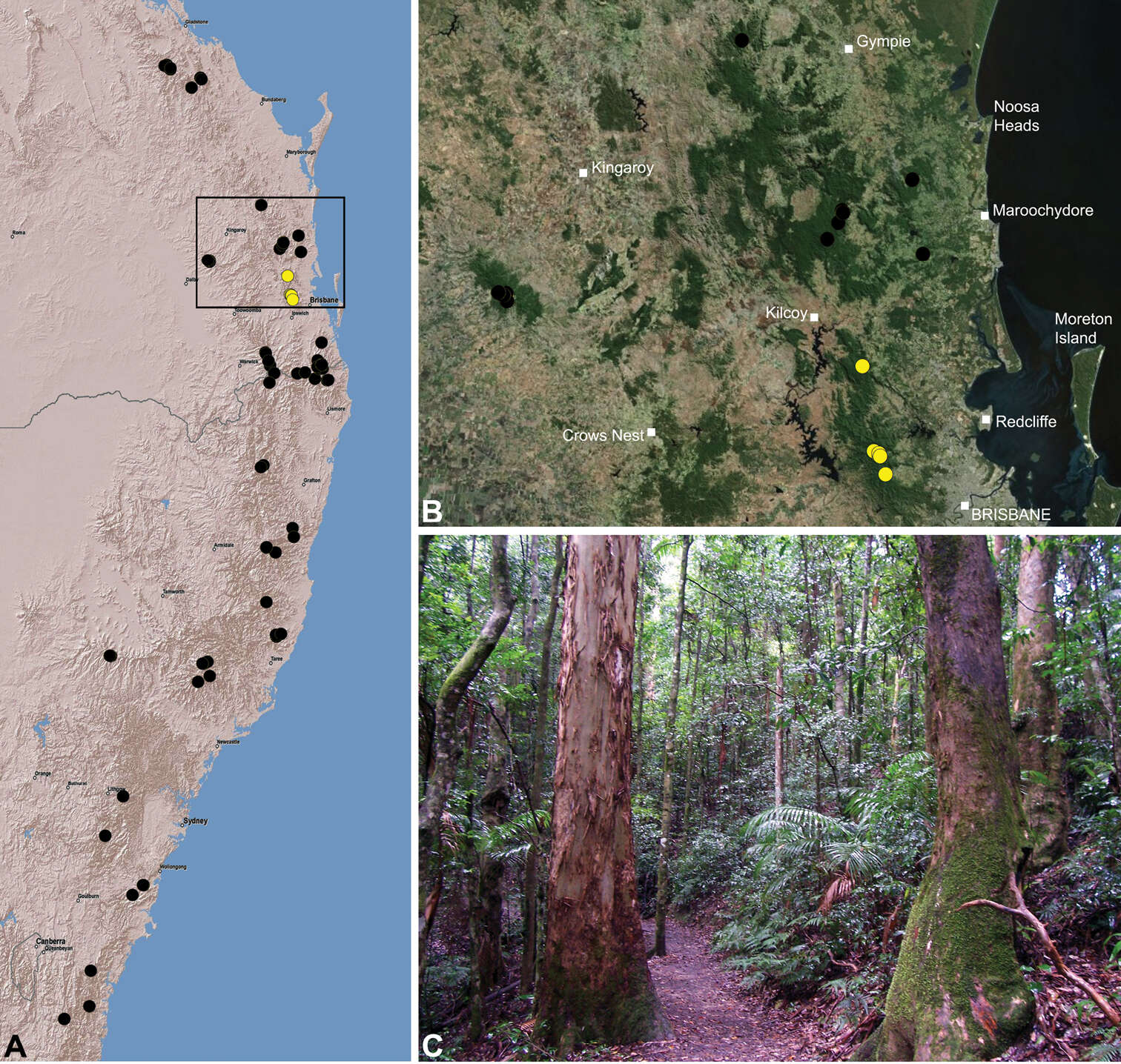
Figure 30.Austrarchaea cunninghami sp. n., distribution and habitat: A, topographic map showing the known distribution of Archaeidae in south-eastern Queensland and eastern New South Wales, with collection localities for A. cunninghami highlighted in yellow (red highlighted localities denote juvenile specimens of tentative identification); B, satellite image showing detail of inset (A); C, subtropical rainforest at the type locality – Cunningham’s Gap, Main Range National Park, Queensland (April 2010). Image (C) by M. Rix.
-
Michael G. Rix, Mark S. Harvey
Zookeys
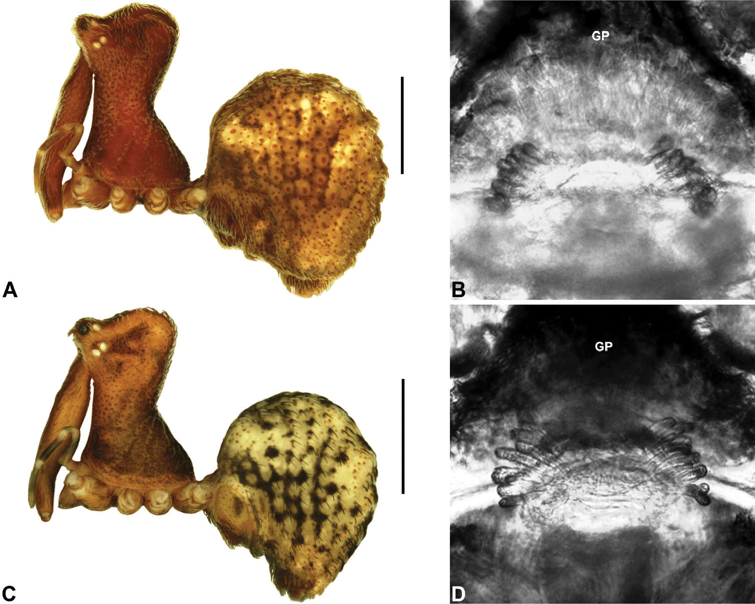
Figure 19.Zephyrarchaea grayi sp. n. and Zephyrarchaea austini sp. n. A–B, holotype female Zephyrarchaea grayi sp. n. (AMS KS109448) from Delley’s Dell, Grampians National Park, Victoria: A, cephalothorax and abdomen, lateral view; B, internal genitalia, antero-dorsal view. C–D, holotype female Zephyrarchaea austini (SAM NN28000) from Western River Wilderness Protection Area, Kangaroo Island, South Australia: C, cephalothorax and abdomen, lateral view; D, internal genitalia, antero-dorsal view. GP = genital plate. Scale bars: A, C = 1.0 mm.
-
Michael G. Rix, Mark S. Harvey
Zookeys
Figure 8.Lateral ‘head’ profiles of males of species of Austrarchaea from south-eastern Queensland and extreme north-eastern New South Wales (including the Border Ranges), showing variation in carapace shape as quantified by the post-ocular ratio (P.O. Ratio) and ratio of highest point of carapace relative to post-ocular length (HPC Ratio): A, holotype A. alani sp. n.; B, holotype A. aleenae sp. n.; C, holotype A. judyae sp. n.; D, holotype A. raveni sp. n.; E, holotype A. harmsi sp. n.; F, holotype A. clyneae sp. n.; G, holotype A. cunninghami sp. n.; H, holotype A. dianneae sp. n.; I, A. nodosa (Forster, 1956) (QMB S75416). Asterisks (*) denote concave depressions.
-
Michael G. Rix, Mark S. Harvey
Zookeys
Figure 29.Zephyrarchaea grayi sp. n., distribution and habitat: A, topographic map showing the known distribution of Archaeidae in Victoria and South Australia, with collection localities for Zephyrarchaea grayi highlighted in yellow; B, satellite image showing detail of inset (A); C, wet sclerophyll forest at the type locality – Delley’s Dell, Grampians National Park, Victoria (March 2010). Image (C) by M. Rix.
-
Michael G. Rix, Mark S. Harvey
Zookeys
Figure 13.Austrarchaea clyneae sp. n. A–E, Holotype male (QMB S20425) from Mount Clunie National Park, New South Wales: A, cephalothorax and abdomen, lateral view; B, chelicerae, lateral view, showing accessory setae; C–D, pedipalpal bulb, retrolateral view; E, detail of distal tegular sclerites, prodistal view. C = conductor; E = embolus; Es = embolic sclerite; T = tegulum; (TS)1–3 = tegular sclerites 1–3. Scale bars: A = 1.0 mm; D = 0.2 mm.
-
Michael G. Rix, Mark S. Harvey
Zookeys
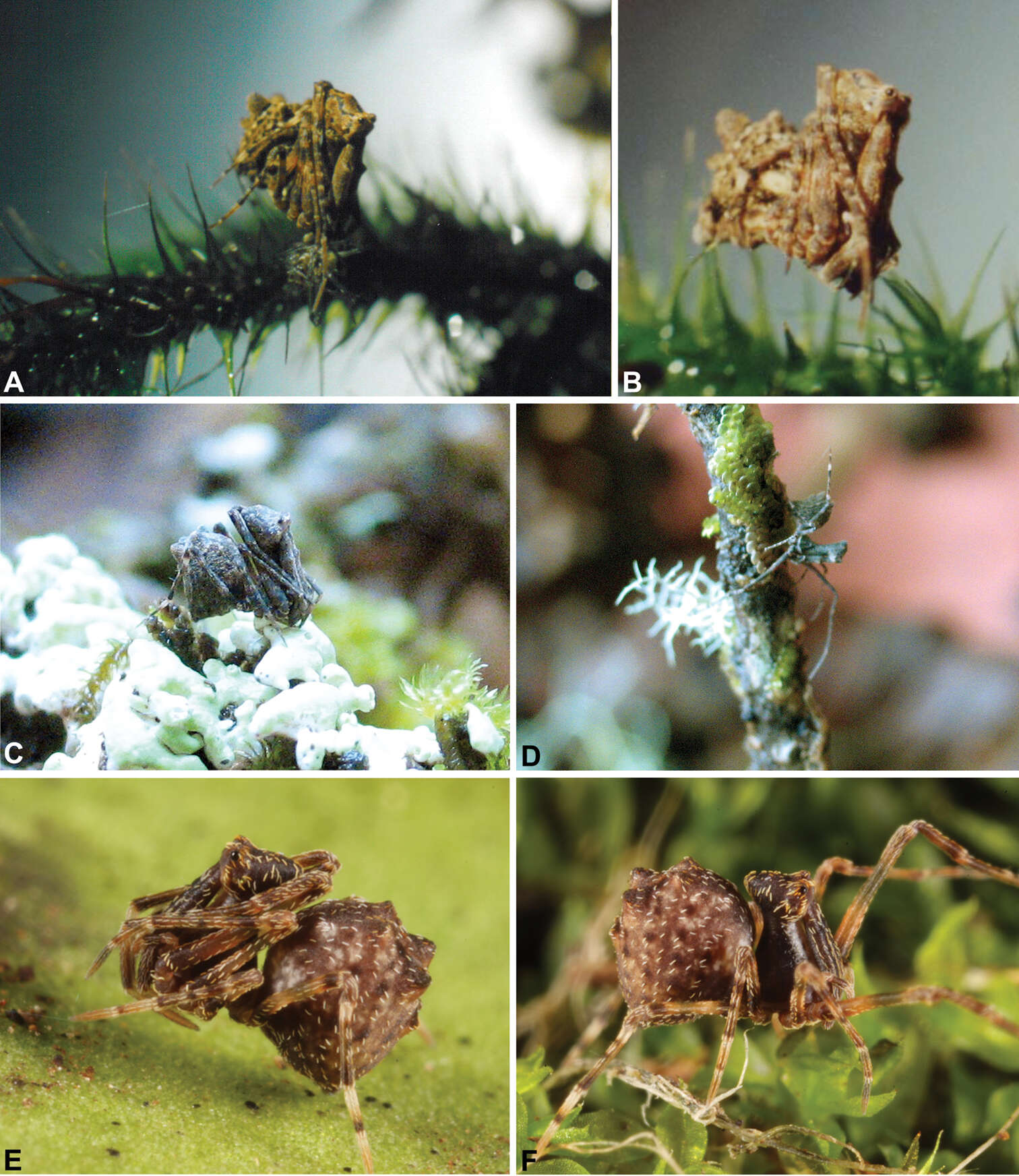
Figure 1.Habitus images of live Archaeidae from mid-eastern Australia: A–B, female Austrarchaea nodosa (Forster, 1956) from Binna Burra, Lamington National Park, Queensland; C–D, female A. mascordi sp. n. from Coolah Tops National Park, New South Wales; E–F, juvenile A. raveni sp. n. from Mount Glorious, Queensland. Images A–D by M. Rix; images E–F by Greg Anderson, used with permission.
-
Michael G. Rix, Mark S. Harvey
Zookeys
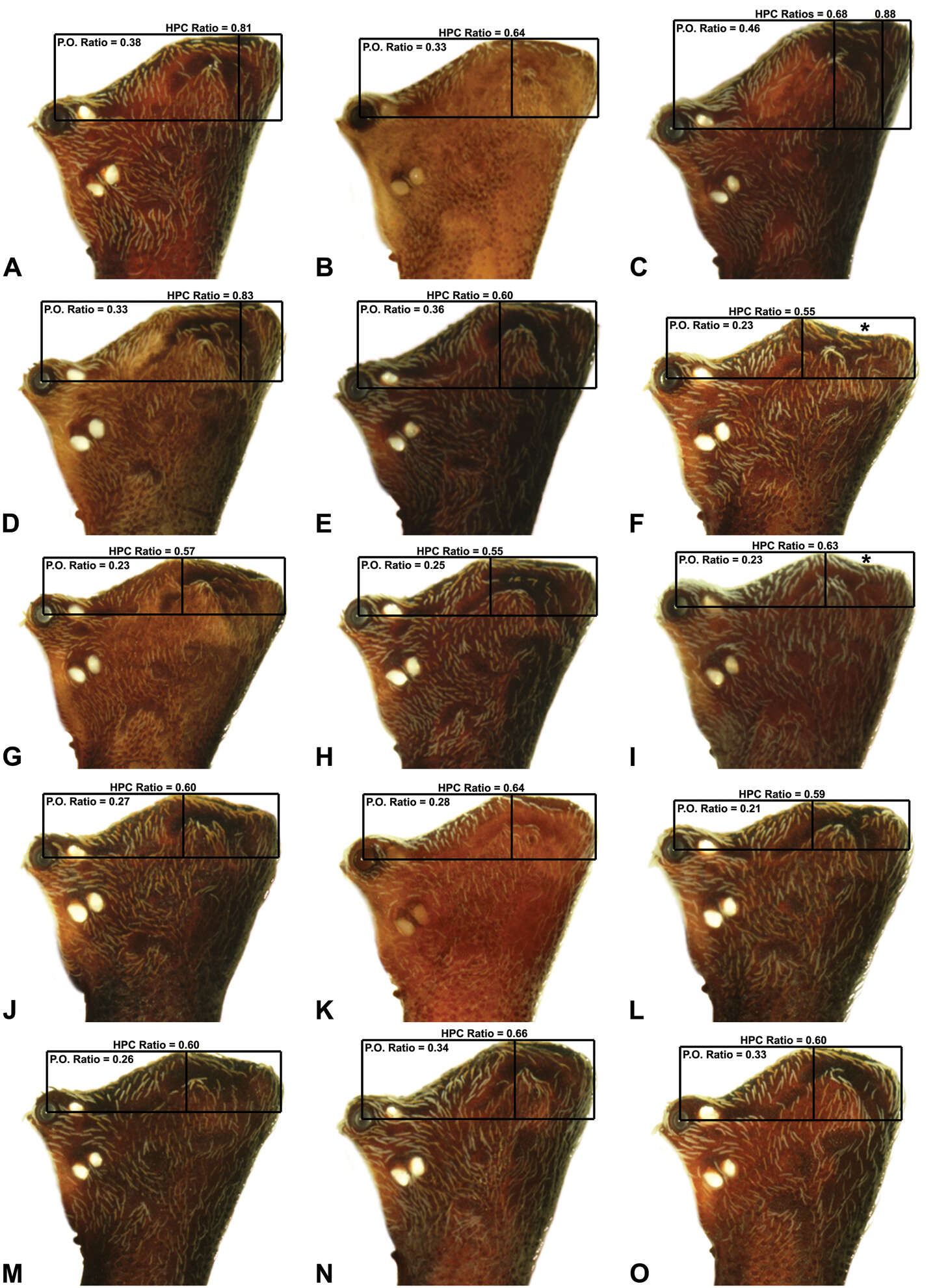
Figure 7.Lateral ‘head’ profiles of females of species of Austrarchaea from mid-eastern Australia, showing variation in carapace shape as quantified by the post-ocular ratio (P.O. Ratio) and ratio of highest point of carapace relative to post-ocular length (HPC Ratio): A, allotype A. alani sp. n.; B, allotype A. aleenae sp. n.; C, allotype A. judyae sp. n.; D, allotype A. raveni sp. n.; E, allotype A. harmsi sp. n.; F, allotype A. monteithi sp. n.; G, allotype A. cunninghami sp. n.; H, allotype A. dianneae sp. n.; I, A. nodosa (Forster, 1956); J, allotype A. platnickorum sp. n.; K, allotype A. binfordae sp. n.; L, A. milledgei sp. n. (WAM T112568); M, allotype A. mascordi sp. n.; N, allotype A. smithae sp. n.; O, allotype A. mcguiganae sp. n. Asterisks (*) denote concave depressions.
-
Michael G. Rix, Mark S. Harvey
Zookeys
Figure 8.Lateral ‘head’ profiles of males of species of Austrarchaea from south-eastern Queensland and extreme north-eastern New South Wales (including the Border Ranges), showing variation in carapace shape as quantified by the post-ocular ratio (P.O. Ratio) and ratio of highest point of carapace relative to post-ocular length (HPC Ratio): A, holotype A. alani sp. n.; B, holotype A. aleenae sp. n.; C, holotype A. judyae sp. n.; D, holotype A. raveni sp. n.; E, holotype A. harmsi sp. n.; F, holotype A. clyneae sp. n.; G, holotype A. cunninghami sp. n.; H, holotype A. dianneae sp. n.; I, A. nodosa (Forster, 1956) (QMB S75416). Asterisks (*) denote concave depressions.
-
Michael G. Rix, Mark S. Harvey
Zookeys
Figure 14.Austrarchaea raveni sp. n. A–B, Cephalothorax and abdomen, lateral view: A, allotype female (QMB S90192) from D’Aguilar National Park, Queensland; B, holotype male (QMB S90193) from D’Aguilar National Park, Queensland. C, Holotype male chelicerae, lateral view, showing accessory setae. D–F, Holotype male pedipalp (partially expanded): D–E, bulb, retrolateral view (inset shows conductor and embolus on unexpanded pedipalp of male from Mt Mee Forest Reserve, Queensland); F, detail of distal tegular sclerites, prodistal view. G, Allotype female internal genitalia, dorsal view. C = conductor; E = embolus; Es = embolic sclerite; T = tegulum; (TS)1–3 = tegular sclerites 1–3. Scale bars: A–B = 1.0 mm; E = 0.2 mm.
-
Michael G. Rix, Mark S. Harvey
Zookeys
Figure 32.Austrarchaea raveni sp. n., distribution and habitat: A, topographic map showing the known distribution of Archaeidae in south-eastern Queensland and eastern New South Wales, with collection localities for A. raveni highlighted in yellow; B, satellite image showing detail of inset (A); C, subtropical rainforest near the type locality – Mount Glorious, D’Aguilar National Park, Queensland (May 2010). Image (C) by M. Rix.
-
Michael G. Rix, Mark S. Harvey
Zookeys
Figure 4.Carapace morphology of Austrarchaea species. A–E, A. alani sp. n.: A, male pars cephalica, frontal view, showing dorsal ‘head’ region, posterior horns (H) and cheliceral foramen (CF); B, female pars cephalica, antero-lateral view, showing ocular bulge (OB), cheliceral foramen (CF) and division of pars cephalica into ‘head’ and ‘neck’ regions; C, male pars thoracica, ‘neck’ and fused cheliceral diastema (fCD), antero-lateral view; D, female chelicerae and peg teeth, frontal view; E, male ‘neck’, lateral view, showing setose tubercles (sT). F–G, A. judyae sp. n.: F, male chelicerae, lateral view, showing accessory setae (AS) and ectal stridulatory file (SF); G, detail of female posterior pars cephalica, lateral view, showing field of densely granulate cuticle.
-
Michael G. Rix, Mark S. Harvey
Zookeys
Figure 5.Abdominal morphology of Austrarchaea species. A–C, A. judyae sp. n.: A, male abdomen, antero-lateral view, showing dorsal scute (S) and additional dorsal sclerites (ds); B, detail of female abdomen, lateral view, showing subcuticular guanine crystals (GC) and concentric arrangements of setae around sclerotic spots (ss); C, female epigastric region, ventral view, showing setose book lung covers (BL) and genital plate (GP). D, Cleared epigastric region of female A. nodosa (Forster), postero-ventral view, showing position of clustered spermathecae under posterior rim of genital plate. E–G, Female abdomens, postero-lateral view, showing arrangement of dorsal hump-like tubercles (HT) in different taxa: E, A. sp. nr. daviesae (QMB S72989, from Mount Bartle Frere, NE. Queensland); F, A. monteithi sp. n.; G, A. aleenae sp. n. Note the presence of only a single posterior hump-like tubercle (HT 5) in A. monteithi.
-
Michael G. Rix, Mark S. Harvey
Zookeys
Figure 7.Lateral ‘head’ profiles of females of species of Austrarchaea from mid-eastern Australia, showing variation in carapace shape as quantified by the post-ocular ratio (P.O. Ratio) and ratio of highest point of carapace relative to post-ocular length (HPC Ratio): A, allotype A. alani sp. n.; B, allotype A. aleenae sp. n.; C, allotype A. judyae sp. n.; D, allotype A. raveni sp. n.; E, allotype A. harmsi sp. n.; F, allotype A. monteithi sp. n.; G, allotype A. cunninghami sp. n.; H, allotype A. dianneae sp. n.; I, A. nodosa (Forster, 1956); J, allotype A. platnickorum sp. n.; K, allotype A. binfordae sp. n.; L, A. milledgei sp. n. (WAM T112568); M, allotype A. mascordi sp. n.; N, allotype A. smithae sp. n.; O, allotype A. mcguiganae sp. n. Asterisks (*) denote concave depressions.
-
Michael G. Rix, Mark S. Harvey
Zookeys
Figure 8.Lateral ‘head’ profiles of males of species of Austrarchaea from south-eastern Queensland and extreme north-eastern New South Wales (including the Border Ranges), showing variation in carapace shape as quantified by the post-ocular ratio (P.O. Ratio) and ratio of highest point of carapace relative to post-ocular length (HPC Ratio): A, holotype A. alani sp. n.; B, holotype A. aleenae sp. n.; C, holotype A. judyae sp. n.; D, holotype A. raveni sp. n.; E, holotype A. harmsi sp. n.; F, holotype A. clyneae sp. n.; G, holotype A. cunninghami sp. n.; H, holotype A. dianneae sp. n.; I, A. nodosa (Forster, 1956) (QMB S75416). Asterisks (*) denote concave depressions.
-
Michael G. Rix, Mark S. Harvey
Zookeys
Figure 15.Austrarchaea judyae sp. n. A–B, Cephalothorax and abdomen, lateral view: A, allotype female (QMB S90191) from Conondale National Park, Queensland; B, holotype male (QMB S90190) from Conondale National Park, Queensland. C, Holotype male chelicerae, lateral view, showing accessory setae. D–F, Holotype male pedipalp: D–E, bulb, retrolateral view; F, detail of distal tegular sclerites, prodistal view. G, Allotype female internal genitalia, dorsal view. C = conductor; E = embolus; Es = embolic sclerite; T = tegulum; (TS)1–3 = tegular sclerites 1–3. Scale bars: A–B = 1.0 mm; E = 0.2 mm.
-
Michael G. Rix, Mark S. Harvey
Zookeys
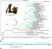
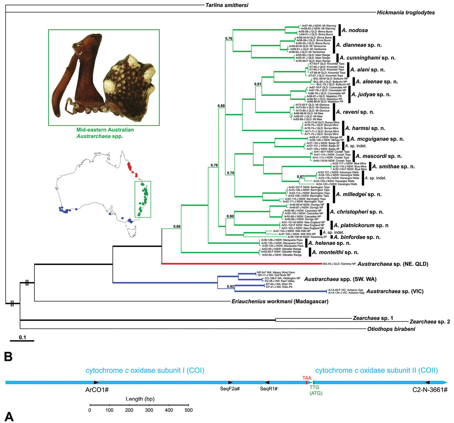
Figure 3.Molecular phylogenetic data analysed as part of this study. A, Schematic map of the mitochondrial cytochrome c oxidase subunit I–II (COI–COII) gene complex in Archaeidae and other basal Araneomorphae, showing (i) the position of primers used to amplify and sequence 1.6 kilobases of mtDNA, and (ii) the inferred stop and initiation codons for COI and COII, respectively. Note the centralised, overlapping position of the two internal sequencing primer sites (SeqF2a/SeqR1), and the TTG initiation codon for COII, present in all but one of the spider species sequenced for this study. B, Majority-rule consensus tree with re-estimated branch lengths, resulting from a combined, gene-partitioned Bayesian analysis of the COI–COII mtDNA data. Thickened branches represent clades with >95% posterior probability support, and individual support values are shown above other nodes.
-
Michael G. Rix, Mark S. Harvey
Zookeys
Figure 1.Habitus images of live Archaeidae from mid-eastern Australia: A–B, female Austrarchaea nodosa (Forster, 1956) from Binna Burra, Lamington National Park, Queensland; C–D, female A. mascordi sp. n. from Coolah Tops National Park, New South Wales; E–F, juvenile A. raveni sp. n. from Mount Glorious, Queensland. Images A–D by M. Rix; images E–F by Greg Anderson, used with permission.
-
Michael G. Rix, Mark S. Harvey
Zookeys
Figure 8.Lateral ‘head’ profiles of males of species of Austrarchaea from south-eastern Queensland and extreme north-eastern New South Wales (including the Border Ranges), showing variation in carapace shape as quantified by the post-ocular ratio (P.O. Ratio) and ratio of highest point of carapace relative to post-ocular length (HPC Ratio): A, holotype A. alani sp. n.; B, holotype A. aleenae sp. n.; C, holotype A. judyae sp. n.; D, holotype A. raveni sp. n.; E, holotype A. harmsi sp. n.; F, holotype A. clyneae sp. n.; G, holotype A. cunninghami sp. n.; H, holotype A. dianneae sp. n.; I, A. nodosa (Forster, 1956) (QMB S75416). Asterisks (*) denote concave depressions.