-
Carlos Perafán, Fernando Pérez-Miles
Zookeys
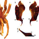
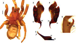
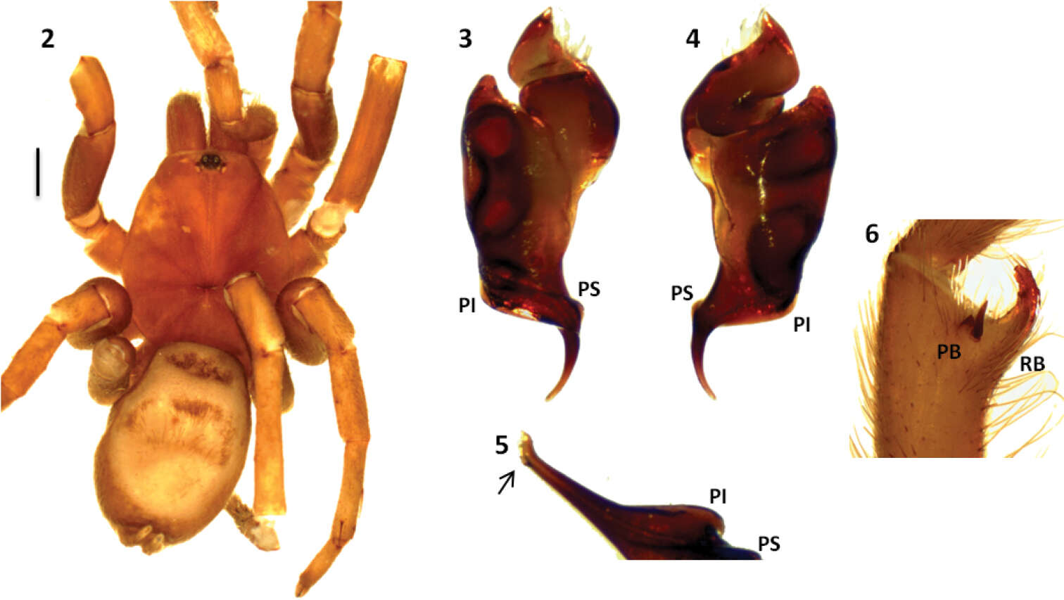
Figures 2–6.Melloleitaoina crassifemur. 2 male holotype, dorsal view 3–5 left palpal bulb, 3 prolateral view 4 retrolateral view 5 detail of apex widened 6 left tibial apophysis (subapical spine on retrolateral branch RB lost). Scale bar = 1 mm.
-
Carlos Perafán, Fernando Pérez-Miles
Zookeys
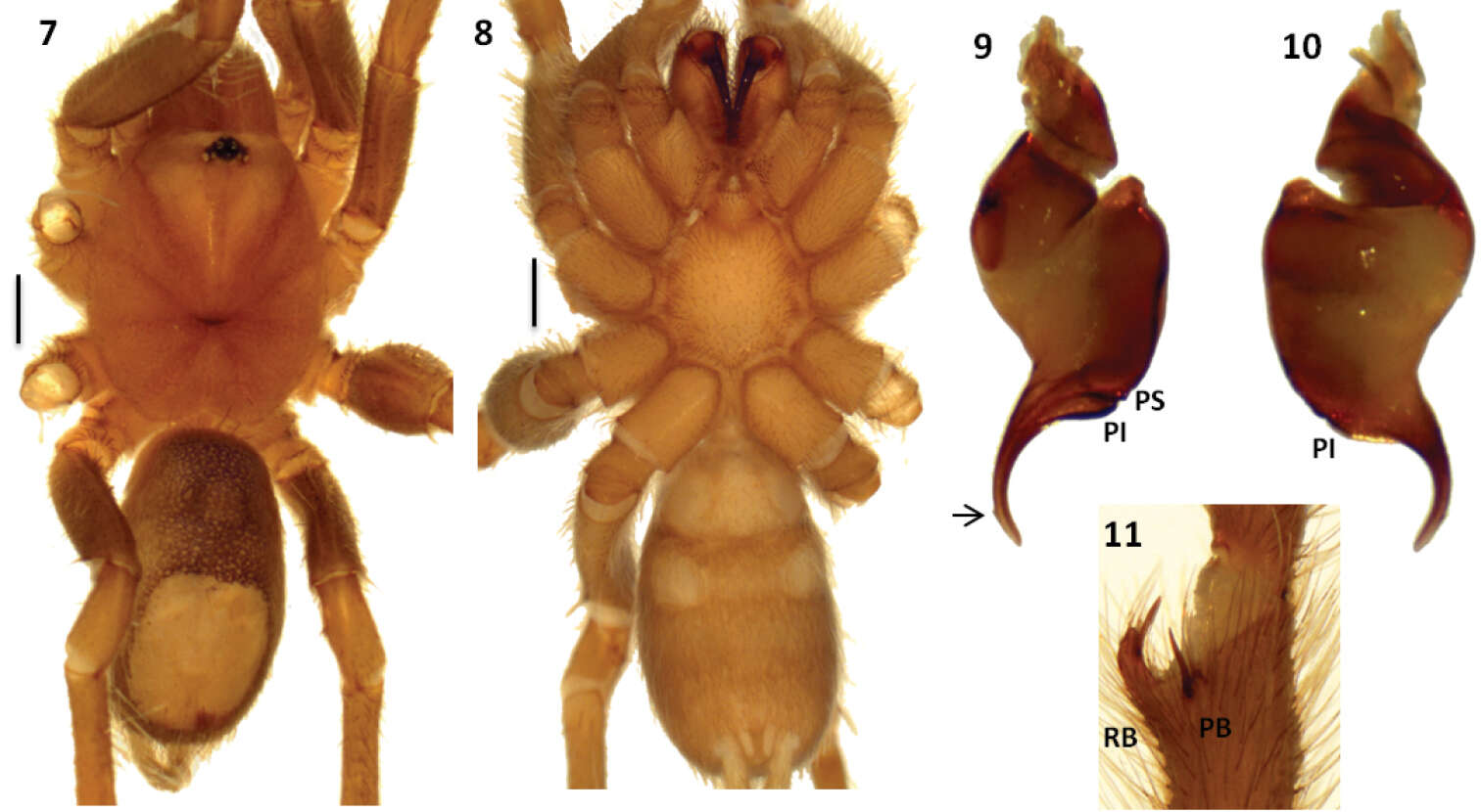
Figures 7–11.Melloleitaoina mutquina. 7–8 male holotype 7 dorsal view 8 ventral view 9–10 right palpal bulb 9 prolateral view 10 retrolateral view 11 right tibial apophysis. Arrow indicates apex widened. Scale bars = 1 mm.
-
Carlos Perafán, Fernando Pérez-Miles
Zookeys
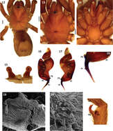
Figures 12–21.Melloleitaoina uru. 12 female, dorsal view 13–14 male holotype 13 cephalotorax 14 sternum, labium, maxillae and quelicerae 15 spermathecae 16–18 left palpal bulb 16 prolateral view 17 retrolateral view 18 detail of triangular tooth on embolus 19–20 coxa III 19 prolateral view 20 detail of spiniform setae 21 right tibial apophysis. Arrow indicates triangular tooth on embolus. Scale bars black = 1 mm.
-
Carlos Perafán, Fernando Pérez-Miles
Zookeys
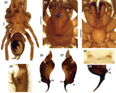
Figures 22–29.Melloleitaoina yupanqui. 22 female, dorsal view 23–24 male holotype 23 cephalothorax 24 sternum, labium, maxillae and quelicerae 25 spermathecae 26–28 left palpal bulb 26 prolateral view 27 retrolateral view 28 detail of embolus showing PS discontinuous 29 right tibial apophysis. Scale bars = 1 mm.
-
Carlos Perafán, Fernando Pérez-Miles
Zookeys
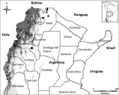
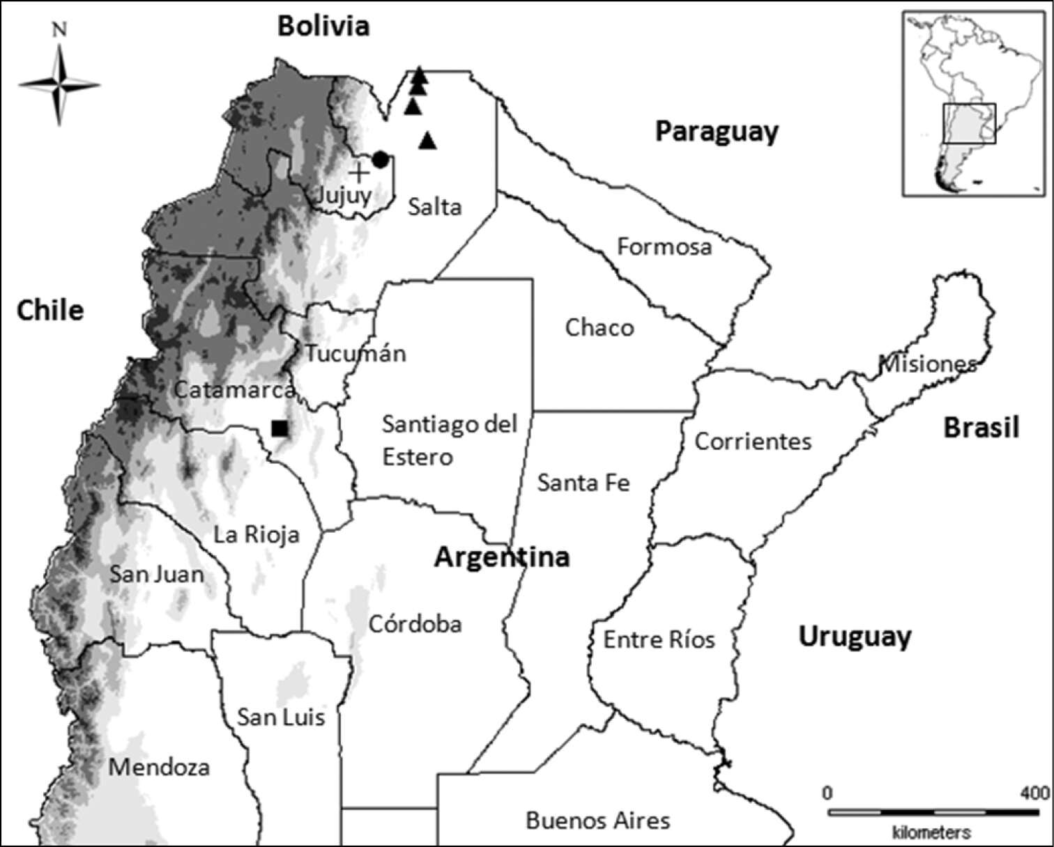
Figure 1.Map of Northern Argentina, geographical distribution of the Melloleitaoina species. Melloleitaoina uru (triangles); Melloleitaoina crassifemur (circle); Melloleitaoina yupanqui (cross); Melloleitaoina mutquina (square).
-
Carlos Perafán, Fernando Pérez-Miles
Zookeys
Figures 2–6.Melloleitaoina crassifemur. 2 male holotype, dorsal view 3–5 left palpal bulb, 3 prolateral view 4 retrolateral view 5 detail of apex widened 6 left tibial apophysis (subapical spine on retrolateral branch RB lost). Scale bar = 1 mm.
-
Carlos Perafán, Fernando Pérez-Miles
Zookeys
Figures 12–21.Melloleitaoina uru. 12 female, dorsal view 13–14 male holotype 13 cephalotorax 14 sternum, labium, maxillae and quelicerae 15 spermathecae 16–18 left palpal bulb 16 prolateral view 17 retrolateral view 18 detail of triangular tooth on embolus 19–20 coxa III 19 prolateral view 20 detail of spiniform setae 21 right tibial apophysis. Arrow indicates triangular tooth on embolus. Scale bars black = 1 mm.
-
Carlos Perafán, Fernando Pérez-Miles
Zookeys
Figures 7–11.Melloleitaoina mutquina. 7–8 male holotype 7 dorsal view 8 ventral view 9–10 right palpal bulb 9 prolateral view 10 retrolateral view 11 right tibial apophysis. Arrow indicates apex widened. Scale bars = 1 mm.
-
Carlos Perafán, Fernando Pérez-Miles
Zookeys
Figures 12–21.Melloleitaoina uru. 12 female, dorsal view 13–14 male holotype 13 cephalotorax 14 sternum, labium, maxillae and quelicerae 15 spermathecae 16–18 left palpal bulb 16 prolateral view 17 retrolateral view 18 detail of triangular tooth on embolus 19–20 coxa III 19 prolateral view 20 detail of spiniform setae 21 right tibial apophysis. Arrow indicates triangular tooth on embolus. Scale bars black = 1 mm.
-
Carlos Perafán, Fernando Pérez-Miles
Zookeys
Figures 22–29.Melloleitaoina yupanqui. 22 female, dorsal view 23–24 male holotype 23 cephalothorax 24 sternum, labium, maxillae and quelicerae 25 spermathecae 26–28 left palpal bulb 26 prolateral view 27 retrolateral view 28 detail of embolus showing PS discontinuous 29 right tibial apophysis. Scale bars = 1 mm.
-
Carlos Perafán, Fernando Pérez-Miles
Zookeys
Figures 12–21.Melloleitaoina uru. 12 female, dorsal view 13–14 male holotype 13 cephalotorax 14 sternum, labium, maxillae and quelicerae 15 spermathecae 16–18 left palpal bulb 16 prolateral view 17 retrolateral view 18 detail of triangular tooth on embolus 19–20 coxa III 19 prolateral view 20 detail of spiniform setae 21 right tibial apophysis. Arrow indicates triangular tooth on embolus. Scale bars black = 1 mm.