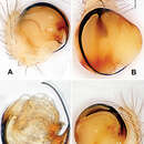
Figure 19.Ogulnius hapalus sp. n., male holotype. A Pedipalp, prolateral view B Pedipalp, ventral view C Embolic division, retrolateral view D Pedipalp, retrolateral view. Co conductor; EA embolic apophysis; MA median apophysis T tegulum. Scale bars: D as A, C as B.
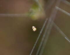
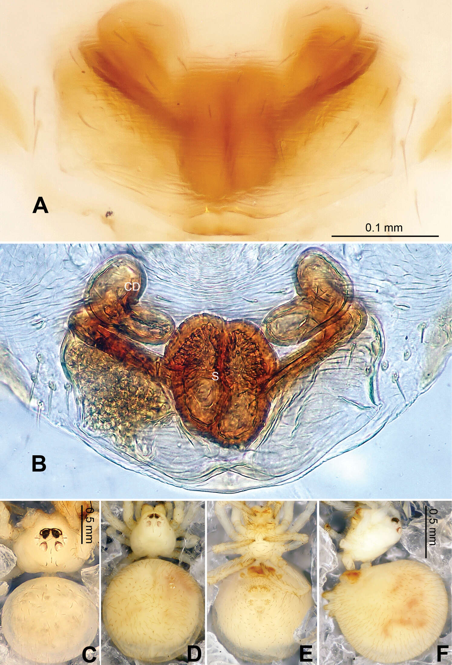
Figure 20.Ogulnius hapalus sp. n., male holotype (C) and female paratype (A–B, D–F). A Epigyne, ventral view B Vulva, dorsal view C Male habitus, dorsal view D Female habitus, dorsal view E Female habitus, ventral view F Female habitus, lateral view. CD copulatory duct; S spermatheca. Scale bars: B as A, D, E as F.
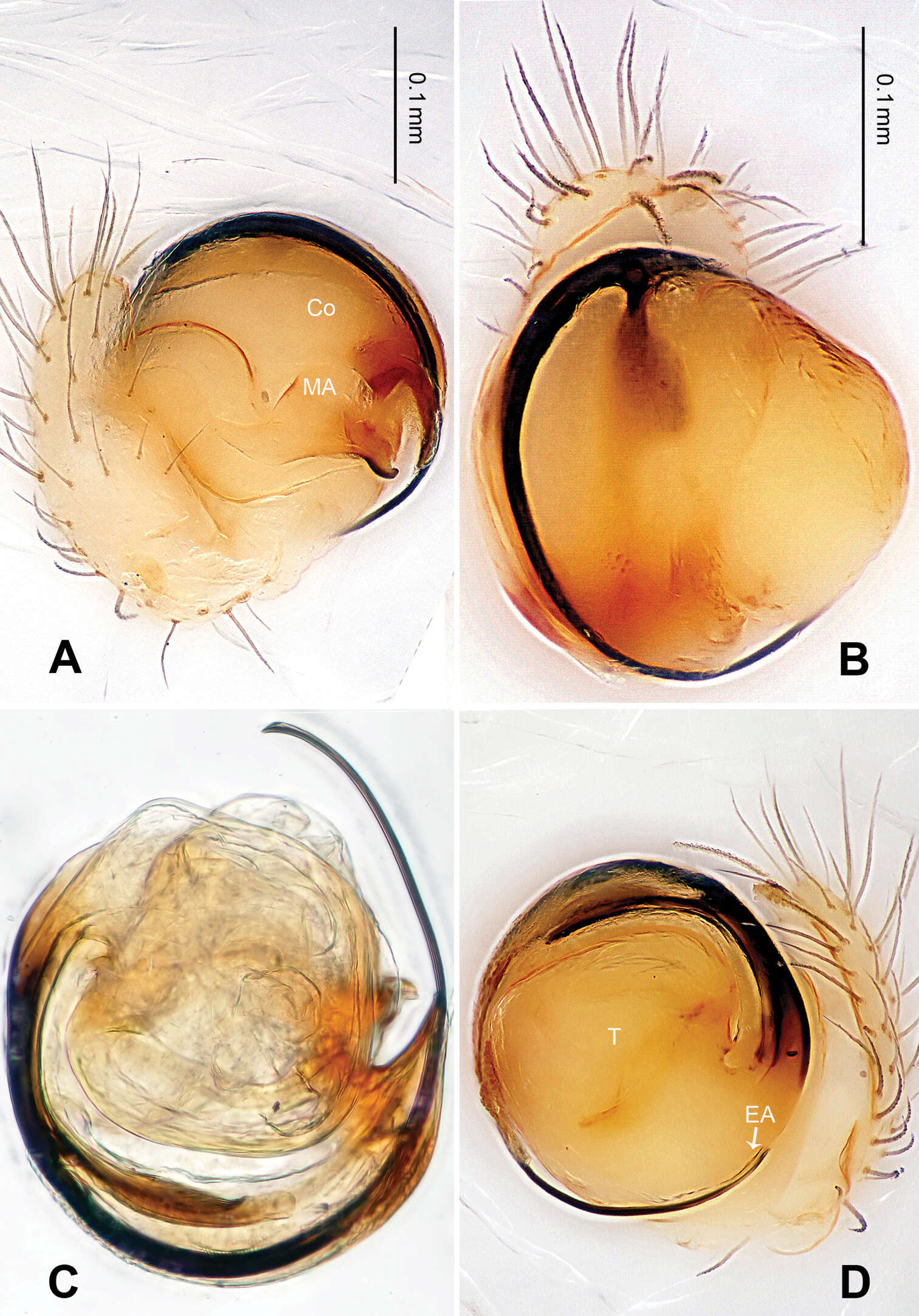
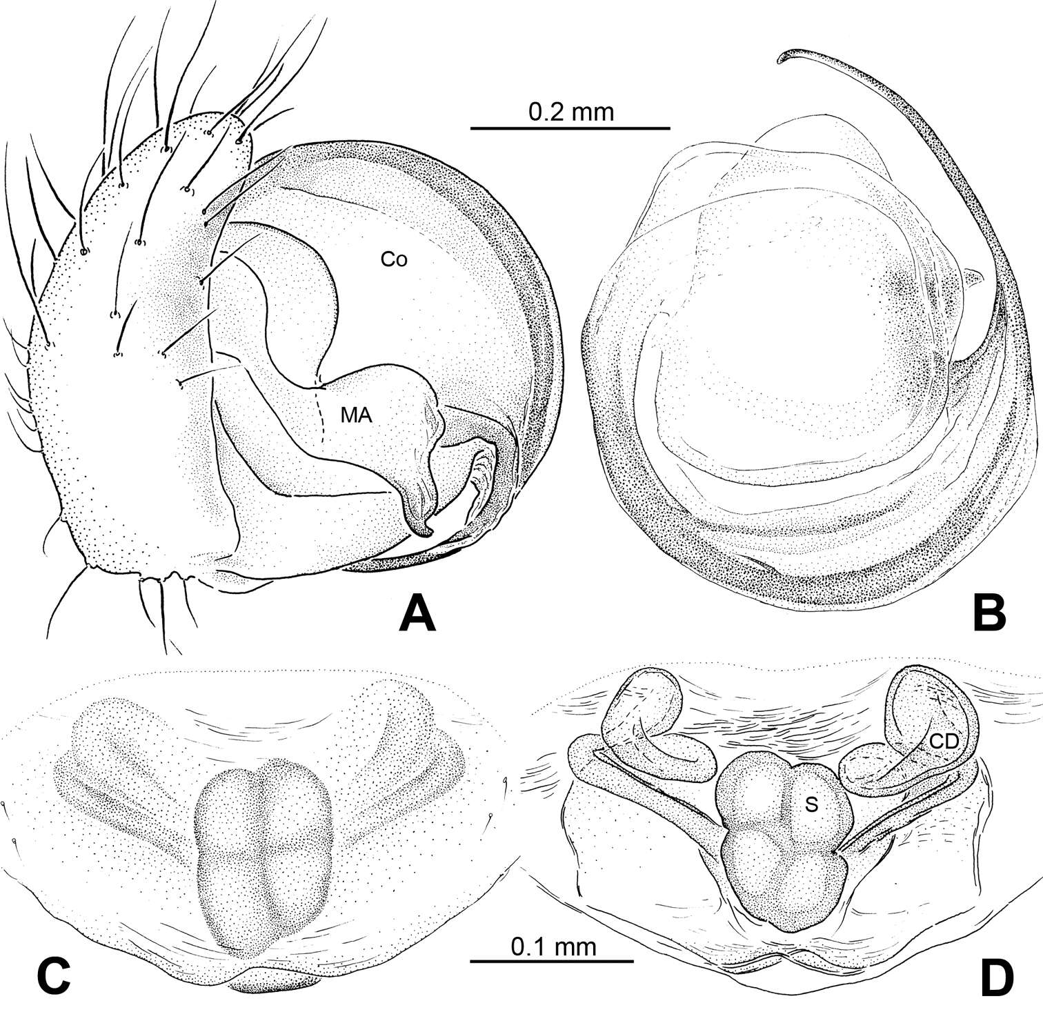
Figure 21.Ogulnius hapalus sp. n., male holotype (A–B) and female paratype (C–D). A Pedipalp, prolateral view B Embolic division, retrolateral view C Epigyne, ventral view D Vulva, dorsal view. CD copulatory duct; Co conductor; MA median apophysis; S spermatheca.