-
-
-
-
Villoslada de Cameros, La Rioja, Spain
-
Rancho de la Herradura, Andalusia, Spain
-
Pumarejo, Castille and Leon, Spain
-
Villoslada de Cameros, La Rioja, Spain
-
Ribadelago de Franco, Castille and Leon, Spain
-
Vallibona, Comunitat Valenciana, Espaa
-
Lumbreras, La Rioja, Spain
-
Lumbreras, La Rioja, Spain
-
Ribadelago de Franco, Castille and Leon, Spain
-
Lumbreras, La Rioja, Spain
-
Lumbreras, La Rioja, Spain
-
Rancho de la Herradura, Andalusia, Spain
-
Mahide, Castilla y Len, Espaa
-
Rancho de la Herradura, Andalusia, Spain
-
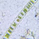
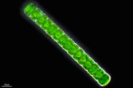
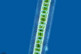
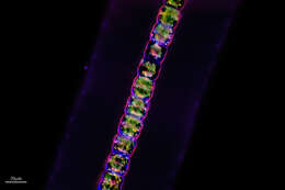
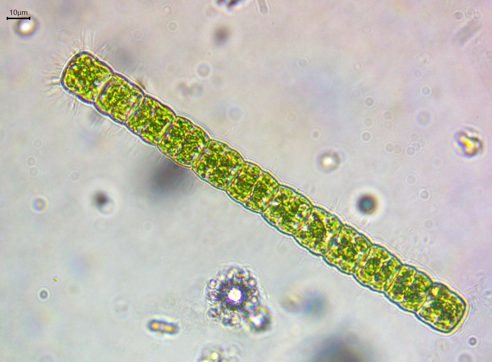
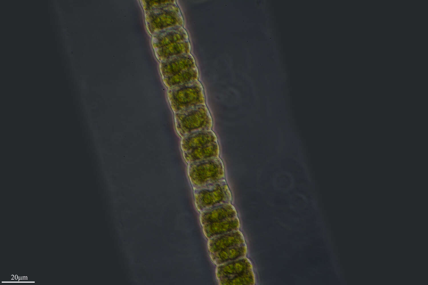
Filemaoentous desmids, cells located within a thick mucus sheath. A green alga, with cellulosic cell walls and bright green chloroplasts. Two forms are shown here. Phase contrast micrograph.
-
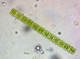
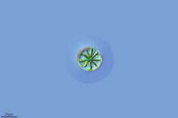
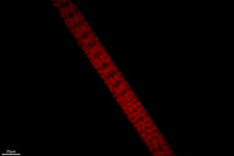
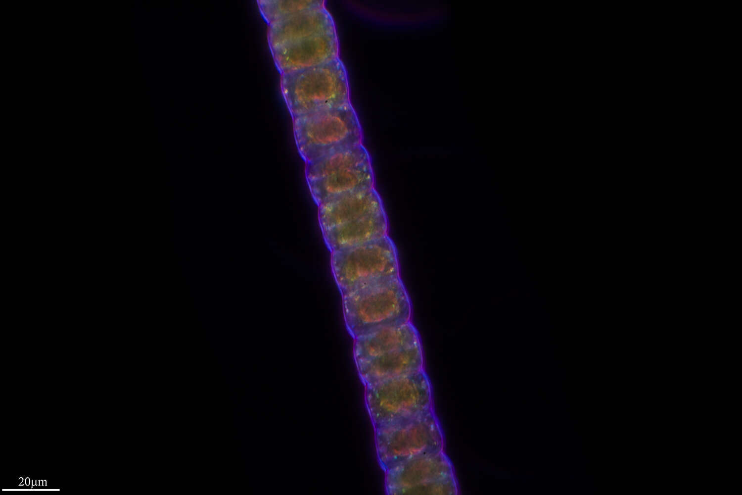
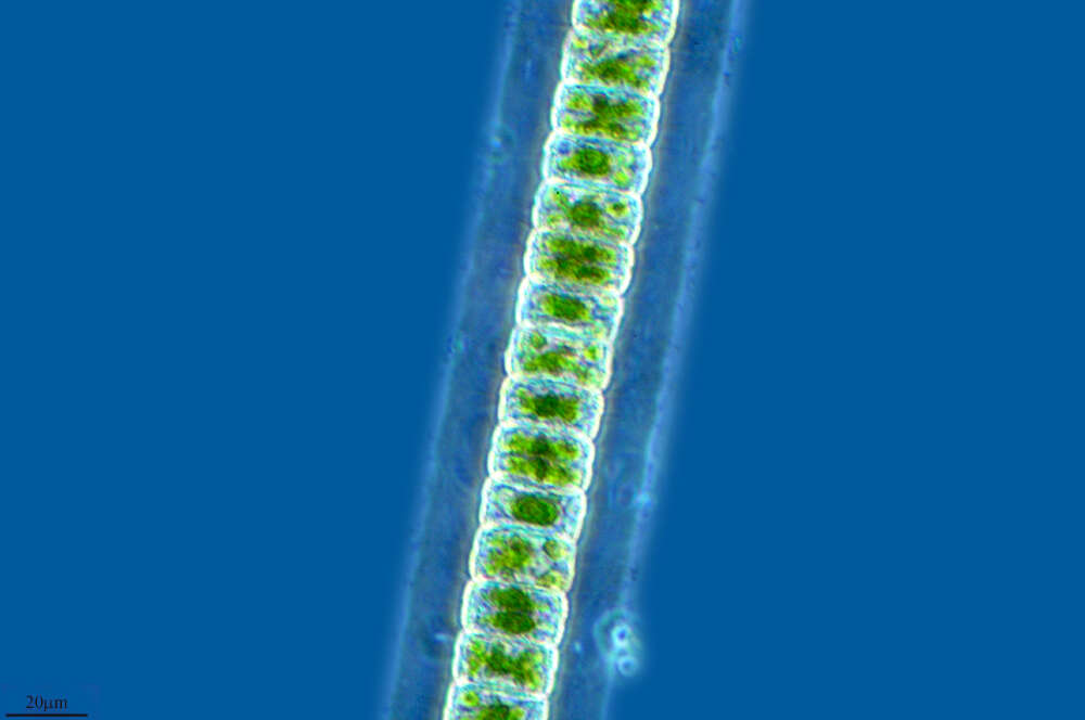
FIlaments (unhappy) observed in freshwater sediments in the vicinity of Broome, Western Australia in September 2003. This image was taken using differential interference contrast optics. Â Â This work was supported by the Australian Biological Resources Study.
-
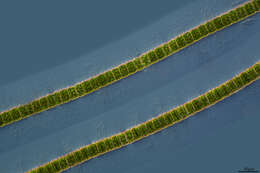
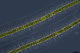
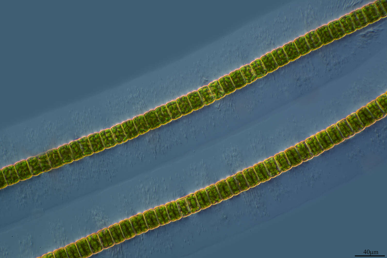
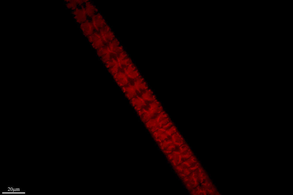
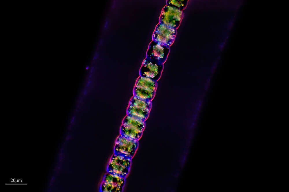
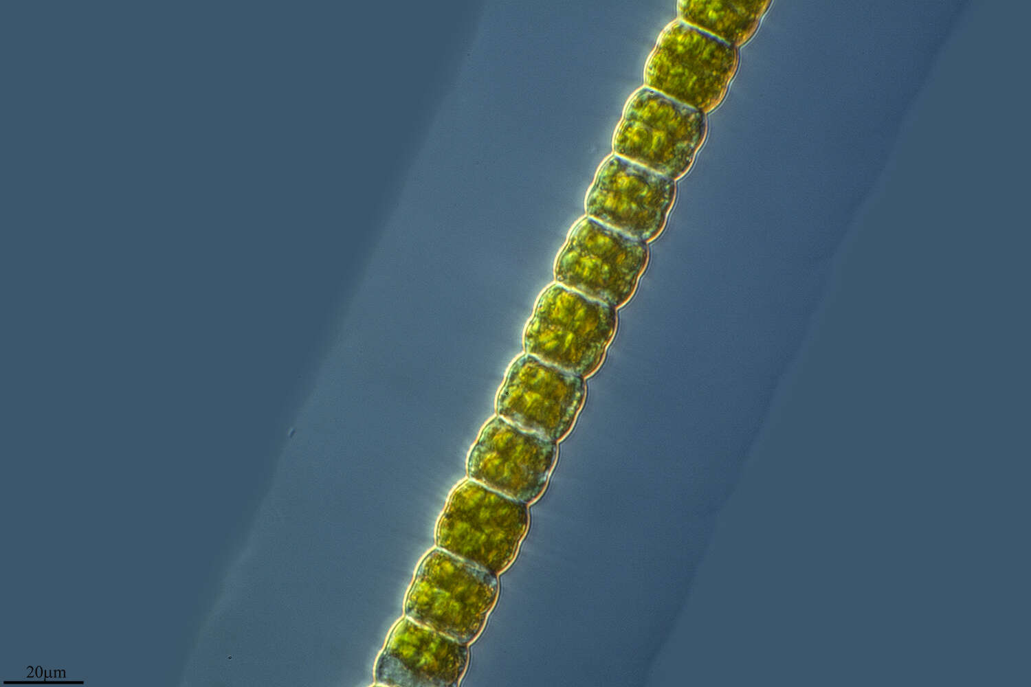
This image of filamentous desmids shows the mucus layer in which the cells are embedded.
-
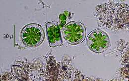
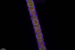
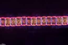
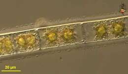
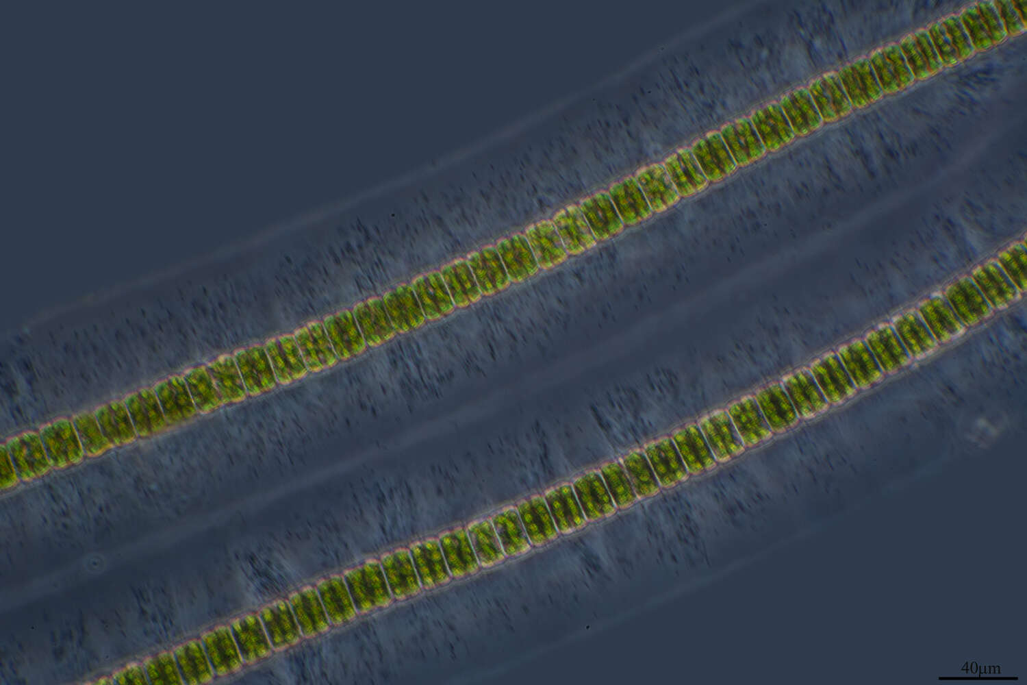
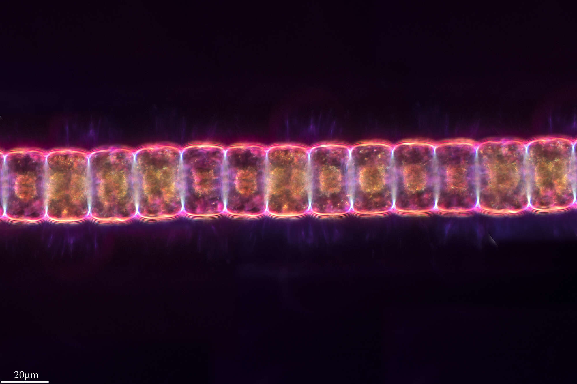
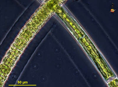
In phase contrast the massive layer of mucilage around the algal filament is clearly visible. On the lower right another filament of H. dissiliens stands perpendicularly providing an apical view which shows the stelloid form of chloroplasts. The scale bar indicates 50 µm. Collected from bottom sediments of a tiny pond at the island of Hiddensee (Baltic Sea, Germany). Images were taken using Zeiss Standard with Olympus C7070 CCD camera.
-
Sampling date 11/2006. Scale bar indicates 50 µm.Place name: Bog Dosenmoor near Neumuenster (Schleswig-Holstein, Germany) Latitude: 54.136219 Longitude: 10.026433Microscope Zeiss Universal, camera Olympus C7070WZ. DOF image.© Wolfgang Bettighofer,images under Creative Commons License V 3.0 (CC BY-NC-SA).For permission to use of (high resolution) images please contact
postmaster@protisten.de.For further information about the image, please click here:
Link to protisten.de page
-
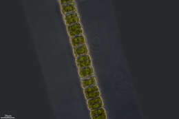
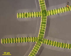
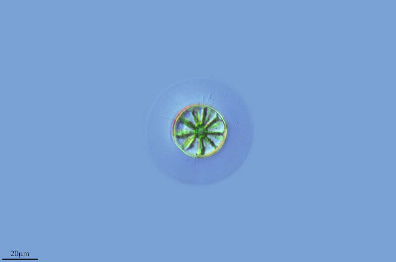
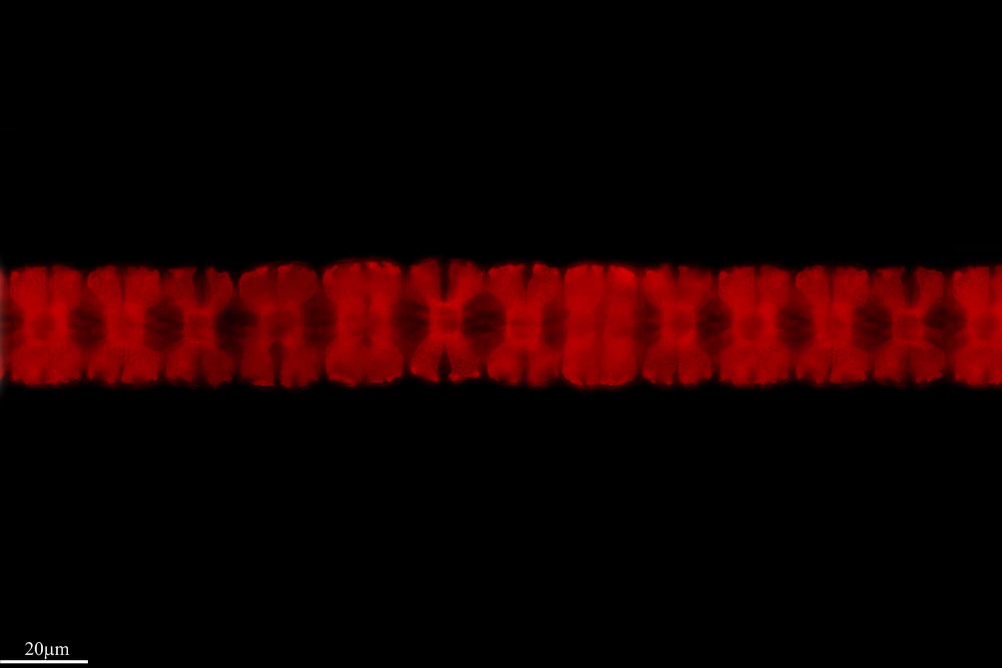
Sampling date 10/2006. Scale bar indicates 50 µm.In phase contrast, the thick mucus layer around the algal filaments is clearly visible. On the right side of the image, another filament of H. dissiliens is perpendicular to the direction of observation, which makes the star-shaped cross-section of the chloroplast particularly clearly visible.Place name: Pond Suploch, Hiddensee (Germany) Latitude: 54.538638 Longitude: 13.097802Microscope Zeiss Universal, camera Olympus C7070WZ.© Wolfgang Bettighofer,images under Creative Commons License V 3.0 (CC BY-NC-SA).For permission to use of (high resolution) images please contact
postmaster@protisten.de.For further information about the image, please click here:
Link to protisten.de page
-
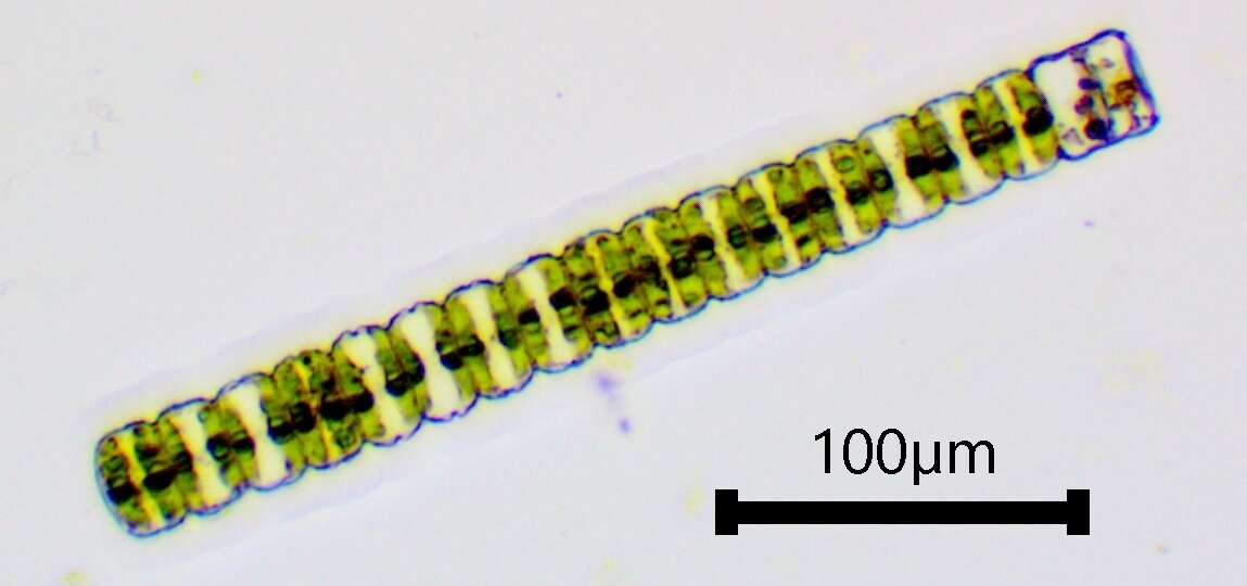
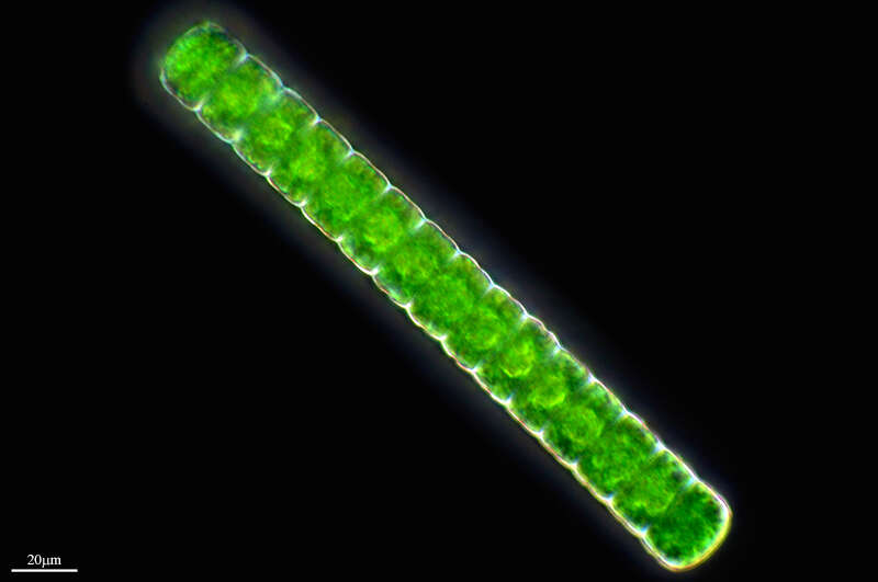
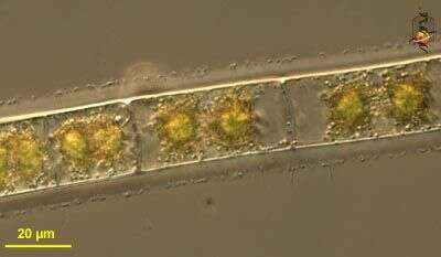
Sampling date 06/2024. Scale bars indicate 100 µm (1), 25 µm (2, 3).Three images.First:Multicellular filament, one can clearly see the thick gelatinous sheath around the cells and the many spherical assimilates (oleosomes) in the cell lumen.Second:Short filament, some chloroplast wings are visible.Third:Short filament showing pyrenoids.Please click on < or > on the image edges or on the dots at the bottom edge of the images to browse through the slides!Place name: Wetland at the Pillersee (Tyrol, Austria)Latitude: 47.531785 Longitude: 12.573095Microscope Zeiss Universal, camera Olympus OM-D M5 MKII. DOF images.© Wolfgang Bettighofer,images under Creative Commons License V 3.0 (CC BY-NC-SA).For permission to use of (high resolution) images please contact
postmaster@protisten.de.For further information about the image, please click here:
Link to protisten.de page