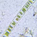
Sampling date 06/2024. Scale bars indicate 100 µm (1), 25 µm (2, 3).Three images.First:Multicellular filament, one can clearly see the thick gelatinous sheath around the cells and the many spherical assimilates (oleosomes) in the cell lumen.Second:Short filament, some chloroplast wings are visible.Third:Short filament showing pyrenoids.Please click on < or > on the image edges or on the dots at the bottom edge of the images to browse through the slides!Place name: Wetland at the Pillersee (Tyrol, Austria)Latitude: 47.531785 Longitude: 12.573095Microscope Zeiss Universal, camera Olympus OM-D M5 MKII. DOF images.© Wolfgang Bettighofer,images under Creative Commons License V 3.0 (CC BY-NC-SA).For permission to use of (high resolution) images please contact
postmaster@protisten.de.For further information about the image, please click here:
Link to protisten.de page