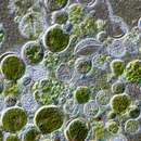
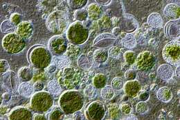
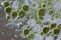
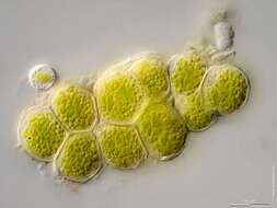
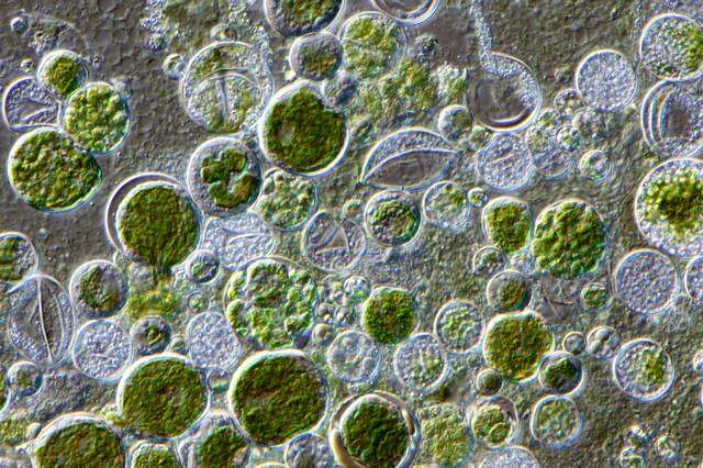
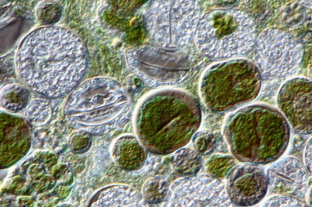
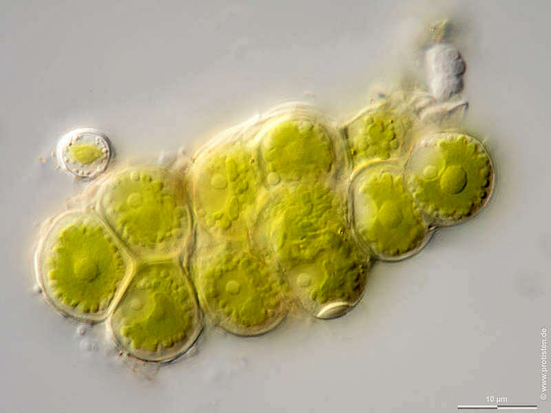
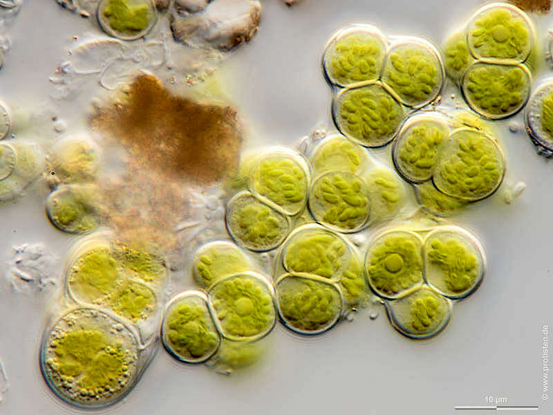
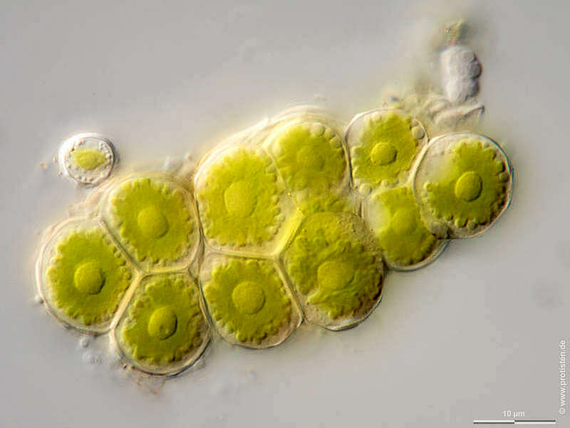
Sampling date 02/2020. Scale bars indicate 10 µm.Three images. First:Synoptic representation of the cell wall and the chloroplast.Second:The optical cross section shows pyrenoids and cell nuclei.Third:Optical cross section showing pyrenoids.Please click on < or > on the image edges or on the dots at the bottom edge of the images to browse through the slides!Place name: Sample from a tree bark near the pond in the forest of Altenholz-Stift (Schleswig-Holstein, Germany) Latitude: 54.384913 Longitude: 10.125691Microscope Zeiss Axioplan, camera Olympus OM-D M5 MKII. DOF images.© Wolfgang Bettighofer,images under Creative Commons License V 3.0 (CC BY-NC-SA).For permission to use of (high resolution) images please contact
postmaster@protisten.de.For further information about the image, please click here:
Link to protisten.de page