-
All Biocode files are based on field identifications to the best of the researcher’s ability at the time.
-
All Biocode files are based on field identifications to the best of the researcher’s ability at the time.
-
All Biocode files are based on field identifications to the best of the researcher’s ability at the time.
-
All Biocode files are based on field identifications to the best of the researcher’s ability at the time.
-
All Biocode files are based on field identifications to the best of the researcher’s ability at the time.
-
All Biocode files are based on field identifications to the best of the researcher’s ability at the time.
-
All Biocode files are based on field identifications to the best of the researcher’s ability at the time.
-
All Biocode files are based on field identifications to the best of the researcher’s ability at the time.
-
All Biocode files are based on field identifications to the best of the researcher’s ability at the time.
-
All Biocode files are based on field identifications to the best of the researcher’s ability at the time.
-
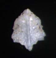
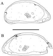
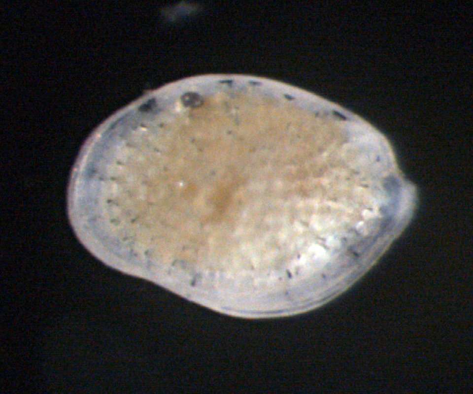
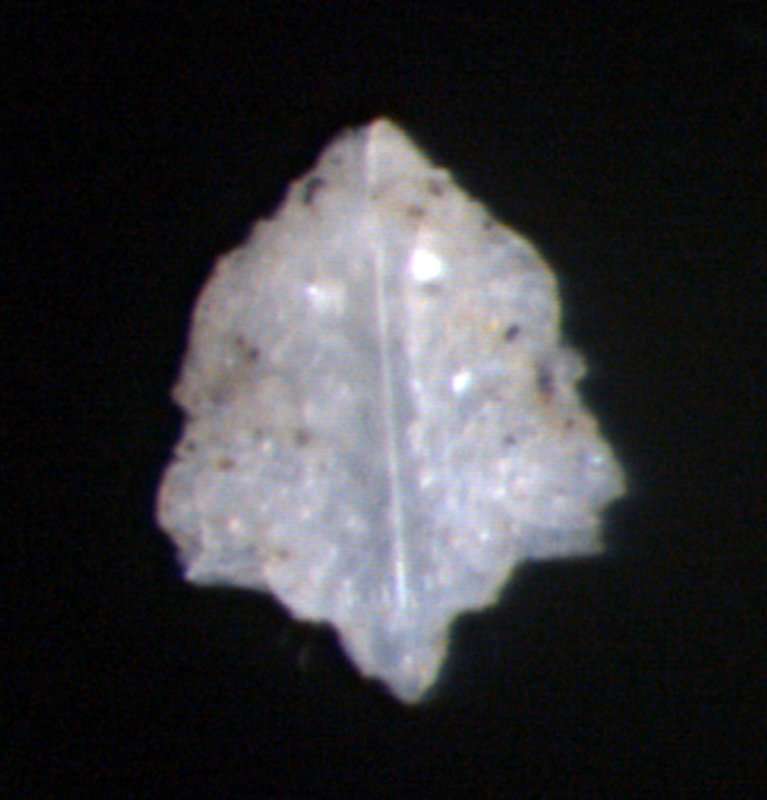
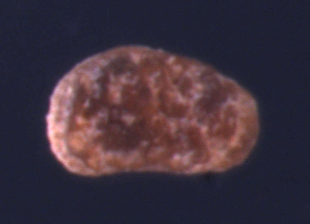
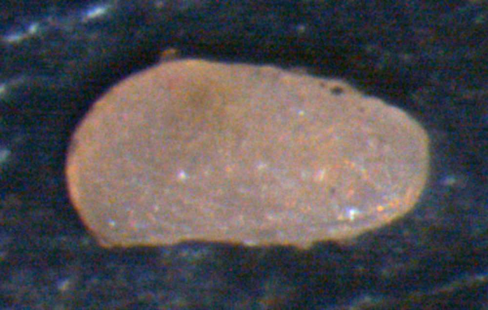
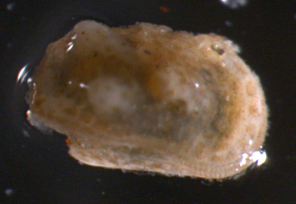
Ryouichi Higashi, Akira Tsukagoshi
Zookeys
Figure 2. Carapaces of Parvocythere gottwaldi sp. n. Holotype (SUM-CO-2023). A right external view B left external view. Each of the carapace structures are transmitted images. Scale bar indicates 100 µm.
-
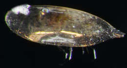
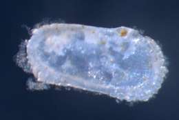
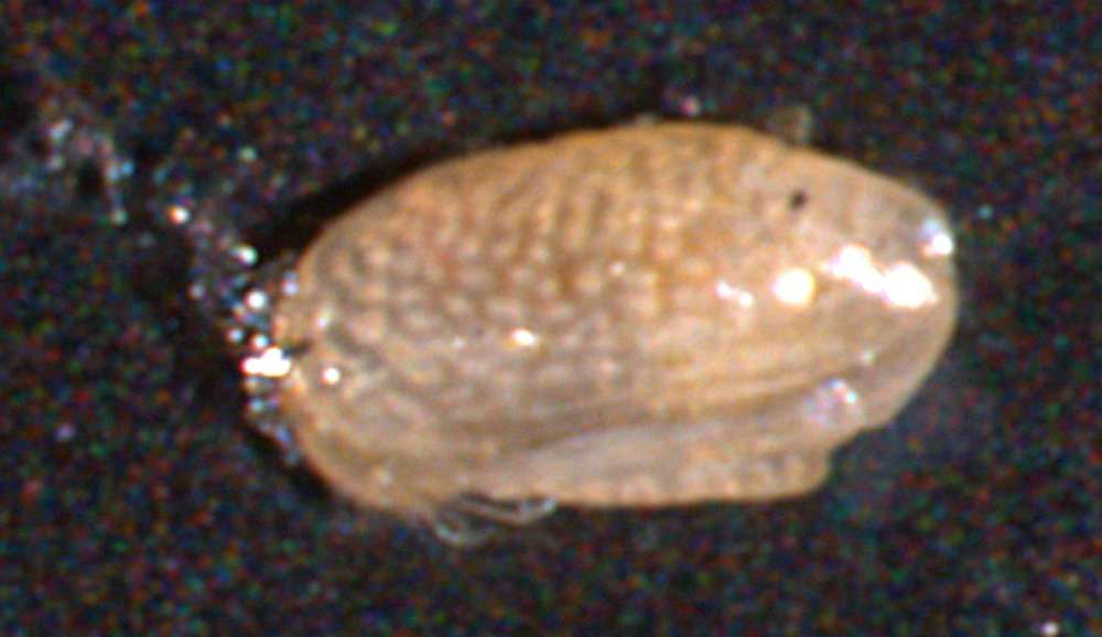
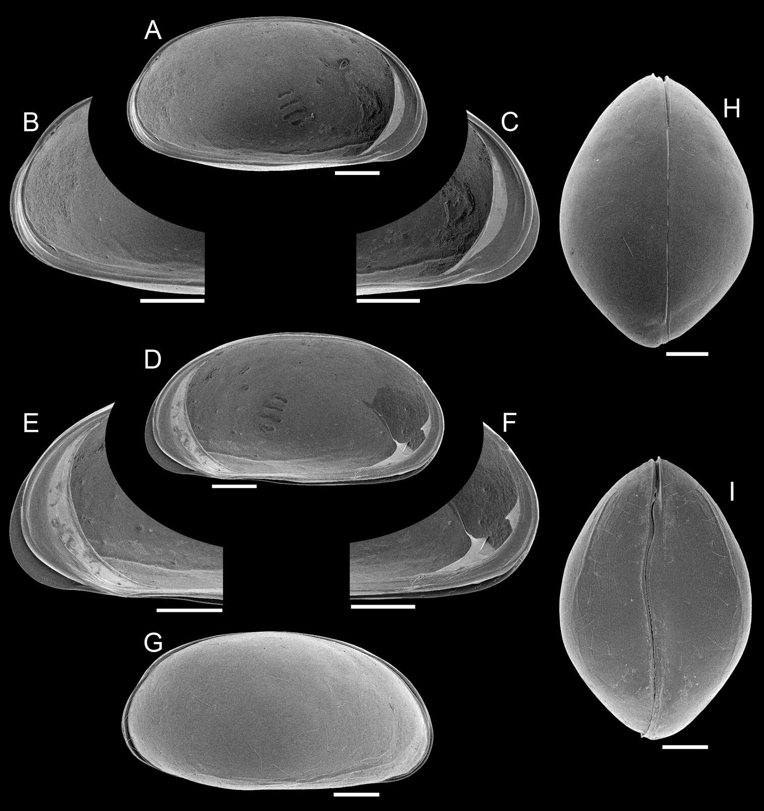
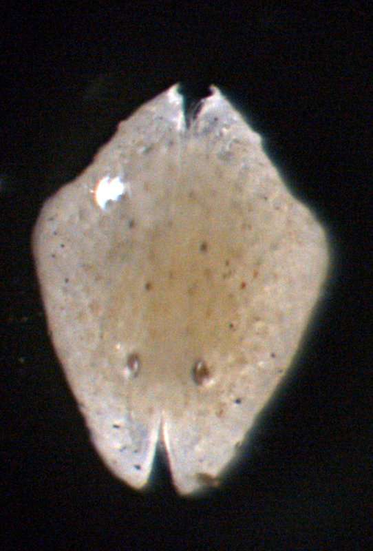
Ricardo L. Pinto, Merlijn Jocqué
Zookeys
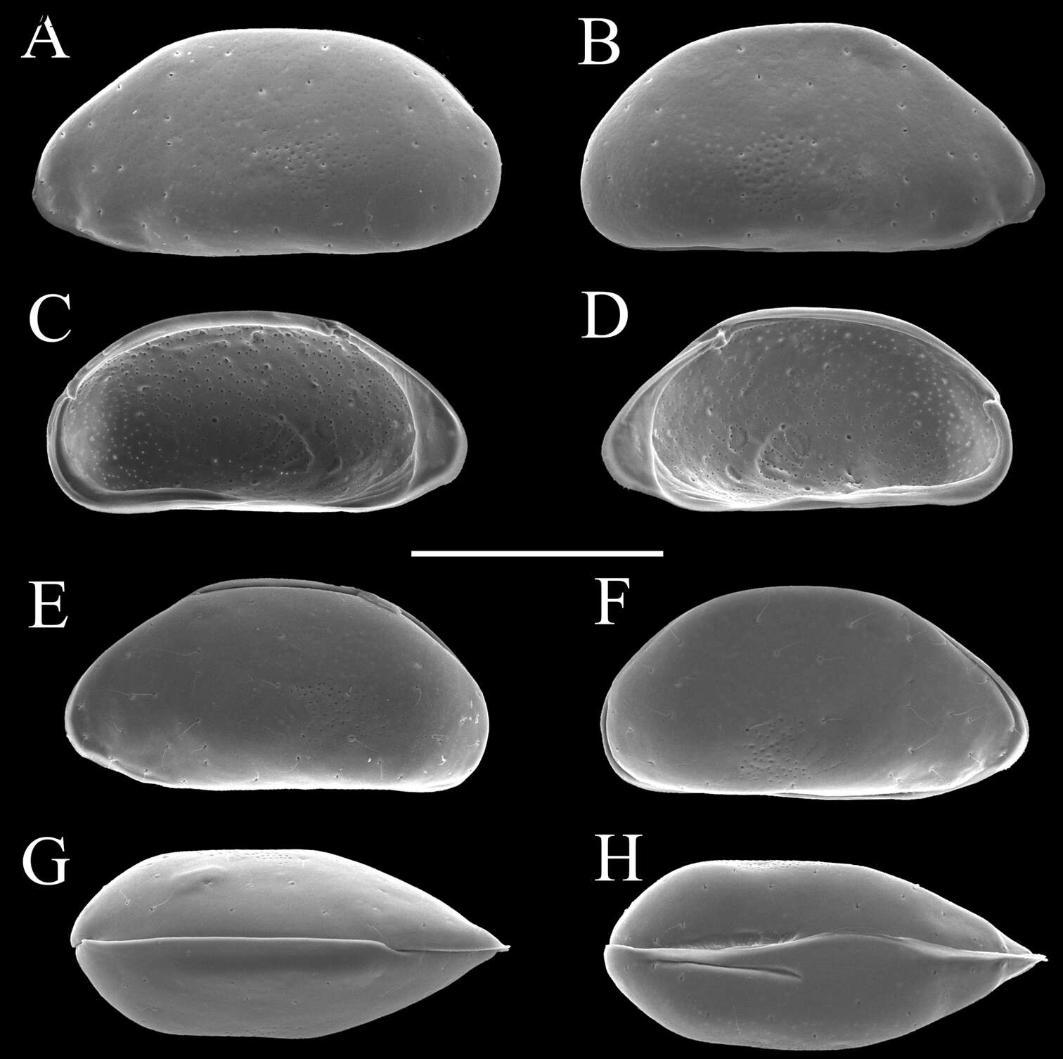
Figure 1.Elpidium merendonense sp. n., male. A Left valve, internal view B left valve, internal view, detail of postero-ventral margin C left valve, internal view, detail of antero-ventral margin D right valve, internal view E right valve, internal view, detail of antero-ventral margin F right valve, internal view, detail of postero-ventral margin G right lateral view H dorsal view I ventral view. A–F holotype, MZUSP 29072; G paratype, MZUSP 29077; H paratype, MZUSP 29078; I paratype, MZUSP 29079. Scale bars: 100 µm.
-
All Biocode files are based on field identifications to the best of the researcher’s ability at the time.
-
All Biocode files are based on field identifications to the best of the researcher’s ability at the time.
-
All Biocode files are based on field identifications to the best of the researcher’s ability at the time.
-
All Biocode files are based on field identifications to the best of the researcher’s ability at the time.
-
All Biocode files are based on field identifications to the best of the researcher’s ability at the time.
-
All Biocode files are based on field identifications to the best of the researcher’s ability at the time.
-
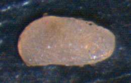
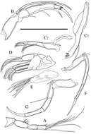
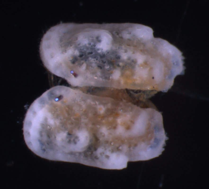
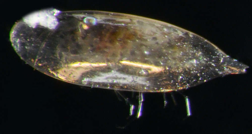
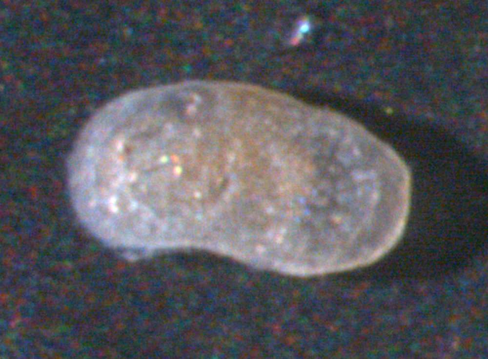
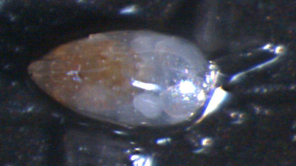
Ryouichi Higashi, Akira Tsukagoshi
Zookeys
Figure 3. Carapaces of Parvocythere gottwaldi sp. n. A–D, G and H male specimens: A and B paratype (SUM-CO-2025) C and D paratype (SUM-CO-2026) G paratype (SUM-CO-2027) H paratype (SUM-CO-2028). A left external lateral view B right external lateral view C internal view of left valve D internal view of right valve G dorsal view H ventral view. E and F female specimens: E paratype (SUM-CO-2039) F paratype (SUM-CO-2040). E left external view F right external view. Scale bar indicates 100 μm.
-
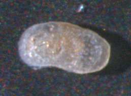
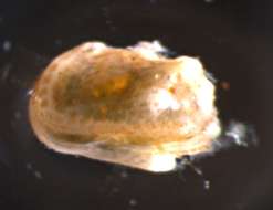
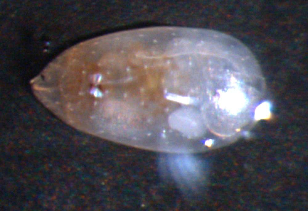
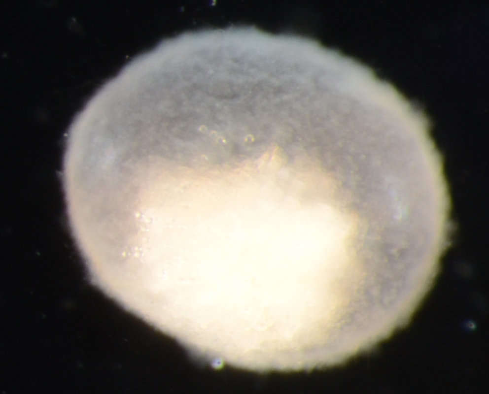
Ricardo L. Pinto, Merlijn Jocqué
Zookeys
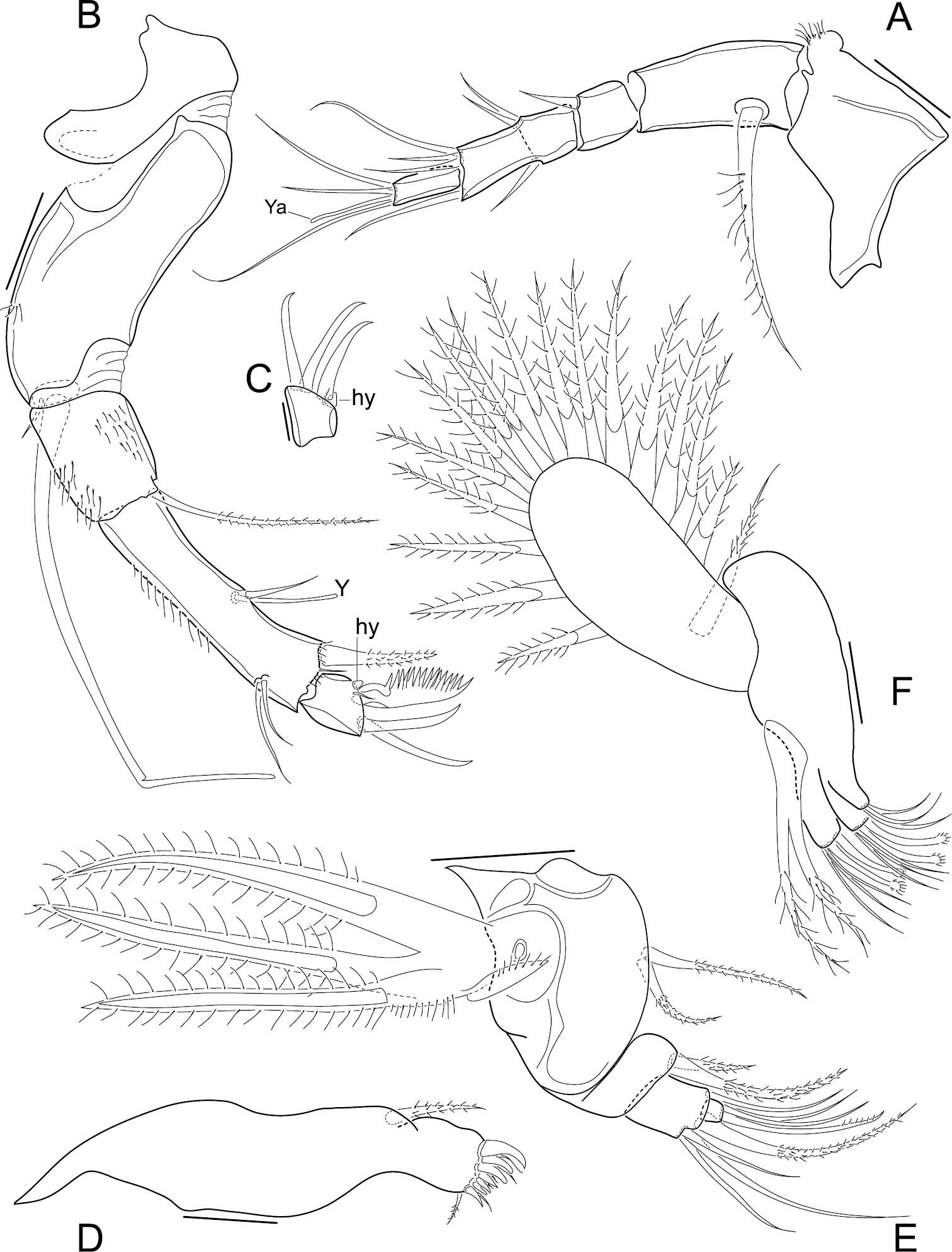
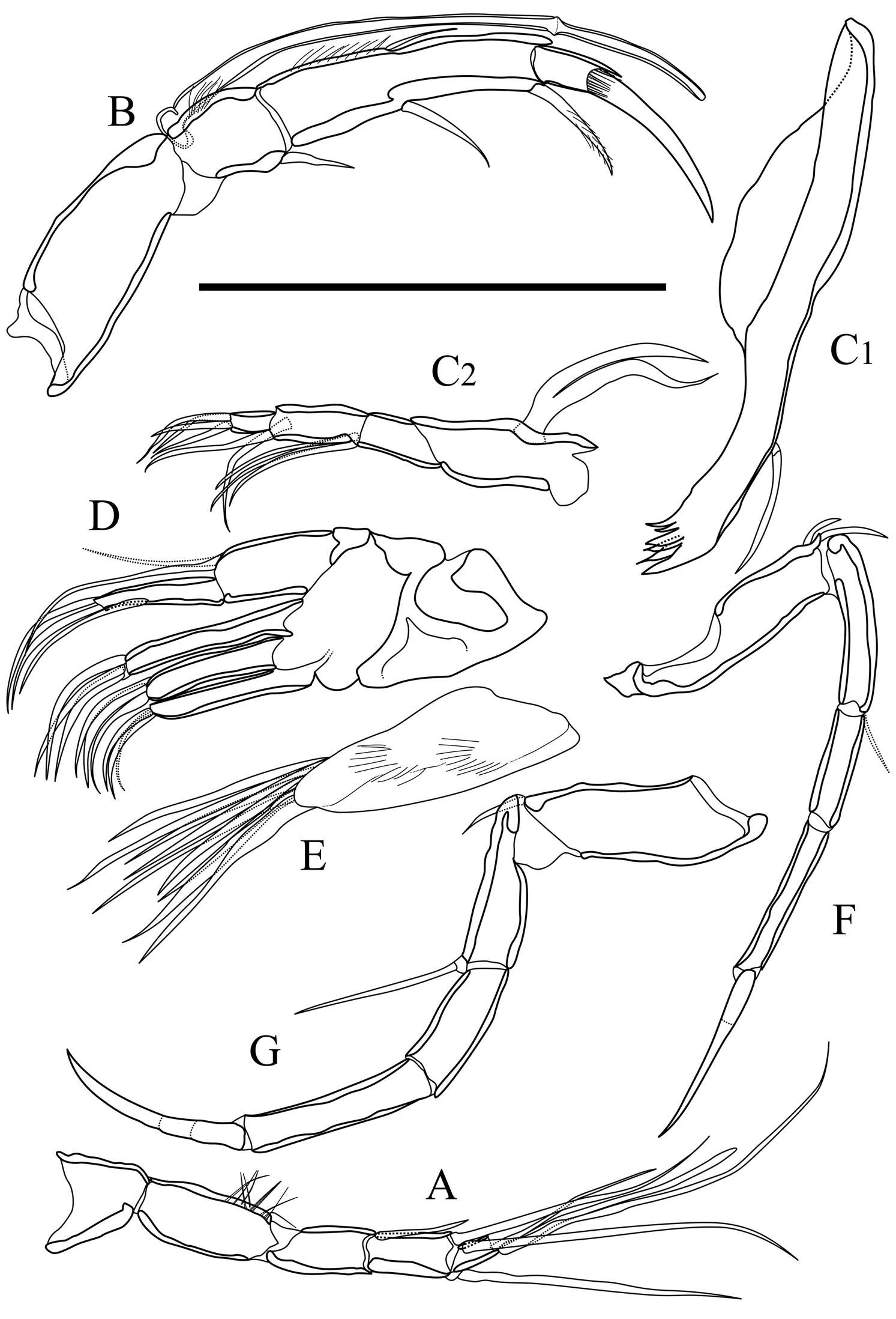
Figure 2.Elpidium merendonense sp. n. A Antennula B Antenna C terminal segment of antenna D Mandibula E Mandibular palp F Maxillula A–B, F male specimen, holotype, MZUSP 29072 C female specimen, allotype, MZUSP 29073 D–E male specimen, paratype, MZUSP 29075. Scale bars: A–B, D–F: 50 µm; C: 20 µm. Ya = aesthetasc on terminal segment of A1. Y = aesthetasc on penultimate segment of antennae. hy = hyaline organ on second antennae.
-
All Biocode files are based on field identifications to the best of the researcher’s ability at the time.
-
All Biocode files are based on field identifications to the best of the researcher’s ability at the time.
-
All Biocode files are based on field identifications to the best of the researcher’s ability at the time.
-
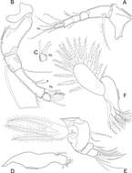
Ryouichi Higashi, Akira Tsukagoshi
Zookeys
Figure 4. Appendages of Parvocythere gottwaldi sp. n. A–D, F and G holotype (SUM-CO-2023) E paratype (SUM-CO-2024). A antennula B antenna C1 coxa of mandibula C2 palp of mandibula D palp and endites of maxillula E branchial plate of maxillula F fifth limb G sixth limb. Scale bar indicates 50 µm.