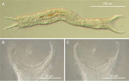
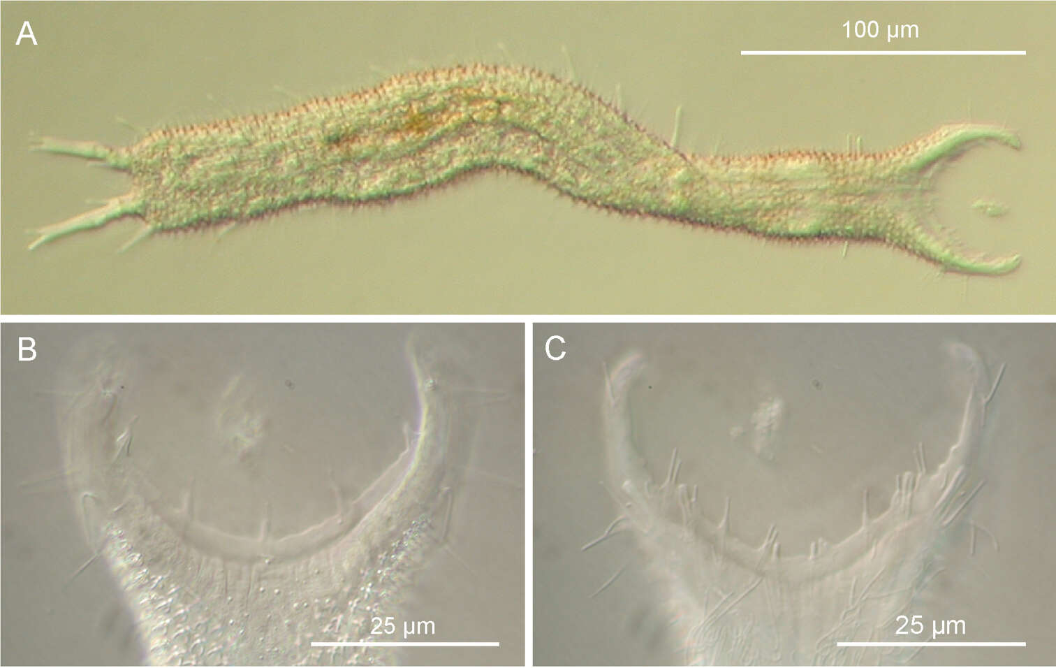
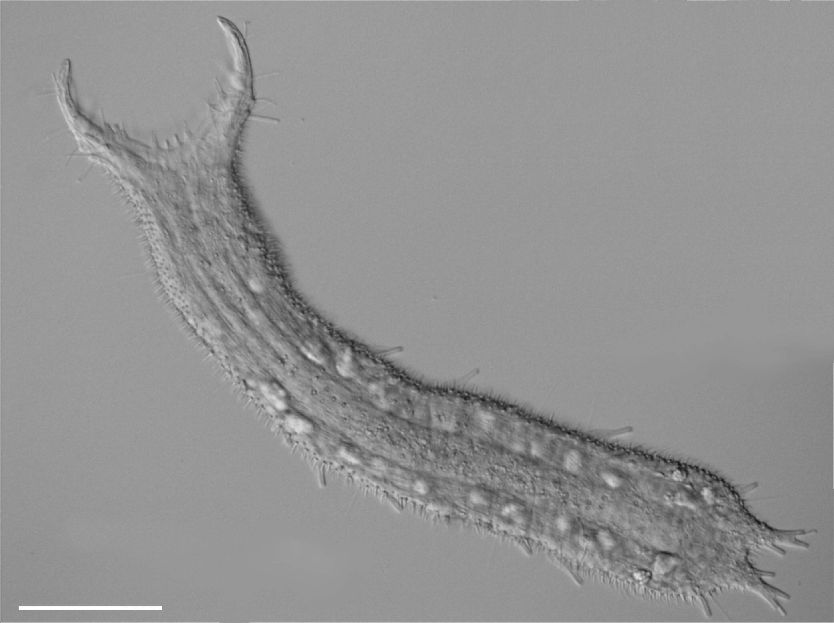
Figure 3.Pseudostomella dolichopoda sp. n. DIC photomicrographs. A habitus B close-up of the anterior region, dorsal view C Close-up of the anterior region, ventral view.
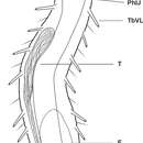
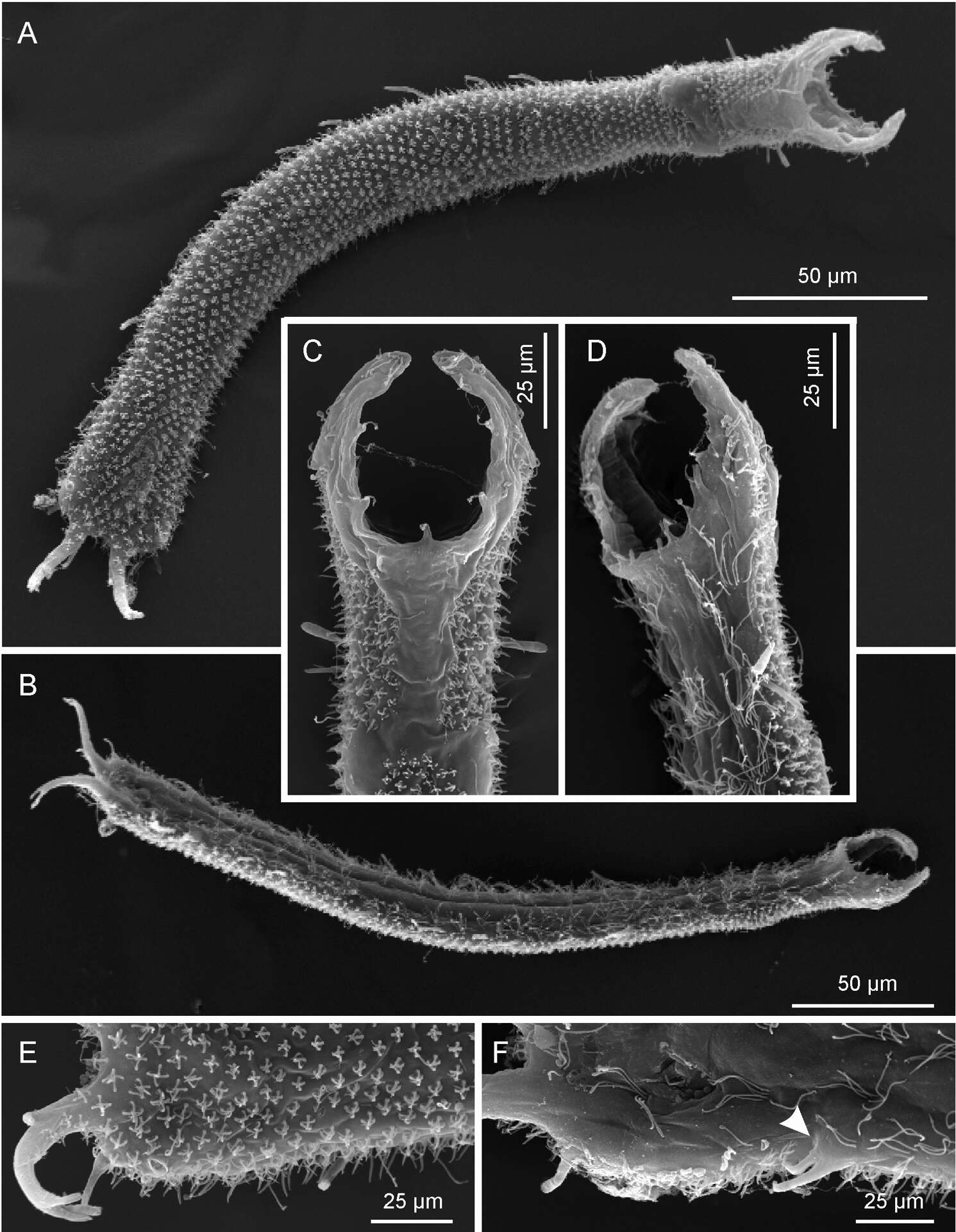
Figure 4.Pseudostomella dolichopoda sp. n. SEM photomicrographs. A habitus, dorsal view B habitus, ventral view; C close-up of the anterior region, dorsal view D close-up of the anterior region, ventrolateral view E close-up of the posterior region, dorsal view F close-up of the posterior region, ventral view, arrow shows the two ventrolateral adhesive tubes borne from a common base.
M. Antonio Todaro, Tobias Kånneby, Matteo Dal Zotto, Ulf Jondelius
Wikimedia Commons
Description: English: DIC photomicrographs showing the general body shape and aspects of the cuticular covering of Pseudostomella etrusca. Scale bars: 50 µm. Русский: Микрофотографии брюхоресничного червя Pseudostomella etrusca. Дифференциальная интерференционно-контрастная микроскопия. Масштабная черта — 50 мкм. Date: March 2011. Source: Todaro, M. A., Kånneby, T., Dal Zotto, M., Jondelius, U. (2011). Phylogeny of Thaumastodermatidae (Gastrotricha: Macrodasyida) inferred from nuclear and mitochondrial sequence data. PLoS ONE 6 (3): e17892. doi:10.1371/journal.pone.0017892. Author: M. Antonio Todaro, Tobias Kånneby, Matteo Dal Zotto, Ulf Jondelius. Other versions:.