-
-
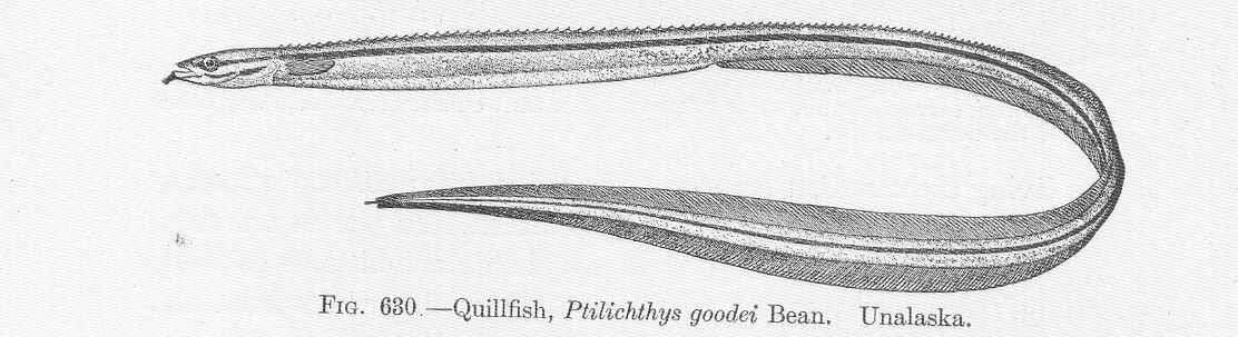
Quillfish, Philichthys goodei Bean. Unalaska.
-
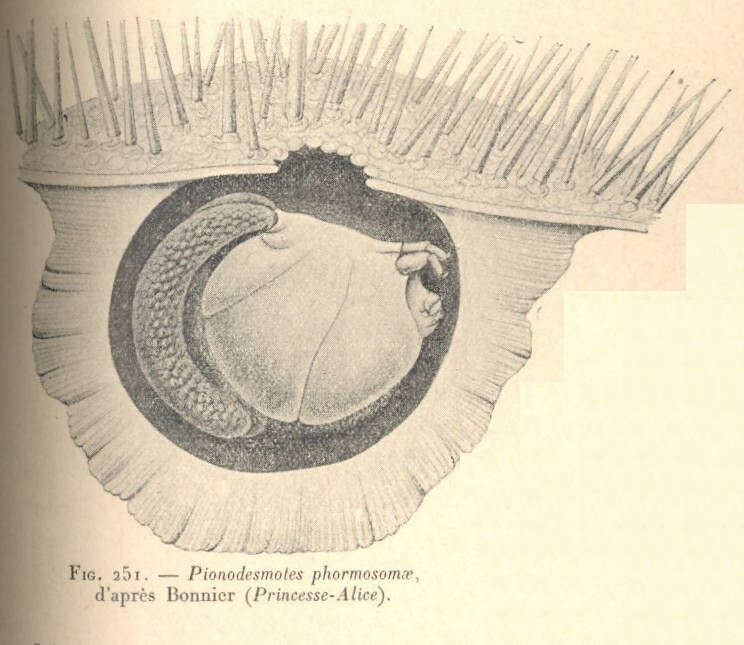
Pionodesmotes phormosomae, d'apres Bonnier (Princesse-Alice).
-
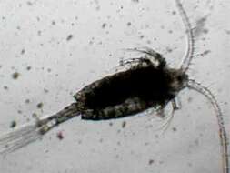
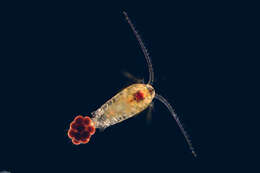
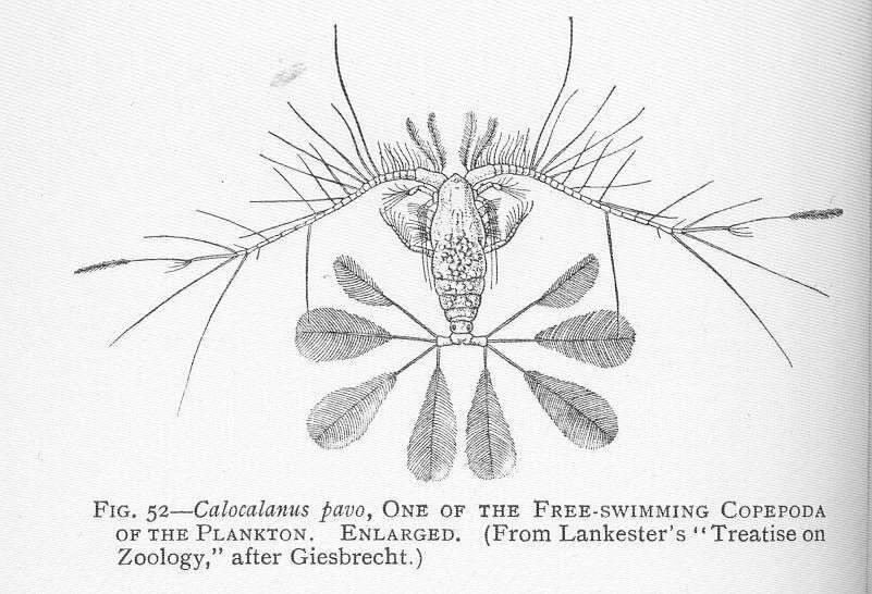
Calocalanus pavo, one of the Free-swimming copepoda of the Plankton.
-
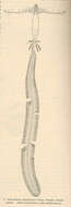
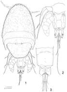
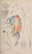
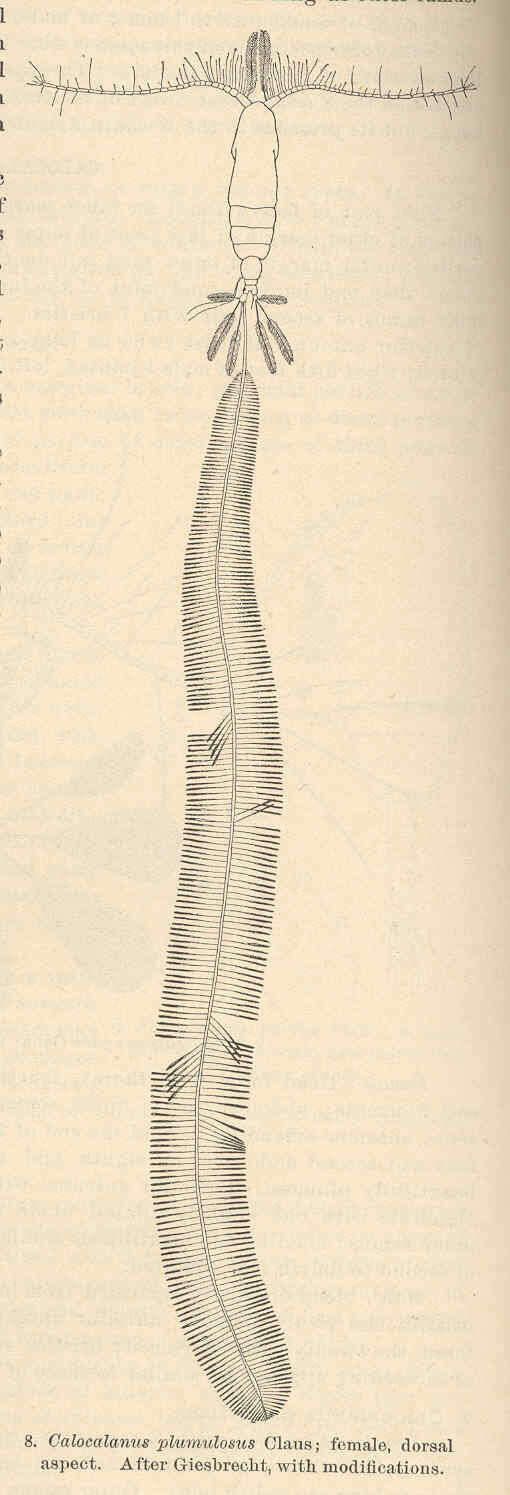
Calocalanus plumulosus Claus; female, dorsal aspect.
-
Diana M. P. Galassi, Paola De Laurentiis, Barbara Fiasca
Zookeys
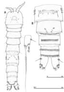
Figure 10.Phyllognathopus inexspectatus sp. n. (♀). A habitus, dorsal view B abdomen, ventral view C caudal ramus, ventral view (scale bars in μm).
-
Susumu Ohtsuka, Michitaka Shimomura, Kota Kitazawa
Zookeys
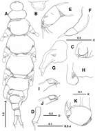
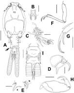
Figure 1. Enterognathus inabai sp. n. holotype female: A Habitus, dorsal view B Rostrum, dorsal view C Genital opening, right, dorsal view D Caudal ramus, left, dorsal view E Antennule F Antenna G Mandible H Labrum and paragnath, ventral view I Maxillule J Other maxillule K Maxilla. Scales in mm.
-
Eduardo Suárez-Morales, Humberto Camisotti, Alberto Martín
Zookeys
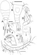
Figure 1.Caligus evelynae sp.n., adult female from Venezuela: A habitus, dorsal view B sternal furca, ventral view C antennule D antenna E postantennal process (b) and maxillule (a) F maxilla G detail of calamus and canna H maxilliped I genital complex and abdomen, ventral view. Scale bars: A, I=0.5 mm, B–F, H =0.1 mm, G=0.05 mm.
-
Samuel Gómez, Nicola K. Carrasco, Francisco Neptalí Morales-Serna
Zookeys
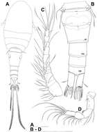
Figure 2.Nitocra taylori sp. n. Female. A habitus B rostrum, dorsal C urosome, dorsal. Scale bar: A=300 µm; B=75 µm; C=150 µm.
-
Tomislav Karanovic, Mark J. Grygier, Wonchoel Lee
Zookeys
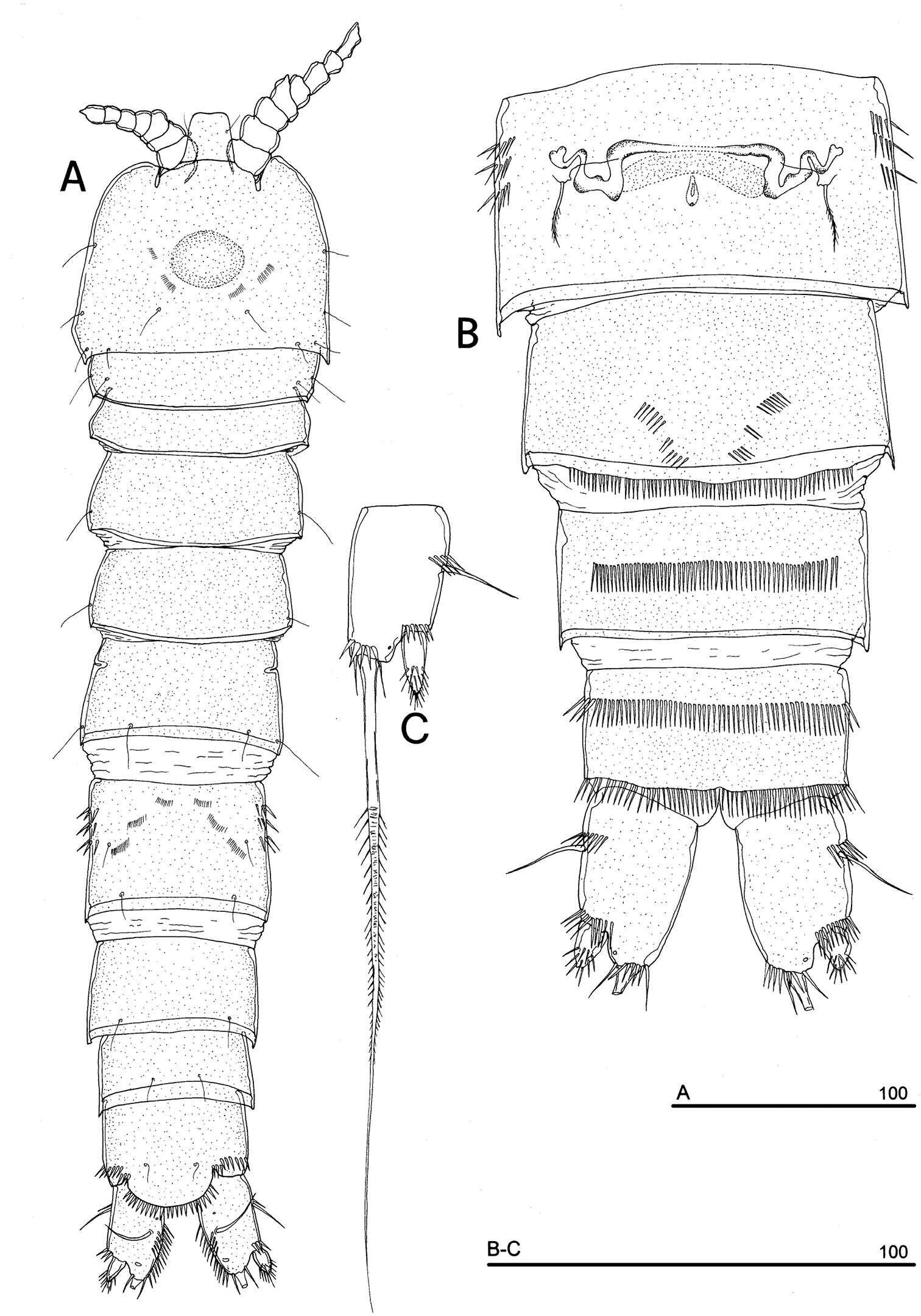
Figure 1.Diacyclops ishidai sp. n., holotype female: A habitus, dorsal view B urosome, ventral view C antennula, dorsal view D antenna, dorsal view. Arabic numerals numbering sensilla and pores consecutively from anterior to posterior end of body, and from dorsal to ventral side (excluding appendages). Scale bars 100 μm.
-
Nancy F. Mercado-Salas, Eduardo Suárez-Morales, Alejandro M. Maeda-Martínez, Marcelo Silva-Briano
Zookeys
Figure 1.Metacyclops deserticus sp. n., female holotype from Coahuila, Mexico. A habitus, dorsal view B urosome, ventral view C antennule D antenna E mandible F maxillule G maxilla H maxilliped I anal operculum. Scales bars A–B= 100µm; C–I= 50 µm.
-
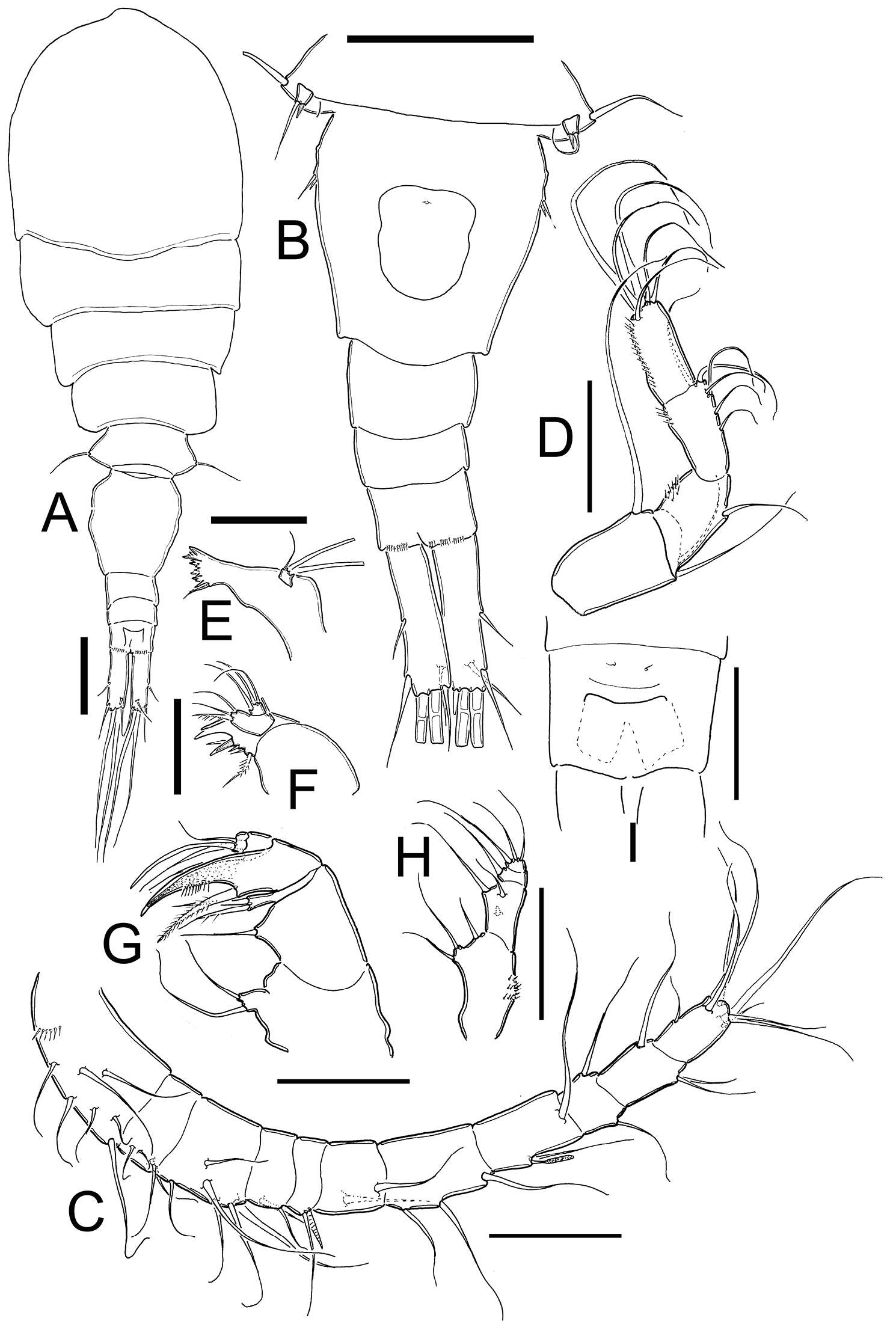
Eduardo Suárez-Morales, Jani Jarquín-González
Zookeys
Figure 2.Peltidium nayarit sp. n., from Playa Careyeros, Nayarit, Mexican Pacific. A adult female, habitus, dorsal view, showing detail of ornamentation of epimeral processes of cephalothorax B antennule C antenna D mandible E maxillule F maxilla. Scales bars: A= 250 μm, B–F=100 μm.
-
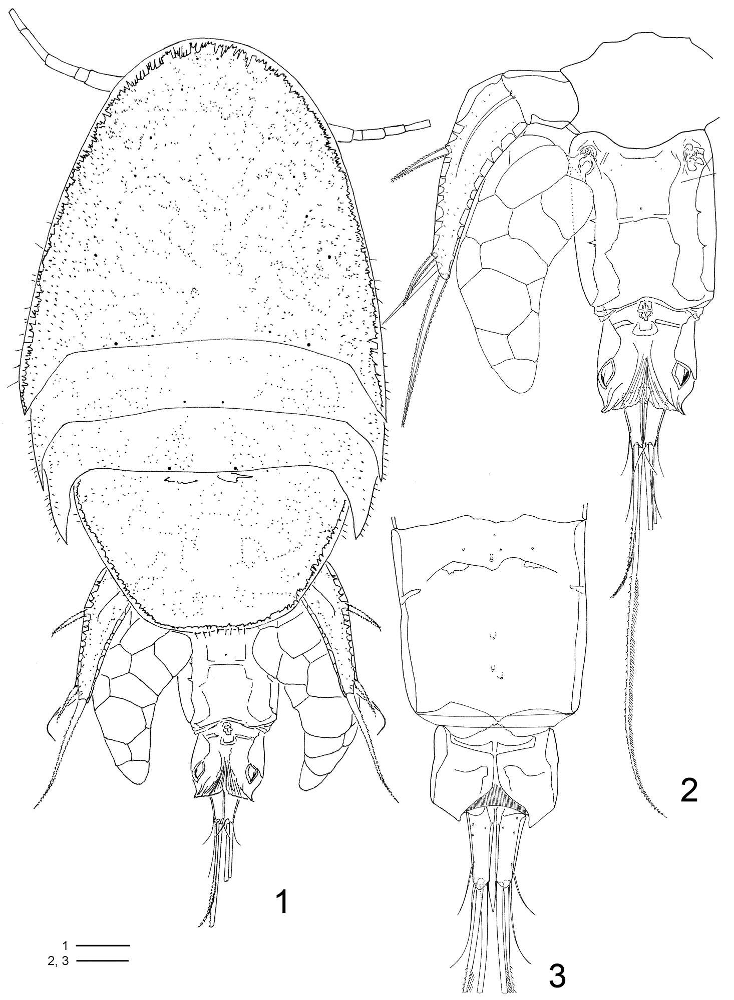
Terue C. Kihara, Carlos E. F. Rocha
Zookeys
Figures 1–3.Clausidium rodriguesi sp. n. Female: 1 habitus, dorsal 2 urosome, dorsal 3 urosome lacking somite bearing P5, ventral. Scale bars: 1 = 100 μm; 2, 3 = 50 μm.
-
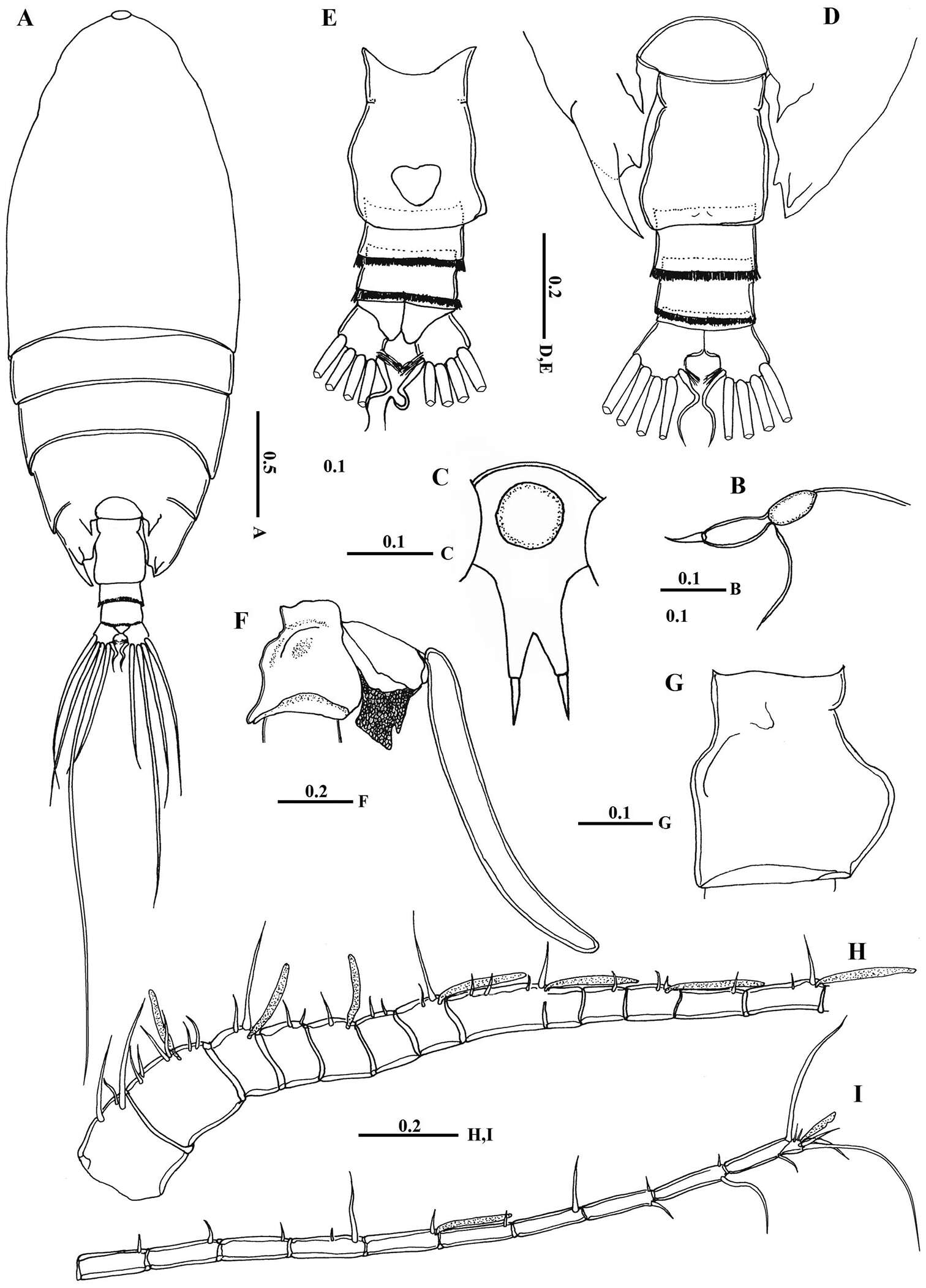
Mohsen M. El-Sherbiny, Ali M. Al-Aidaroos
Zookeys
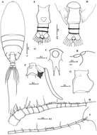
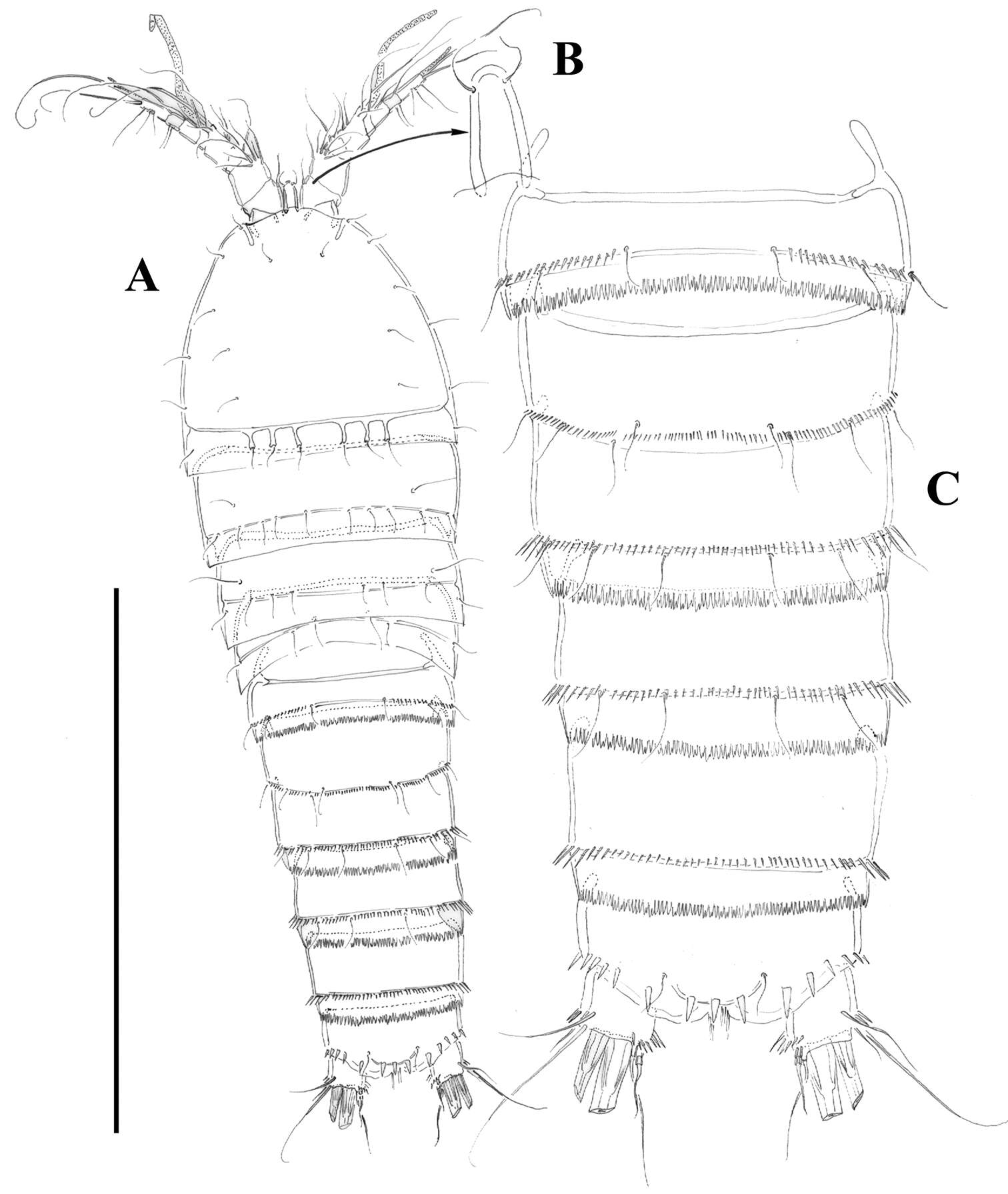
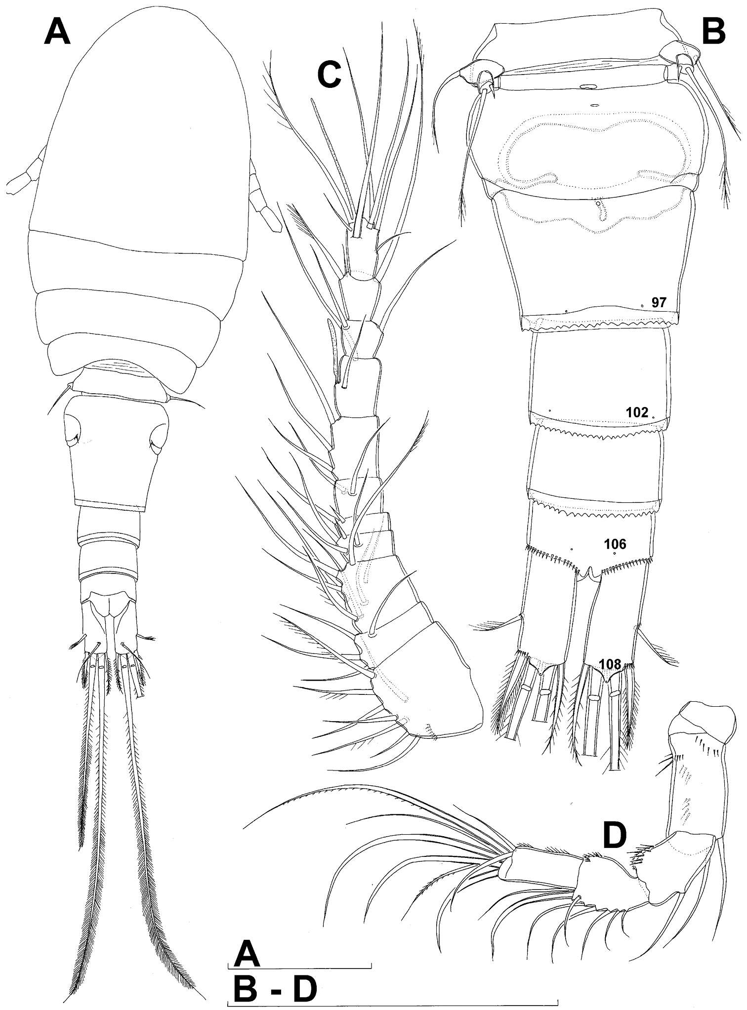
Figure 2.Macandrewella cochinensis female from the northern Red Sea. A habitus, dorsal view B rostrum, lateral view C rostrum, ventral view D posterior prosome and urosome, dorsal view E urosome, ventral view F genital double-somite with spermatophore, lateral view (right) G genital double-somite, lateral view (right) H–I antennules. All scale bars in mm.
-
Juan M. Fuentes-Reinés, Eduardo Suárez-Morales
Zookeys
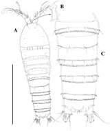
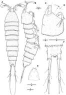
Figure 1.Nitokra affinis colombiensis ssp. n., from northern Colombia. A adult female, habitus, lateral view B adult male, habitus, lateral view C female, anal somite showing ornamentation of anal operculum D male, urosome, ventral view E male, third urosomite, ventral view F male, caudal rami, ventral view. Scale bars: A, B = 100 μm, C, E, F = 10 μm, D = 50 μm.
-
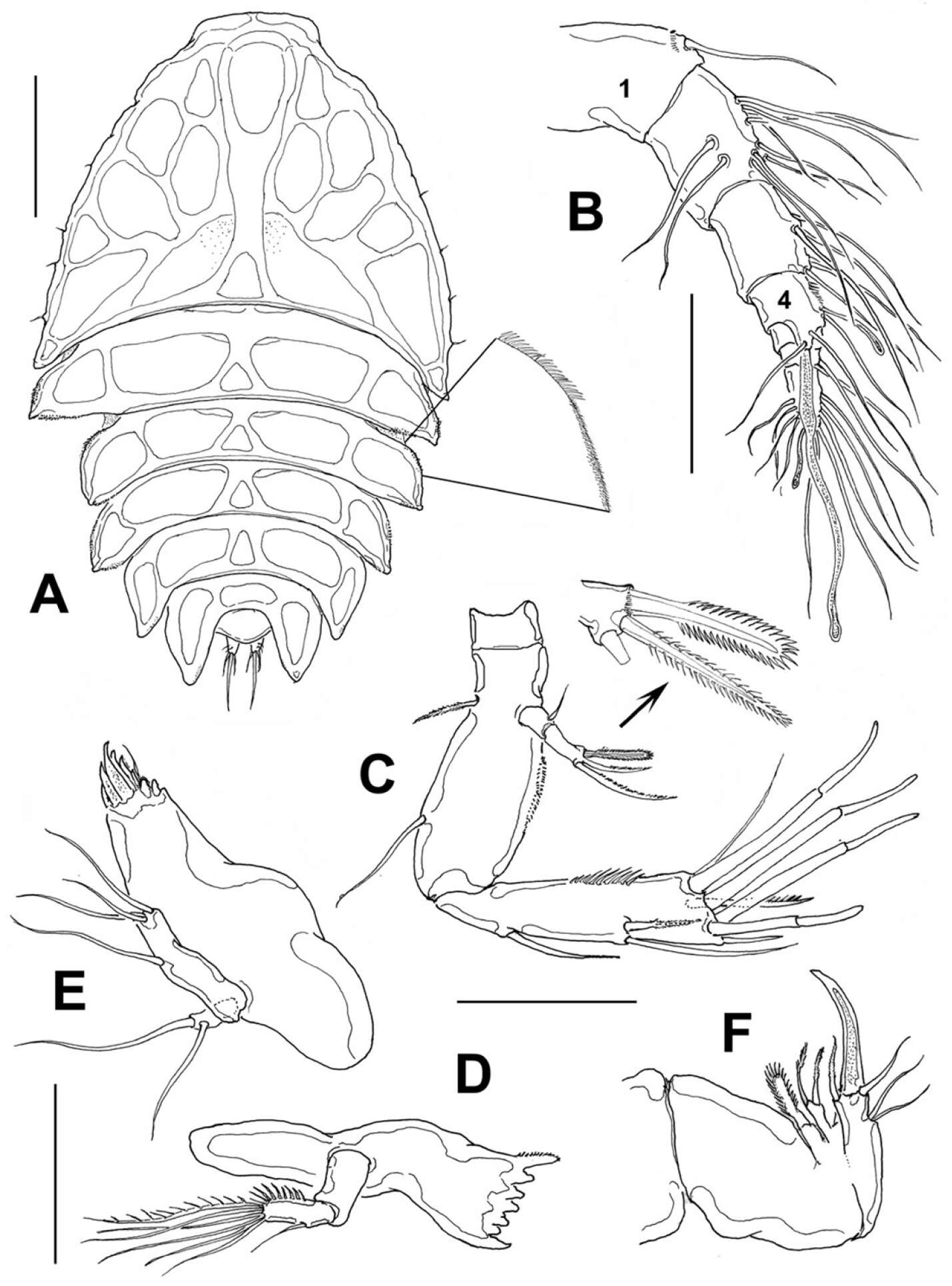
Hyun Woo Bang, Jeffrey G. Baguley, Heejin Moon
Zookeys
Figure 1.Pentacletopsyllus montagni gen. et sp. n. female: A habitus, dorsal B habitus, lateral C cephalothorax, lateral D tooth-like process of cephalothorax lateral anterior margin E rostrum, dorsal F caudal ramus, dorsal.
-
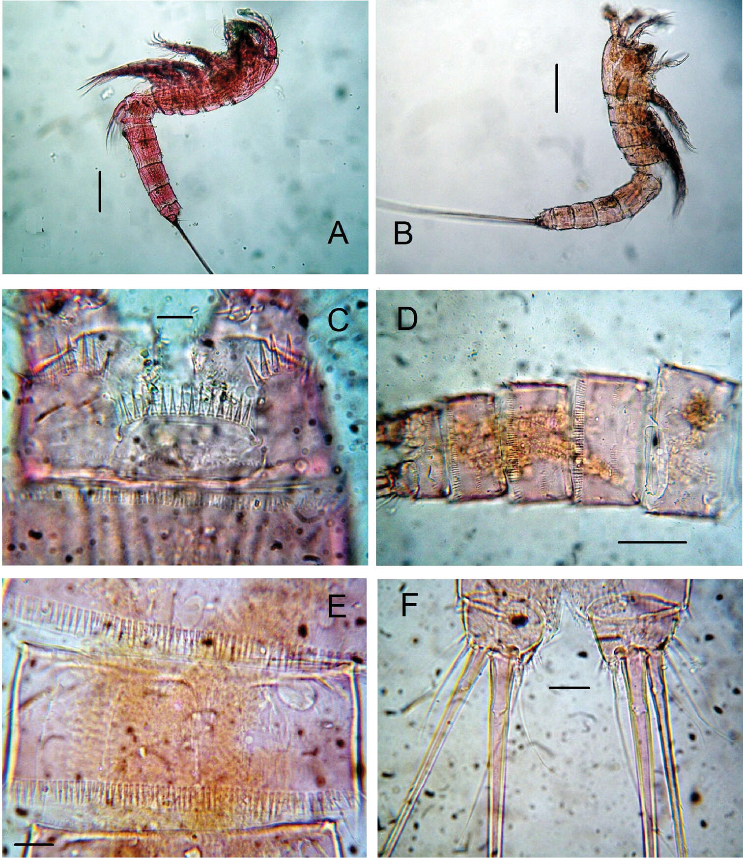
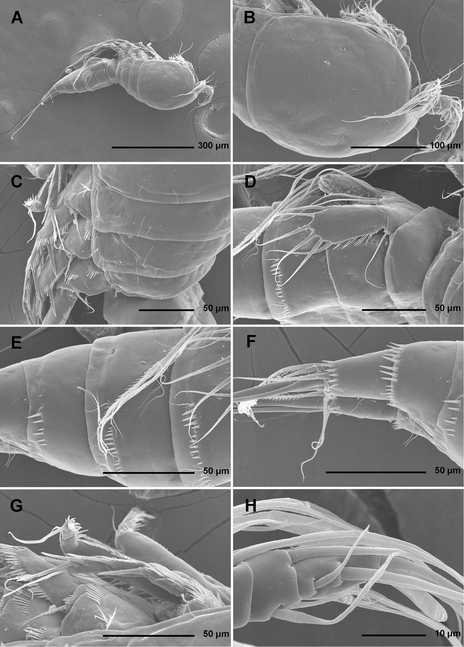
Tomislav Karanovic, Kichoon Kim, Wonchoel Lee
Zookeys
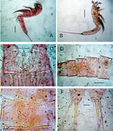
Figure 1.Stenhelia pubescens Chislenko, 1978, scanning electron micrographs, female 1: A habitus, lateral B cephalothorax, lateral C free thoracic somites, lateral D fifth pedigerous somite and genital double-somite, lateral, with one spermatophore attached on ventral side E fourth and fifth urosomites, lateral F anal somite and caudal rami, lateral G first legs and proximal part of second and third legs, lateral H distal part of right antennula, dorsal.
-
Figure 2.Mesocletodes elmari sp. n., adult female, paratype 2. CLSM photograph of a Congo-red stained specimen, lateral view. Scale bar: 100 µm
-
Ribadelago, Castilla y Len, Espaa
-
Guitiriz, Galicia, Spain
-
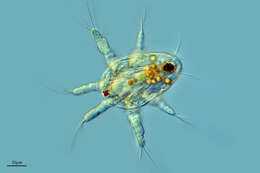
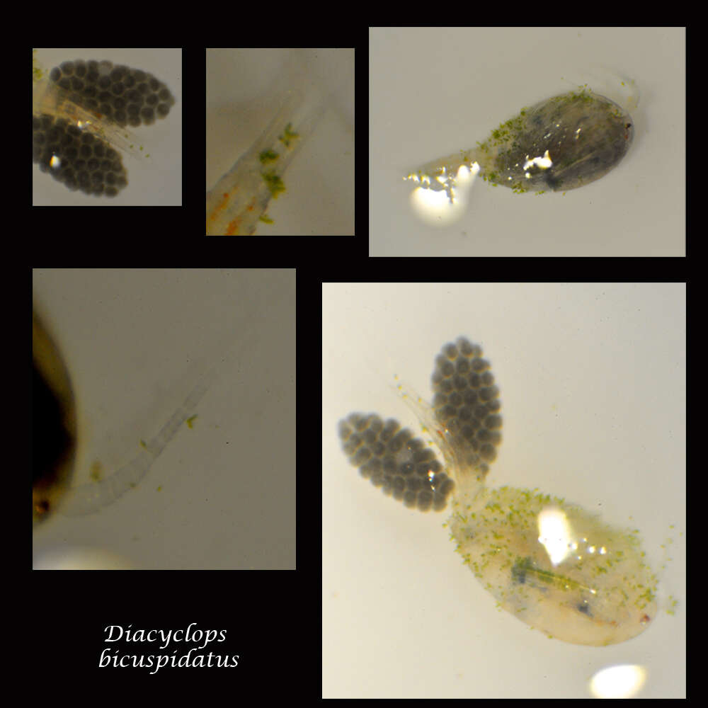
Found in medium sized beaver pond, Hudson NH.Tentative ID.Eleven antennal segments, stout body, short antennae, and small egg sacs on female.
-
-
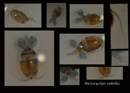
Found in medium sized beaver pond, Hudson NH.Tentative ID.14 Antennal segments, medium sized.
-