-
Terue C. Kihara, Carlos E. F. Rocha
Zookeys
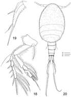
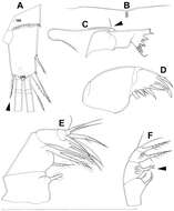
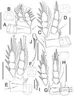
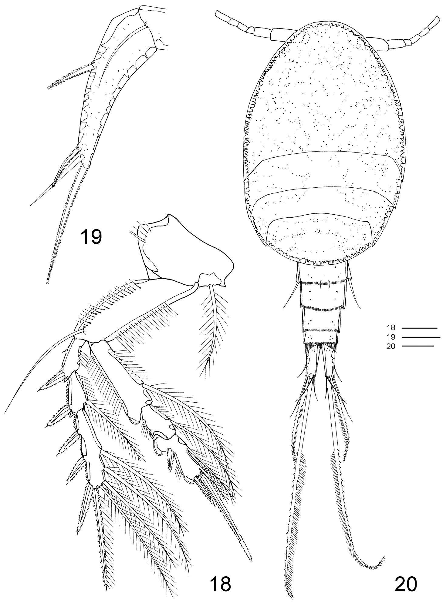
Figures 18–20.Clausidium rodriguesi sp. n. Female: 18 P4, anterior 19 P5, anterior. Male: 20 habitus, dorsal. Scale bars: 18 = 20 μm; 19 = 50 μm; 20 = 100 μm.
-
Mohsen M. El-Sherbiny, Ali M. Al-Aidaroos
Zookeys
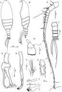
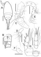
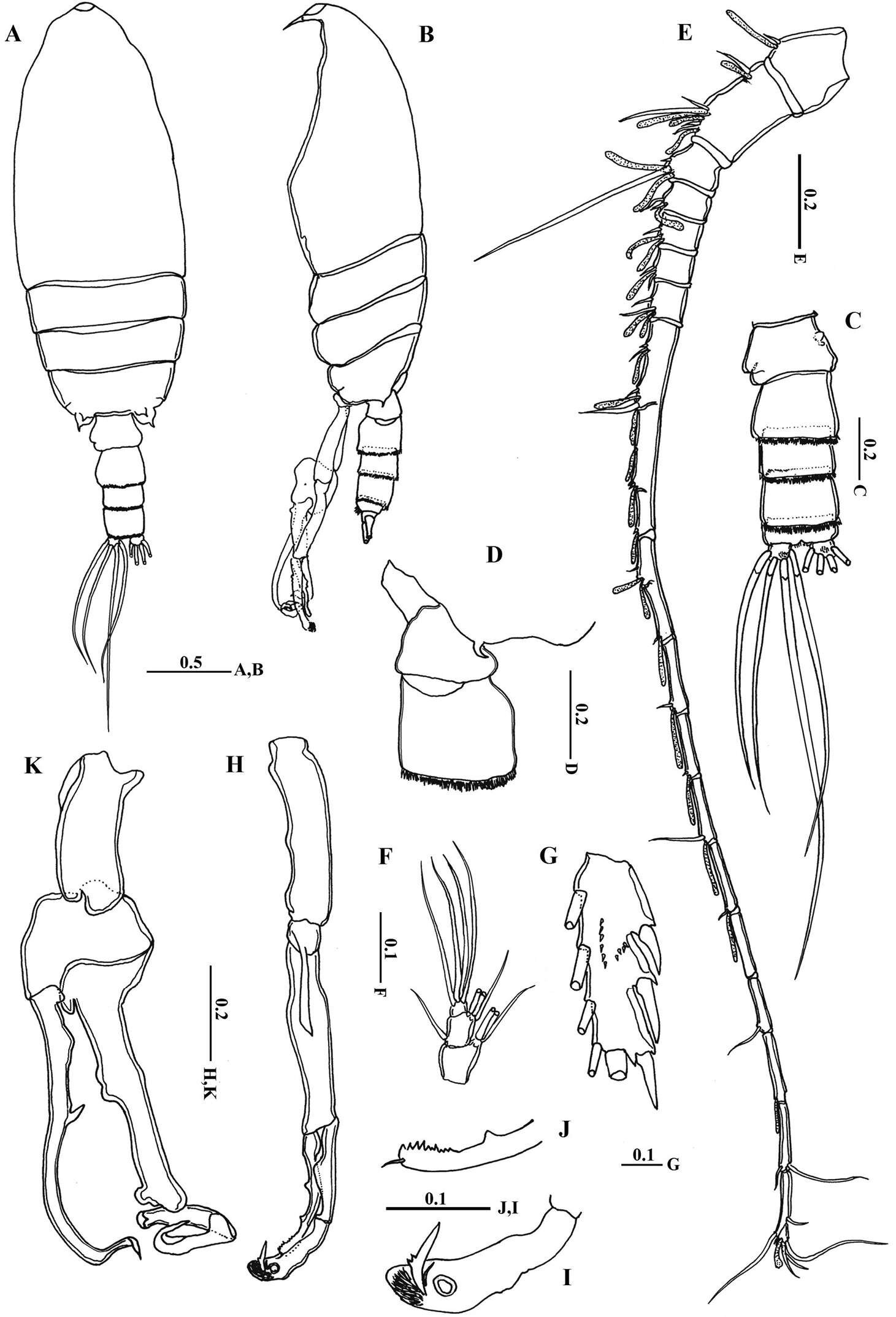
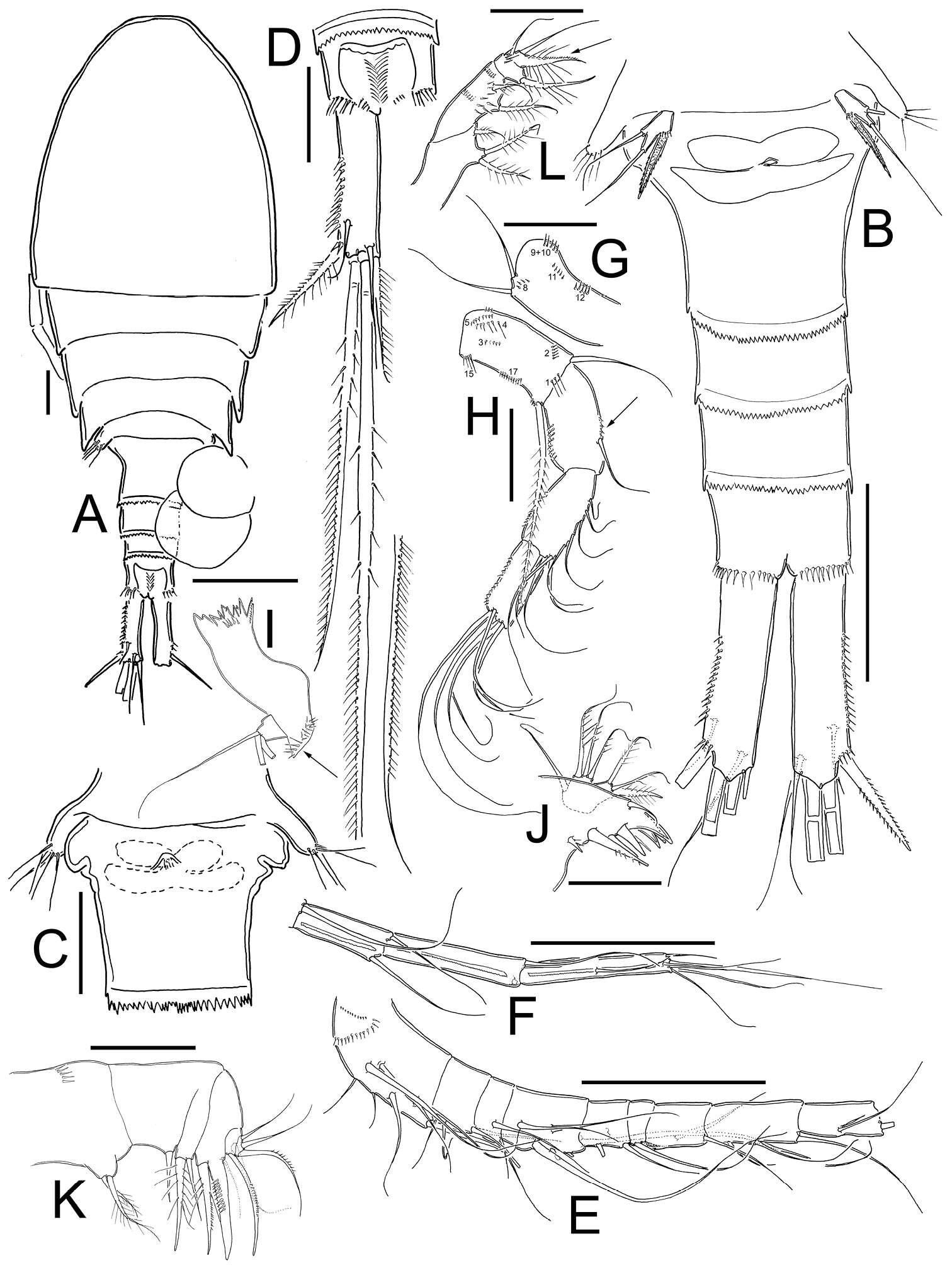
Figure 6.Macandrewella cochinensis male from the northern Red Sea. A habitus, dorsal view B habitus, lateral view C urosome, dorsal view D first and second urosomal segment, lateral view (right) E left antennule F maxilliped, terminal endopod segments G Exopod segment 3 of leg 2 H left leg 5 I terminal portion of left exopodal of leg 5 J terminal portion of left endopod of leg 5 K right leg 5. All scale bars in mm.
-
Juan M. Fuentes-Reinés, Eduardo Suárez-Morales
Zookeys
Figure 5.Nitokra affinis colombiensis ssp. n., adult male from northern Colombia. A first swimming leg (P1) B modified inner basipodal spine of P1 C second swimming leg (P2) D third swimming leg (P3) E fourth swimming leg (P4). Scale bars: A, C–E = 50 μm, B = 10 μm.
-
Hyun Woo Bang, Jeffrey G. Baguley, Heejin Moon
Zookeys
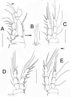
Figure 6.Pentacletopsyllus montagni gen. et sp. n. male: A antennule, ventral B third segment of antennule, anterior C fourth segment of antennule, anterior D fifth segment of antennule, anterior E P4, anterior F P4 endopod 3 (arrow indicating reduced outer seta), anterior G P5, anterior.
-
Tomislav Karanovic, Kichoon Kim, Wonchoel Lee
Zookeys
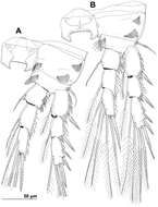
Figure 6.Stenhelia pubescens Chislenko, 1978, line drawings, female 3: A second leg, anterior B third leg, anterior.
-
-
Samuel Gómez, Nicola K. Carrasco, Francisco Neptalí Morales-Serna
Zookeys
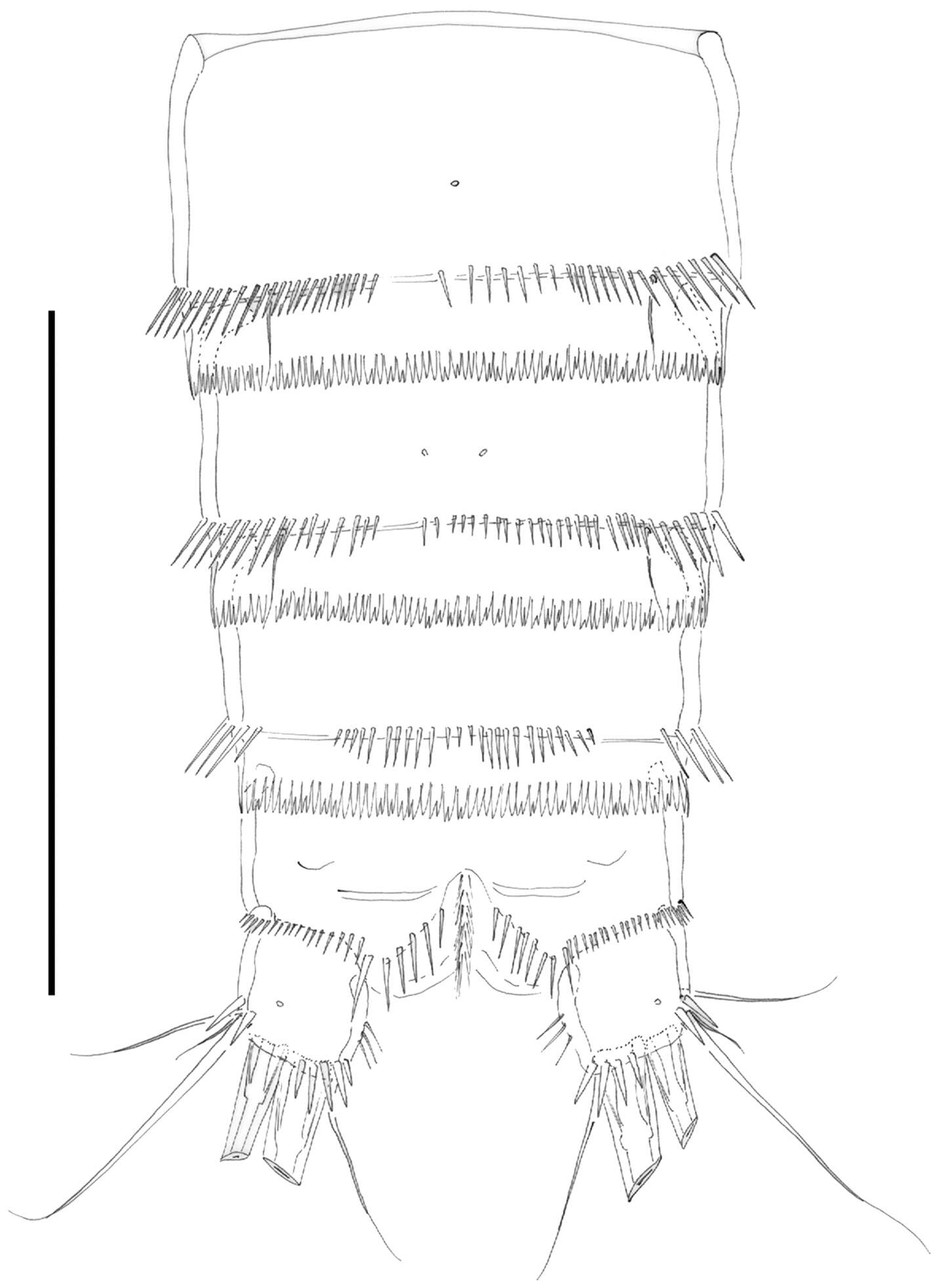
Figure 8.Nitocra taylori sp. n. Male. Urosome, ventral (P5- and P6-bearing somites omitted). Scale bar: 100 µm.
-
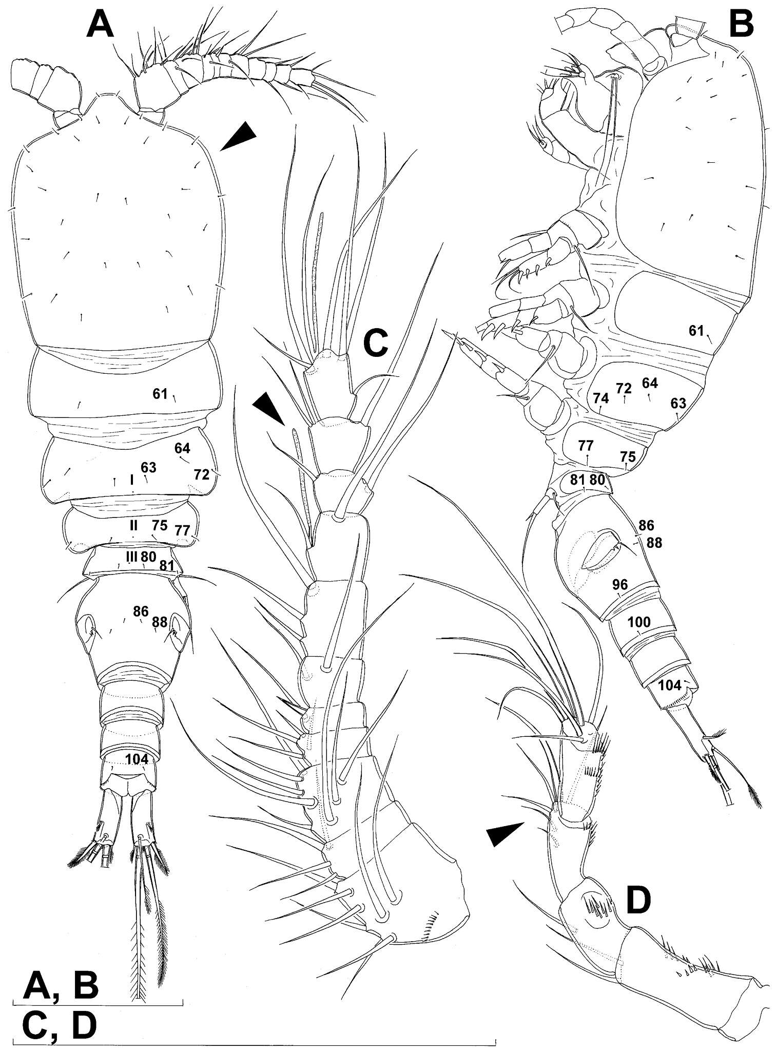
Tomislav Karanovic, Mark J. Grygier, Wonchoel Lee
Zookeys
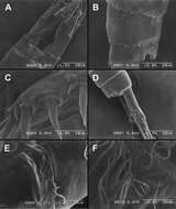
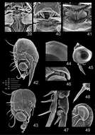
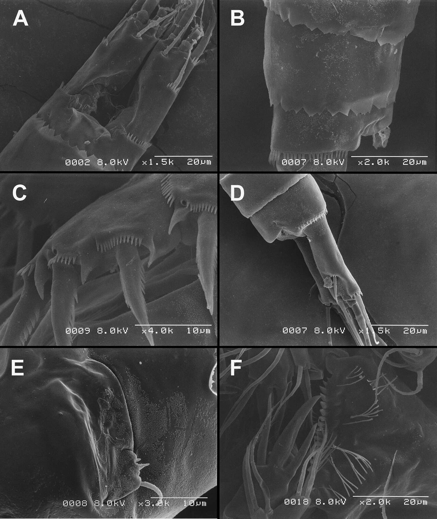
Figure 26.Scanning electron micrographs, A–C Diacyclops ishidai sp. n. D–E Diacyclops parasuoensis sp. n. F Diacyclops suoensis Ito, 1954: A anal somite and caudal rami, dorsal view, paratype female 1 B preanal and anal somites, lateral view, paratype female 2 C last two exopodal segments of second swimming legs, lateral view, paratype female 2 D anal somite and caudal rami, lateral view, paratype female E sixth leg, lateral view, paratype female F labrum and maxillulae, ventral view. Scale bars 20 μm (A, B, D, F) and 10 μm (C, E).
-
Terue C. Kihara, Carlos E. F. Rocha
Zookeys
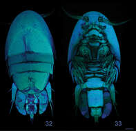
Figures 32–33.Clausidium rodriguesi sp. n. Female: Confocal laser scanning microscopy maximum projections33 habitus, dorsal 34 habitus, ventral. Scale bars: 100 μm.
-
Martha Angélica Gutiérrez-Aguirre, Nancy Fabiola Mercado-Salas, Adrián Cervantes-Martínez
Zookeys
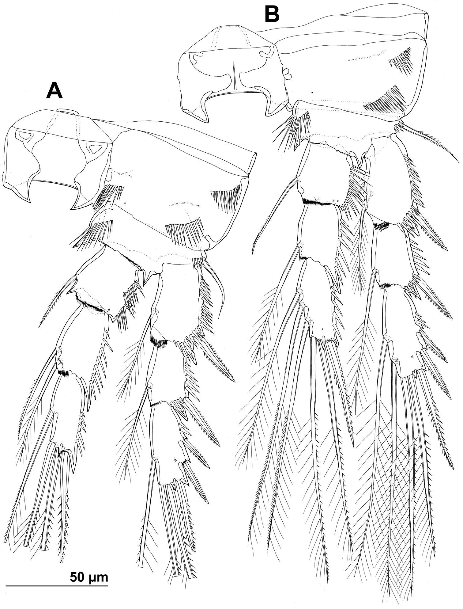
Figure 2.Eucyclops tziscao sp. n. A, C, D paratype B, E–L holotype from Laguna Tziscao, Chiapas. A Habitus, dorsal B Urosome C Genital double-somite, ventral D Anal somite and caudal ramus, dorsal E Antennule, segments 1–9 F Antennule, segments 10–12 G Antenna, caudal H Antenna, frontal I Mandible J Maxillule, caudal K Maxilla, frontal L Maxilliped, frontal. Scales bars: K = 20 µm; A, C, D, G, H, I, J, L = 50 µm; B, E, F = 100 µm.
-
Tomislav Karanovic, Kichoon Kim, Wonchoel Lee
Zookeys
Figure 7.Stenhelia pubescens Chislenko, 1978, line drawings, female 3: A fourth leg, anterior B fifth leg, dissected and flattened, anterior.
-
Samuel Gómez, Nicola K. Carrasco, Francisco Neptalí Morales-Serna
Zookeys
Figure 9.Nitocra taylori sp. n. Male. A antennule B fifth, sixth and seventh segments of the antennule, showing modified setae and blunt processes C eight segment of the antennule D P1 basis, anterior E P3ENP F P5, anterior G P6, anterior. Scale bar: A, E=50 µm; B, C=67 µm; D, F, G=35 µm.
-
Tomislav Karanovic, Mark J. Grygier, Wonchoel Lee
Zookeys
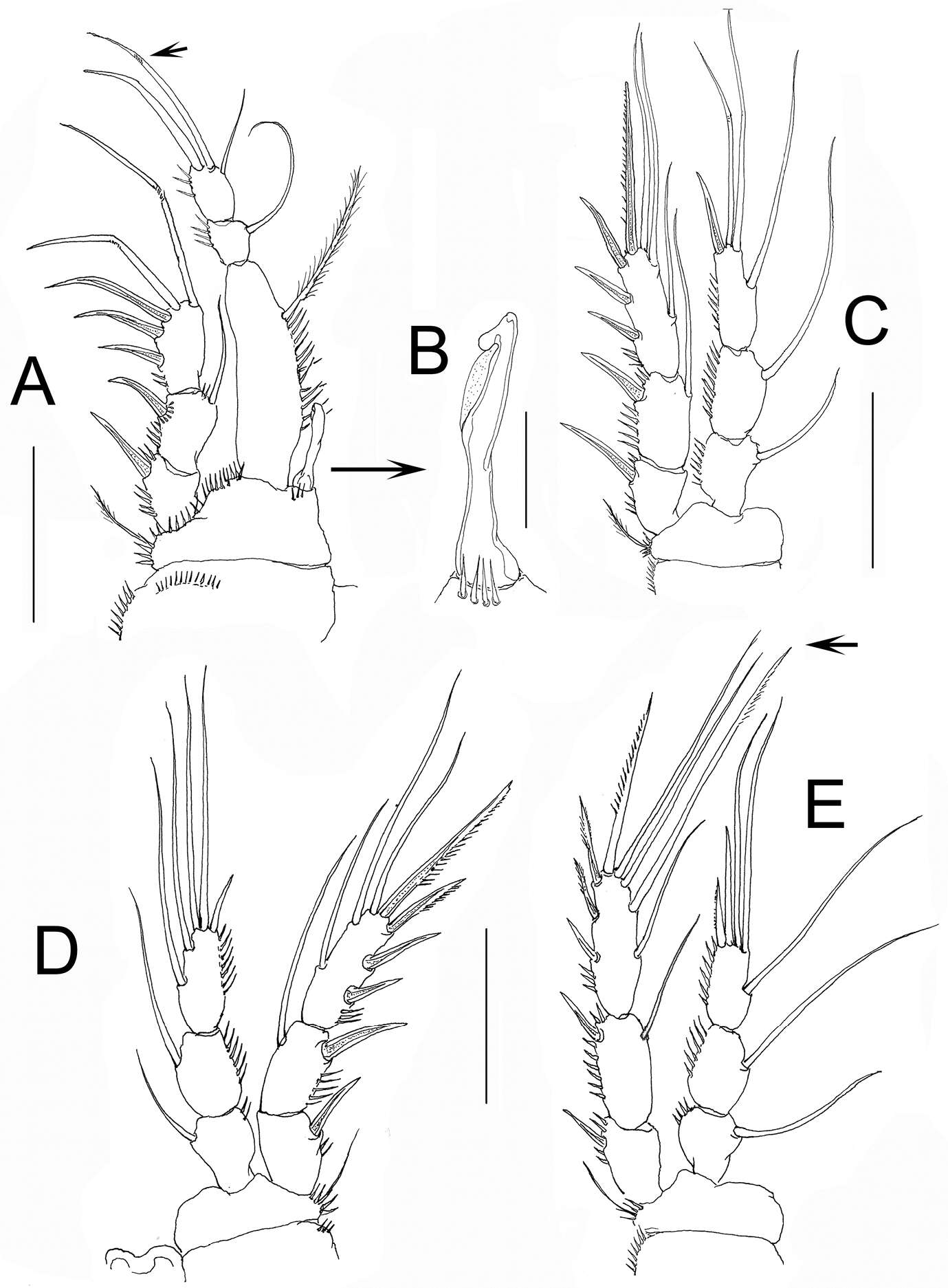
Figure 7.Diacyclops brevifurcus Ishida, 2006, holotype female: A left caudal ramus, ventral view B copulatory pore, ventral view C mandibula, posterior view D maxillula, posterior view (palp broken off) E maxilla, anterior view F maxilliped, anterior view. Arabic numerals indicating sensilla and pores presumably homologous to those in Diacyclops ishidai sp. n. Arrows pointing most prominent specific features. Scale bar 100 μm.
-
Terue C. Kihara, Carlos E. F. Rocha
Zookeys
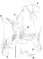
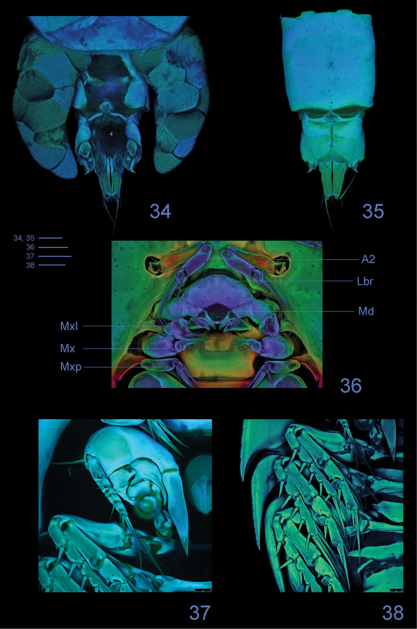
Figures 34–38.Clausidium rodriguesi sp. n. Female: Confocal laser scanning microscopy maximum projections 34 urosome, dorsal 35 urosome lacking somite bearing P5, ventral 36 antenna and oral region 37 P1, anterior 38 P2-P4, anterior. Scale bars: 50 μm.
-
Martha Angélica Gutiérrez-Aguirre, Nancy Fabiola Mercado-Salas, Adrián Cervantes-Martínez
Zookeys
Figure 4.Eucyclops tziscao sp. n. A–B paratype C–G allotype from Laguna Tziscao, Chiapas. A Habitus, dorsal B P5, and P6 C Antennule, segments 1–14 D Antennule, segments 15–16 E Antenna, frontal F Antenna, caudal G P4, caudal. Scales bars: B–G = 50 µm; A = 100 µm.
-
Tomislav Karanovic, Kichoon Kim, Wonchoel Lee
Zookeys
Figure 7.Stenhelia pubescens Chislenko, 1978, line drawings, female 3: A fourth leg, anterior B fifth leg, dissected and flattened, anterior.
-
Daisuke Uyeno, Kaori Wakabayashi, Kazuya Nagasawa
Zookeys
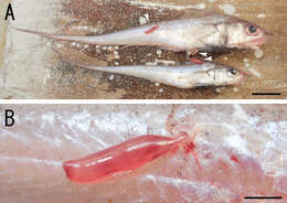
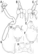
Figure 1.Sarcotretes umitakae sp. n., female on Coelorinchus jordani Smith and Pope. A two specimens of Coelorinchus jordani (181.5 mm TL and 142.8 mm TL) carrying the type series of Sarcotretes sp. n. (arrowheads) B coloration in life of paratype NSMT–Cr 22254 attached to host’s body. Scale bars: A=20 mm; B=3 mm.
-
Tomislav Karanovic, Mark J. Grygier, Wonchoel Lee
Zookeys
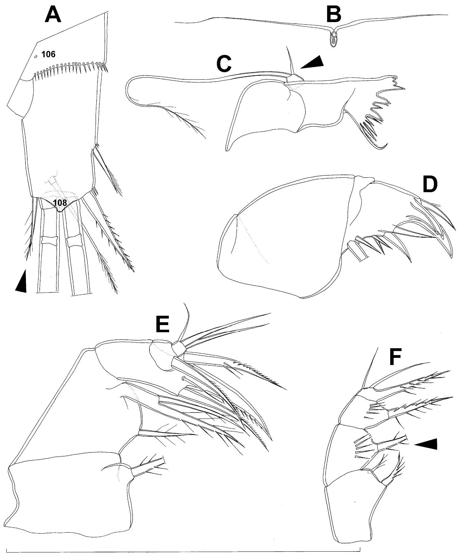
Figure 8.Diacyclops brevifurcus Ishida, 2006, holotype female: A third swimming leg, posterior view B fourth swimming leg, posterior view C fifth leg, anterior view. Arrows pointing most prominent specific features. Scale bars 100 μm.
-
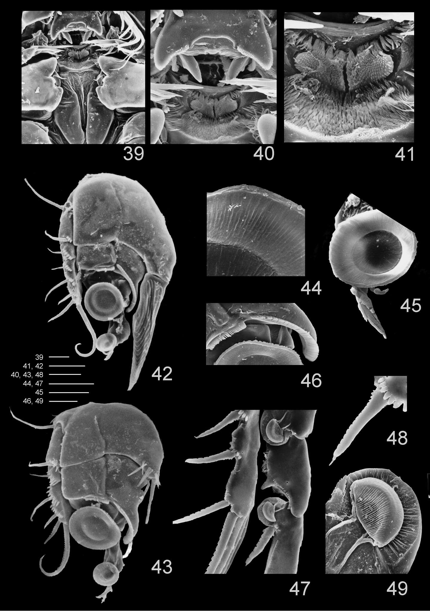
Terue C. Kihara, Carlos E. F. Rocha
Zookeys
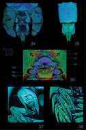
Figures 39–49.Clausidium rodriguesi sp. n.: Scanning electron microscopy photos 39 metastomal area, male 40 detail of metastomal area, male 41 detail of metastomal area, male 42 P1, anterior, female 43 P1, anterior, male 44 detail of sucking disc of P1, male 45 detail of lobe with serrate margin and distal sucking disc of enp-3 of P1, female 46 detail of P1 enp-1 adhesive fringe, male 47 sucking discs of P2, female 48 detail of serrate spine with apical flagellum of P2, female 49 detail of sucking disc of P2, female. Scale bars: 39, 40, 47 = 25 μm; 41, 44–46 = 10 μm; 42 = 35 μm; 43 = 20 μm; 48 = 12.5 μm; 49 = 4 μm.
-
Martha Angélica Gutiérrez-Aguirre, Nancy Fabiola Mercado-Salas, Adrián Cervantes-Martínez
Zookeys
Figure 3.Eucyclops tziscao sp. n. Holotype from Laguna Tziscao, Chiapas. A P1, frontal B Intercoxal sclerite of P1, caudal C P2, frontal D Intercoxal sclerite of P2, caudal E P3, frontal, Exp and Enp separated F Intercoxal sclerite of P3, caudal G P4, caudal H Intercoxal sclerite of P4, frontal I Coxal spine P4 J P5. Scales bars: I= 25µm, J= 50 µm; A–H = 100 µm.
-
Tomislav Karanovic, Kichoon Kim, Wonchoel Lee
Zookeys
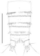
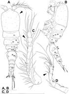
Figure 8.Stenhelia taiae Mu & Huys, 2002, scanning electron micrographs, female: A habitus, lateral B cephalothoracic shield, lateral C free thoracic somites, lateral D fifth pedigerous somite and genital double-somite, lateral E fourth and fifth urosomites, lateral F anal somite and caudal rami, lateral G posterior part of right caudal ramus, lateral H rostrum, lateral. Arrowheads indicate morphological characters different from those in Stenhelia pubescens Chislenko, 1978.
-
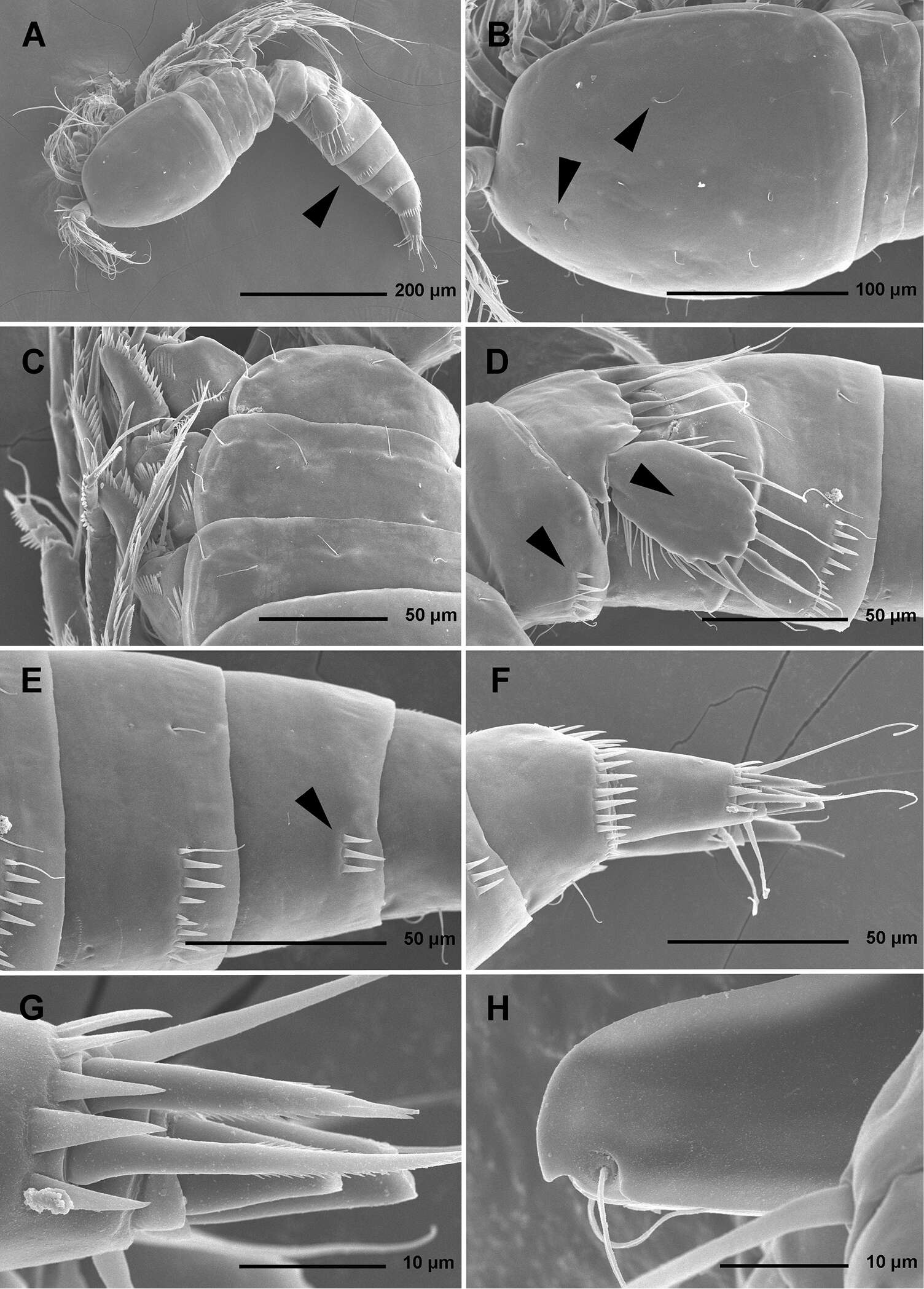
Daisuke Uyeno, Kaori Wakabayashi, Kazuya Nagasawa
Zookeys
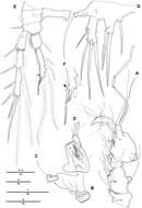
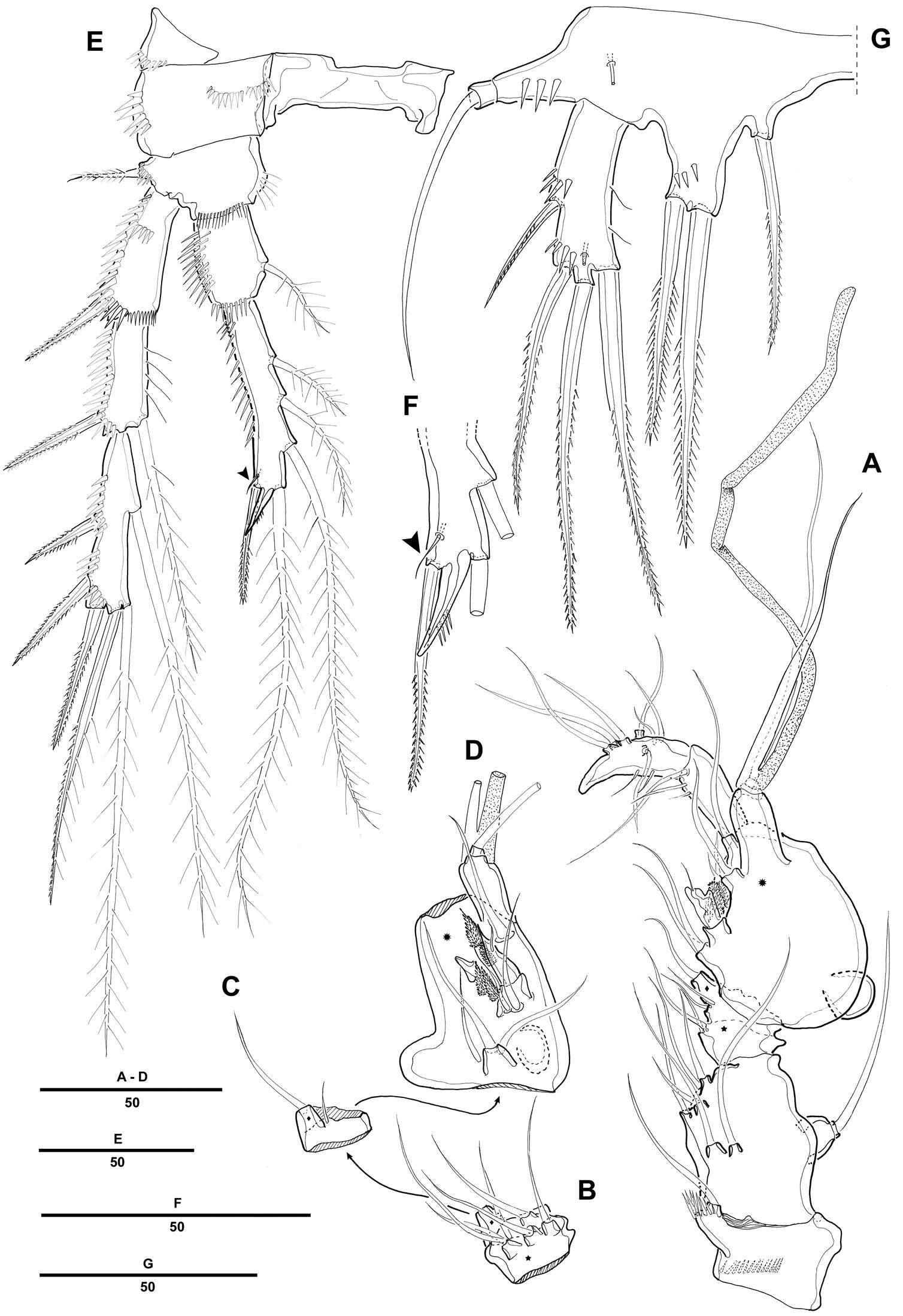
Figure 2.Sarcotretes umitakae sp. n., female, holotype NSMT–Cr 22253. A habitus B anterior portion of body, dorsal, a1 = antennule, a2 = antenna C same, ventral, m1 = maxillule, m2 = maxilla, p1 = leg 1, p2 = leg 2, p3 = leg 3, p4 = vestige of leg 4 D vestige of dorsal cephalothoracic shield E tip of proboscis, lateral F posterior portion of body, ventral G same, lateral H rostral area and antennae, dorsal. Scale bars: A=3 mm; B, C, F, G=1 mm; D=500 μm; E=300 μm; H=150 μm.
-
Tomislav Karanovic, Mark J. Grygier, Wonchoel Lee
Zookeys
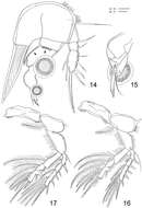
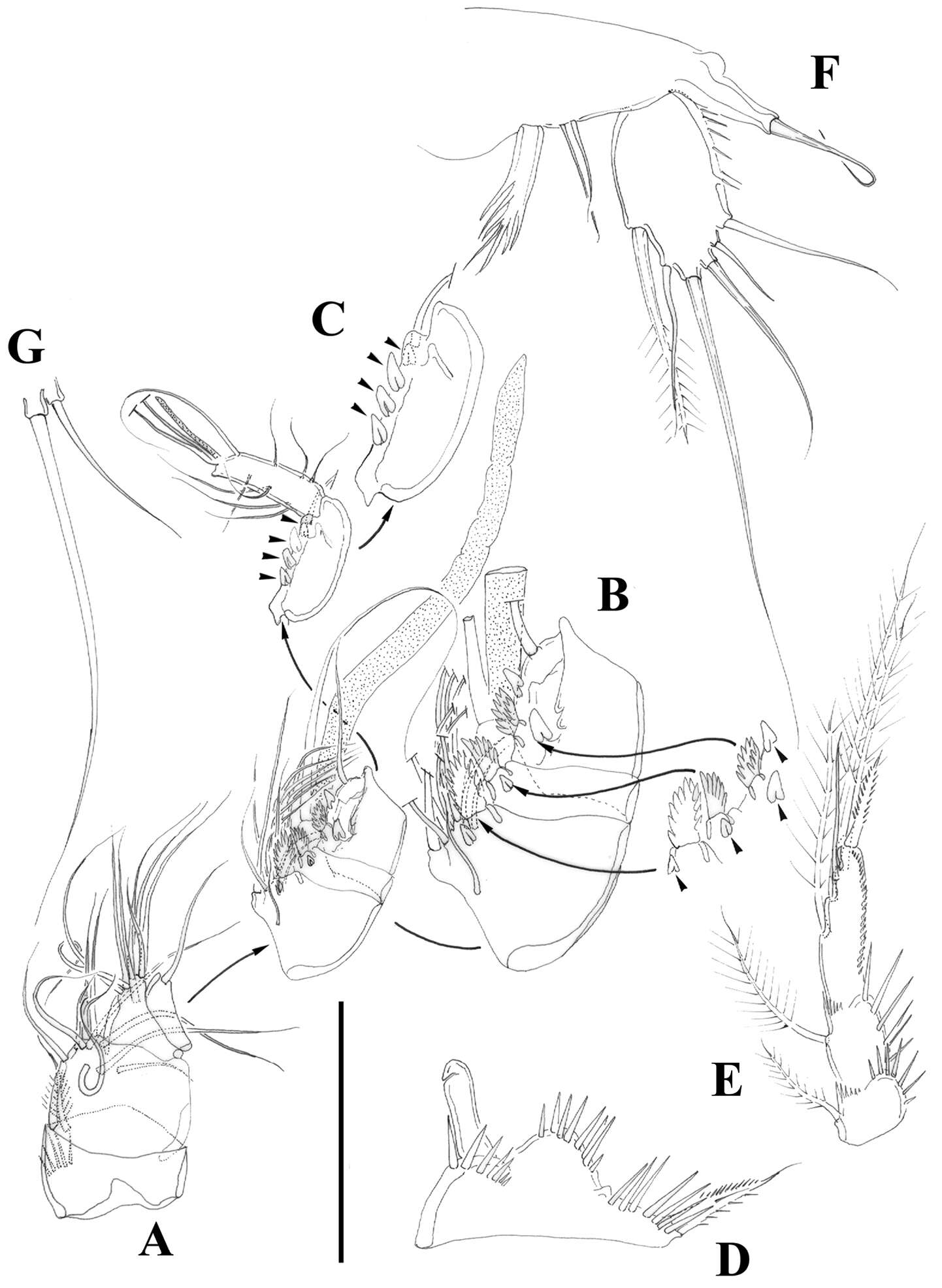
Figure 9.Diacyclops parasuoensis sp. n., holotype female: A habitus, dorsal view B habitus, lateral view C antennula, ventral view D antenna, dorsal view. Arabic numerals indicating sensilla and pores presumably homologous to those in Diacyclops ishidai sp. n. Roman numerals indicating pores not present in Diacyclops ishidai sp. n. Arrows pointing most prominent specific features. Scale bars 100 μm.
-
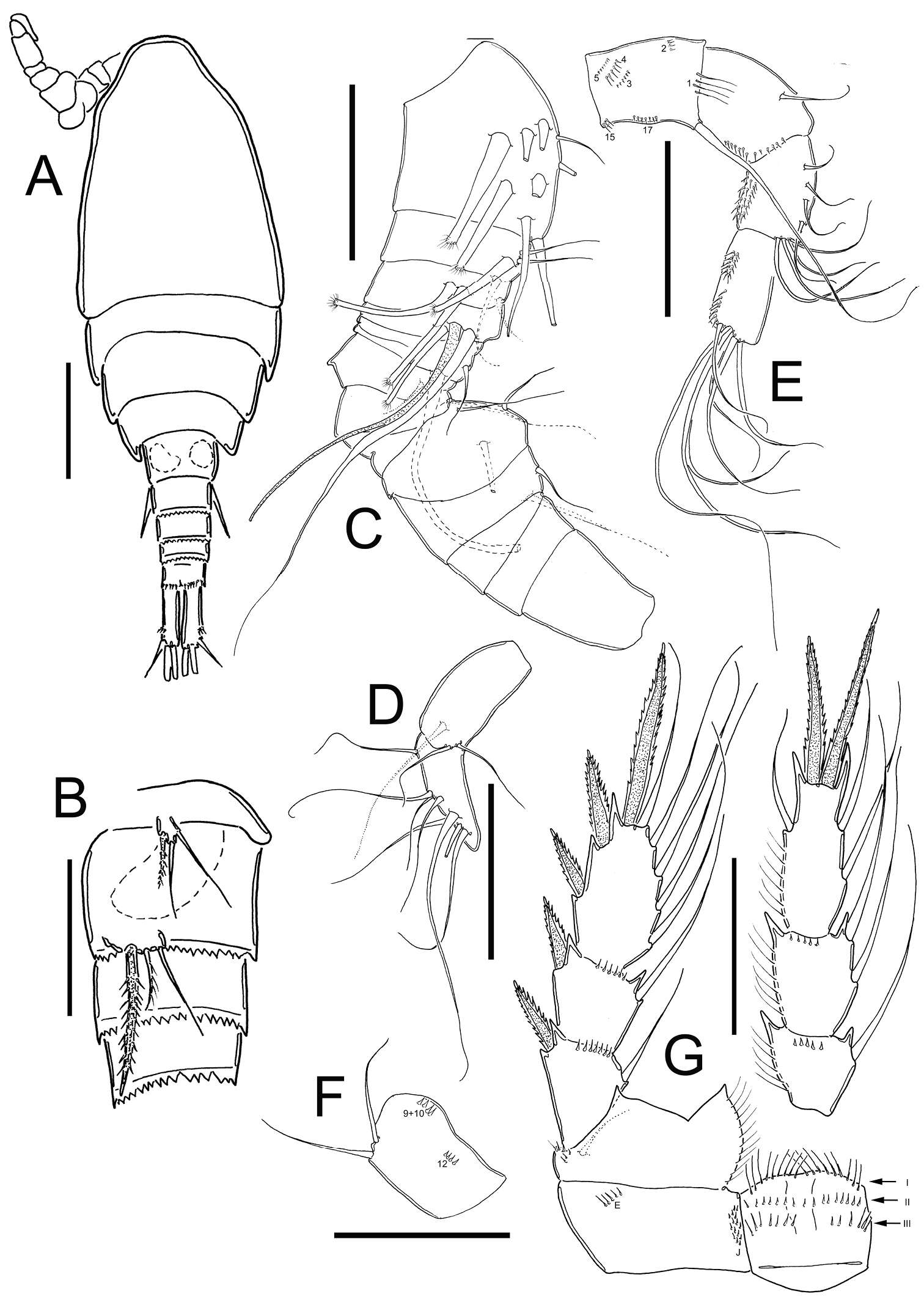
Terue C. Kihara, Carlos E. F. Rocha
Zookeys
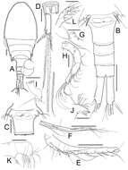
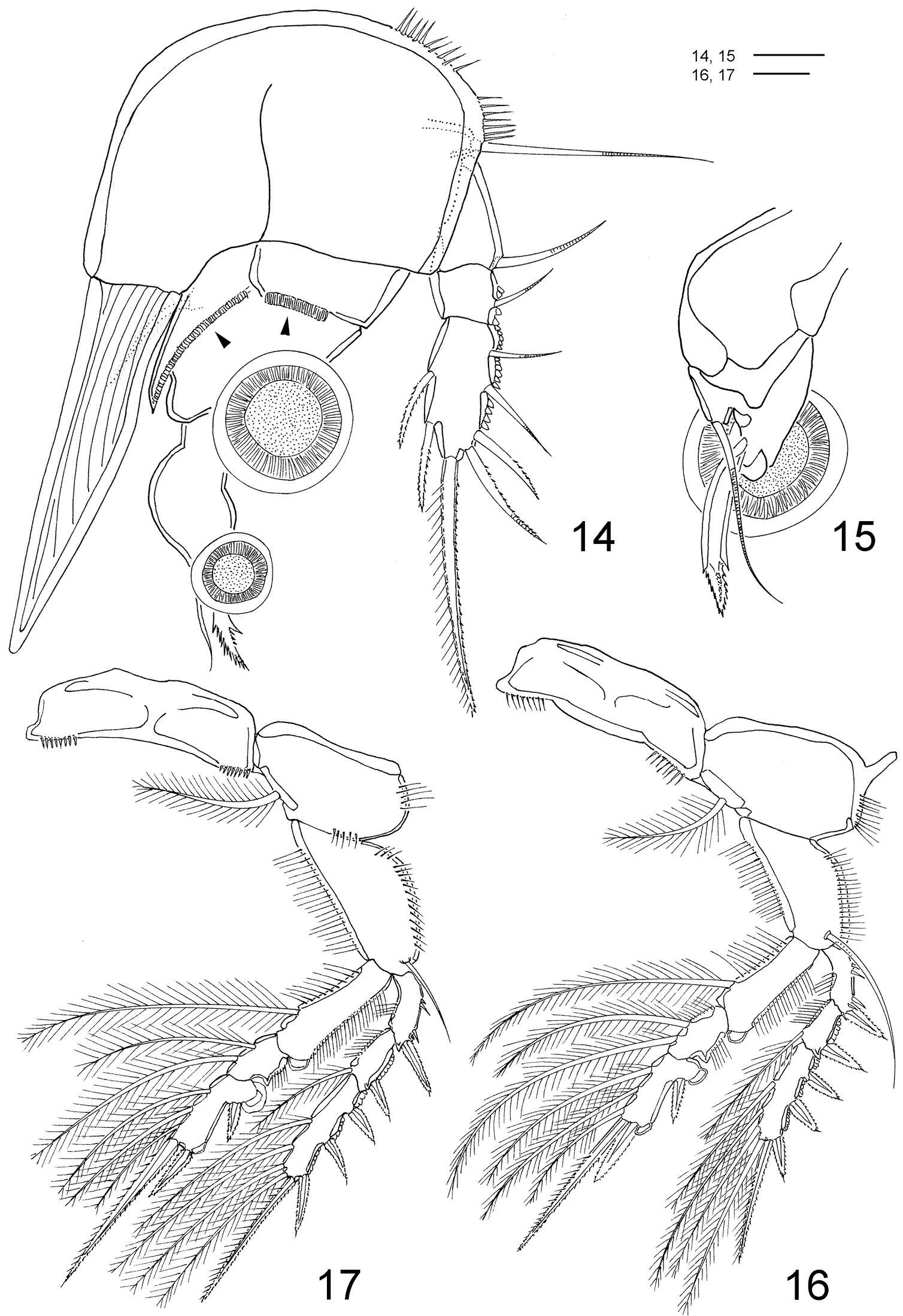
Figures 14–17.Clausidium rodriguesi sp. n. Female: 14 P1, anterior (arrows indicating adhesive fringe) 15 detail of distal area of P1 endopod, posterior 16 P2, anterior 17 P3, anterior. Scale bars: 14 = 20 μm; 15 = 10 μm; 16, 17 = 50 μm.