-
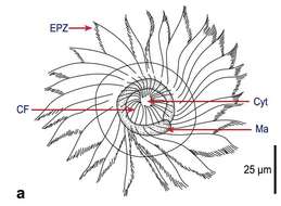
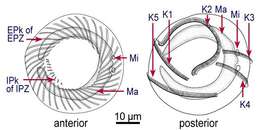
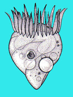
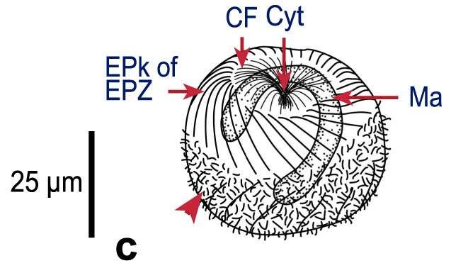
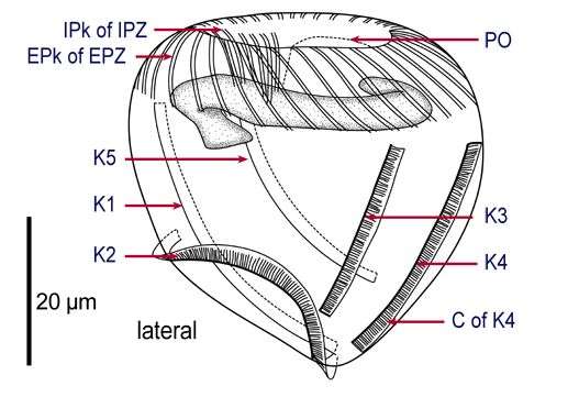
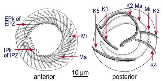
Fig 1a: Strobilidium sphaericum Line drawings of protargol stained cells, showing kineties, oral structures and nucleus: Apical view;
-
-
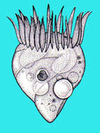
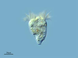
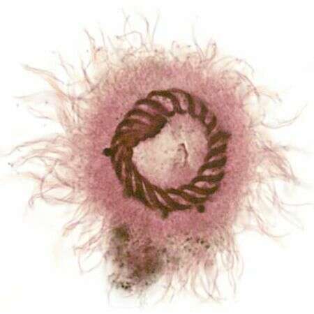
Strobilidium caudatum (Fromental, 1874) Foissner, 1987, a spirotrich ciliate. Synonym of S. gyrans.The cell body is goblet shaped with a complete circular (closed type) wreath of membranelles at the anterior end. The adoral zone of membranelles is well seen in this image. Unlike Strombidium, there is no posterior lorica. There are five very reduced rows of somatic cilia which converge at the posterior end of the cell to form a tight spiral as seen in this image. The terminus of the cell secretes a mucus thread allowing attachment to the substrate. Once attached the cell moves back and forth in pendulum fashion for a time then breaking free to swim away very rapidly. The oral aperture is eccentrically located within the adoral zone of membranelles. A peripheral contractile vacuole is seen posterolaterally in this image. Feeds on diatoms, flagellates and probably bacteria. From freshwater pond near Boise, Idaho. Brightfield illumination.
-

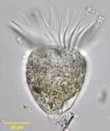
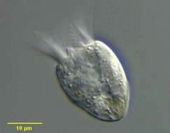
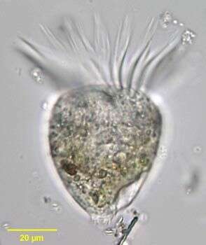
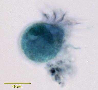
Detail of somatic ciliary row of the oligotrich ciliate, Rimostrombidium hyalinum. This genus is distinguished from the similar Strombidium by the small size and the termination of the spiral somatic ciliary rows before they reach the posterior pole of the cell (in Strombidium they form a terminal spiral). R. hyalinum is colorless. The cell body is transversely truncate anteriorly and bluntly conical posteriorly. The cytostome is at the anterior end. A circular adoral zone of membranelles is present (seen well here). There are six indistinct, slightly spiraling rows of short somatic cilia that extend nearly the length of the body. It is seldom possible to keep more than one row in the focal plane in the lateral view (part of one row is seen here on viewer's left). The C- shaped macronucleus is anterior and transversely oriented (not seen here). The peripheral contractile vacuole is located in the posterior of the cell (not seen here). Collected from freshwater pond near Boise, Idaho September 2003. DIC optics.
-
Rumoroso, Cantabria, Spain
-
Fig 1b: Strobilidium sphaericum Line drawings of protargol stained cells, showing kineties, oral structures and nucleus: View from posterio-lateral
-
Anterior apical view of the adoral zone of membranelles of Strobilidium caudatum (Fromental,1874) Foissner,1987, a spirotrich ciliate. Synonym of S. gyrans.Stained by the silver carbonate technique (see Foissner, W. Europ. J. Protistol., 27:313-330;1991).Brightfield.
-
Methyl green-Pyronin stained preparation of the oligotrich ciliate, Rimostrombidium hyalinum demonstrating the C-shaped transversely oriented anterior macronucleus. The small spherical micronucleus is superimposed on the center of the macronucleus in this image. Collected from freshwater pond near Boise, Idaho September 2003. DIC optics.
-
Vitoria, Euskadi, Espaa
-
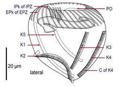
Fig 1c: Strobilidium sphaericum Line drawings of protargol stained cells, showing kineties, oral structures and nucleus: lateral view.
-
Strobilidium caudatum (FROMENTAL, 1874) FOISSNER, 1987, a spirotrich ciliate. Synonym of S. gyrans.The cell body is goblet shaped with a complete circular (closed type) wreath of membranelles at the anterior end. The adoral zone of membranelles is well seen in this image. Unlike Strombidium, there is no posterior lorica. There are five very reduced rows of somatic cilia which converge at the posterior end of the cell to form a tight spiral as seen in this image.Three of these are seen in this image (red arrowheads).The posterior terminus of the cell secretes a mucus thread allowing attachment to the substrate. Once attached the cell moves back and forth in pendulum fashion for a time then breaks free to swim away very rapidly. The cytostome is eccentrically located within the adoral zone of membranelles. From freshwater pond near Boise, Idaho. Protargol (see Foissner, W. Europ. J. Protistol., 27:313-330;1991).Brightfield.
-
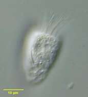
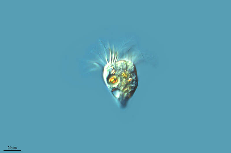
Portrait of the oligotrich ciliate, Romstrombidium hyalinum. This genus is distinguished from the similar Strombidium by the small size and the termination of the spiral somatic ciliary rows before they reach the posterior pole of the cell (in Strombidium they form a terminal spiral). There are several species. R. hyalinum is colorless. The cell body is transversely truncate anteriorly and bluntly conical posteriorly. The cytostome is at the anterior end. A circular adoral zone of membranelles is present. There are six indistinct, slightly spiraling rows of short somatic cilia that extend nearly the length of the body. It is seldom possible to keep more than one row in the focal plane in the lateral view. The C- shaped macronucleus is anterior and transversely oriented The peripheral contractile vacuole is located in the posterior of the cell. A food vacuole is seen here posteriorly. Collected from freshwater pond near Boise, Idaho September 2003. DIC optics.
-
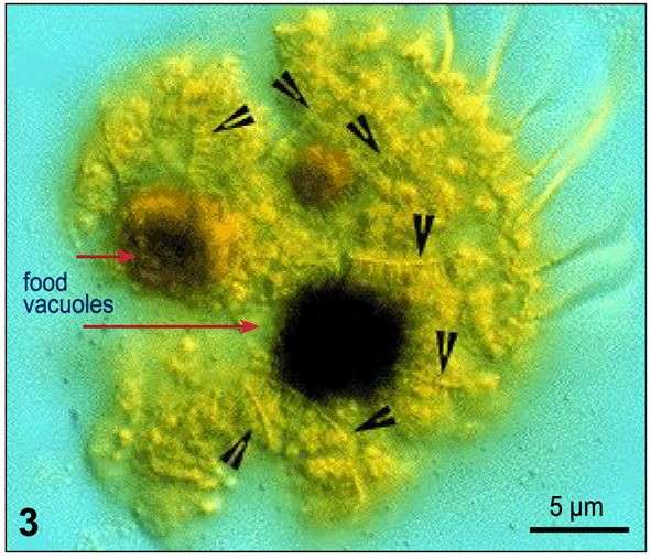
Fig 3: Strobilidium sphaericum Ruptured cell, showing kinety fragments (arrowheads).
-
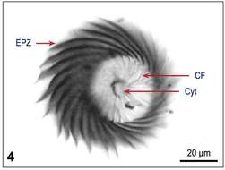
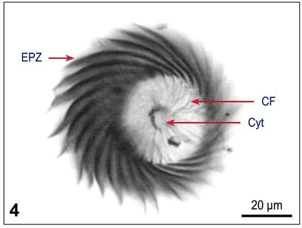
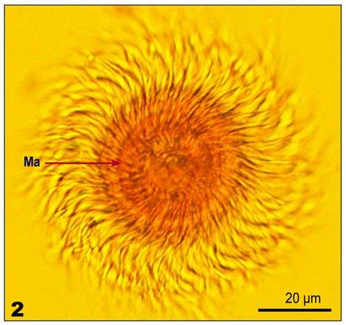
Fig 4: Strobilidium sphaericum Protargol-stained cell: Apical view, showing oral structures
-
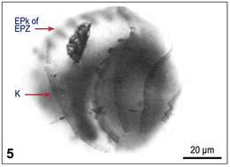
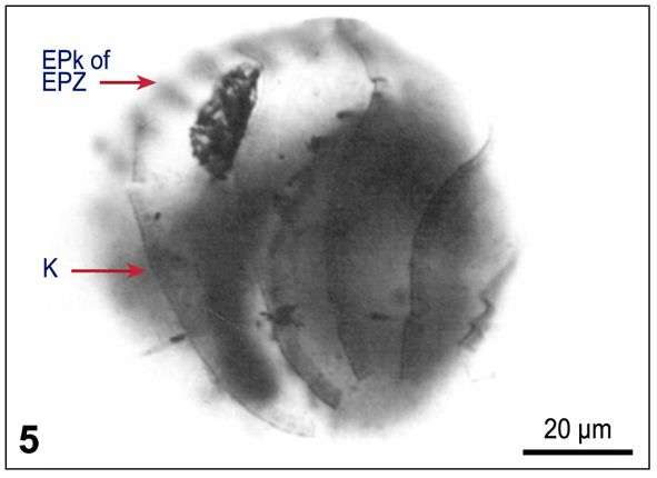
Fig 5: Strobilidium sphaericum Protargol-stained cell: posterio-lateral view showing somatic kineties.
-
Fig 1a: Strobilidium neptuni Line drawing of protargol stained cell
-
Fig 1b: Strobilidium neptuni Line drawing of protargol stained cells, showing kineties, oral structures and nuclei.
-
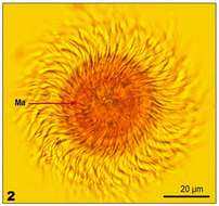
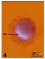
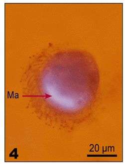
Fig 2: Strobilidium neptuni Lugol's fixed cell. Oral region, viewed from apical end with Ma
-
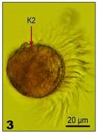
Fig 3: Strobilidium neptuni Lugol's fixed cell. Lateral view, showing the flattened region (arrow)
-
Fig 4: Strobilidium neptuni Lugol's fixed and DAPI stained cell, illustrating nuclear shape
-
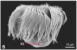
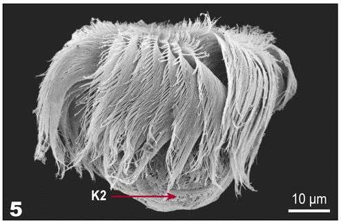
Fig 5: Strobilidium neptuni SEM of Lugol's fixed cell
-
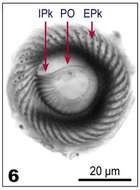
Fig 6: Strobilidium neptuni protargol stained cell, viewed from apical end: Oral region of the cell
-
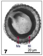
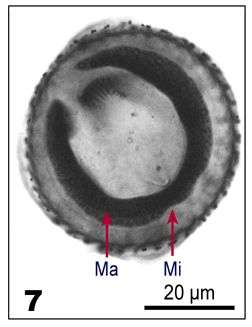
Fig 7: Strobilidium neptuni protargol stained cell, viewed from apical end: Macronucleus, showing indentations in macronucleus where micronuclei sit
-
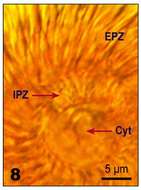
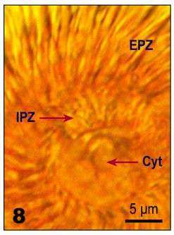
Fig 8: Strobilidium neptuni Lugol's fixed cell, details of the oral structure