James Davis Reimer, Yuka Irei, Takuma Fujii
Zookeys
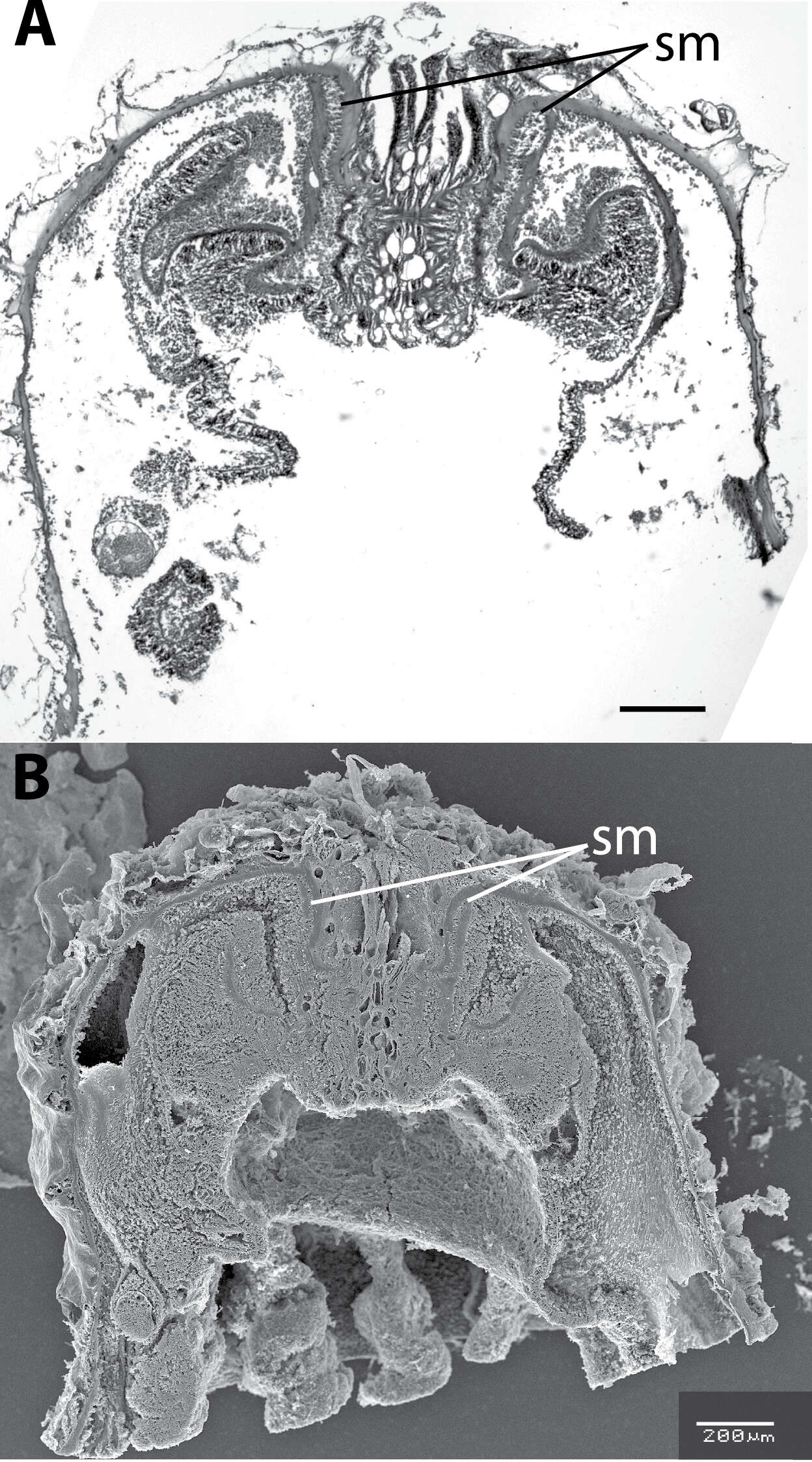
Figure 2.Longitudinal section of Neozoanthus caleyi sp. n. specimen HI225 showing endodermal sphincter muscle (=sm). A Light microscope B scanning electron microscope. Both scales =200 µm.
James Davis Reimer, Yuka Irei, Takuma Fujii
Zookeys
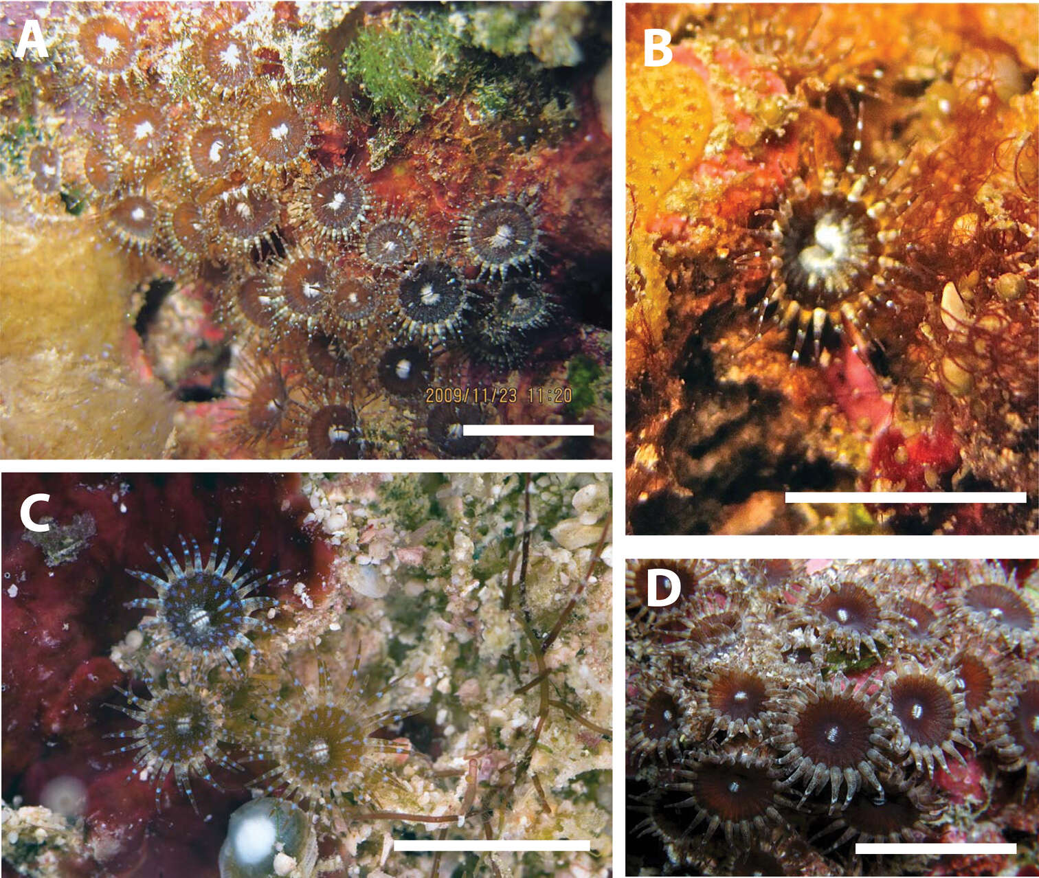
Figure 3.Neozoanthus caleyi sp. n. in situ around Heron Island on the Great Barrier Reef, Queensland, Australia. A Specimen HI214 at Sykes Reef, depth=9 m, November 23, 2009 B Close-up of a single polyp showing yellow coloration at base of tentacles; specimen HI145 at Sykes Reef, depth=18 m, November 18, 2009 C Specimen HI231 at Heron Channel, depth=23 m, November 24, 2009 D Uncollected specimen at Heron Channel, depth=approximately 20 m, November 2011. Scales approximately 1 cm. A, B taken by JD Reimer, C, D taken by Gary Cranitch.
James Davis Reimer, Yuka Irei, Takuma Fujii
Zookeys
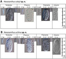
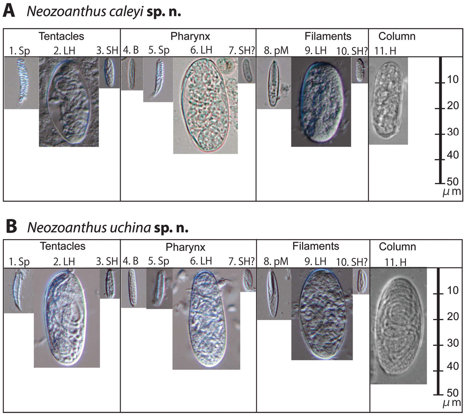
Figure 5.Cnidae of Neozoanthus caleyi sp. n. and Neozoanthus uchina sp. n. from the tentacles, pharynx, and filaments showing their relative size. Type abbreviations: Sp=spirocysts, H=holotrichs, LH=large holotrichs, SH=small holotrichs, B=basitrichs, SH?=potential small holotrichs, pM=p-mastigophores. Size and frequency data are given in Table S1.