Comprehensive Description
provided by Smithsonian Contributions to Zoology
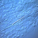
Pliciloricus orphanus
DIAGNOSIS.—Higgins-larva 298 μm long, mouth cone without oral ventral setae, minute hairs surrounding mouth opening. Internal mouth armature hexaradially symmetrical. Each of 8 clavoscalids 3-segmented, only lateroventral pair club-shaped, other clavoscalids more or less spinose. Spinoscalids long (15–35 μm, protoscalids triangular-shaped; collar well defined, but irregular. Lorica with 2 pairs of long anterior setae, branched without hairs; sculpturing indistinct; with mid-transverse constriction. Toes straight and long (106 μm), pointed terminally. Two pairs of sensory setae present at posterior end of lorica in addition to a pair of hollow stiff spines near anus; anal plate triangular.
MATERIAL EXAMINED.—Type Material: No adults were found; the holotype (Figure 47) is a large larva, 298 μm long, from station RH 1834. One small paratype (Figure 55), ~150 μm long, mounted on SEM-stub 78, is from station RH 1839. The holotypic larva (USNM 98562) has been deposited in the National Museum of Natural History, Smithsonian Institution, Washington, DC. The single paratype has been deposited in the Zoological Museum, Copenhagen.
ETYMOLOGY.—From the Latin orphanus (bereft); masculine gender.
DESCRIPTION.—The holotypic larva (Figure 47) is 298 μm long. The mouth cone is tripartite with the terminal portion slightly retracted telescopically. The thorax and abdomen are extended fully. The terminal portion of the mouth cone has six valves, and the mouth opening is surrounded by very fine hairs. The internal mouth armature is hexaradially symmetrical, but its detailed structure could not be resolved by light microscopy.
The appendages of the head (introvert) are arranged in seven rows. The scalids are of the Pliciloricus-type seen in all other larvae of this genus, but the first row consists of eight more or less spine-shaped clavoscalids. These three-segmented appendages are approximately 45–50 μm long. The first two segments of the lateroventral pair are slightly broader than those of the remaining six clavoscalids; the terminal segment is spinous. The basal segment has a few stiff hairs. The second row of head appendages consists of typical two-segmented spinoscalids, ~25–40 μm long, six dorsal, four ventral. The ventral spinoscalids are longer and more robust than the dorsal ones. No hairs were seen on any of the spinoscalids. The basal segment of the spinoscalid is triangular in cross-section and has a slight lateral projection on two lateral surfaces. The spinose terminal segment is more oval in cross-section.
The third row of head appendages consists of spinoscalids ~25 μm long, eight dorsal and seven ventral. The fourth row has 14 spinoscalids, six dorsal and eight ventral; the two midventral spinoscalids have very broad basal segments and articulate with the terminal segments by a well-defined joint. A similar two-segmented spinoscalid is found in the Kinorhyncha. The two ventralmost spinoscalids of the second, third, and fourth row are nearly fused midventrally, but they characteristically diverge from the midline. The fifth row has seven spinoscalids, each with a hooked accessory spine on the basal segment, similar to that noted for the larva of P. gracilis. The middorsal spinoscalid of this same row has a large triangular basal segment with a prominent filiform proximal segment (Figure 47). The sixth row of head appendages consists of eight double protoscalids; each projection has a serrulate margin and central keel that continues beyond the margin as a very small spinose tip, a condition most prominent in the dorsal protoscalids. One of the diagnostic characters of the genus Pliciloricus is the presence of four clusters of protoscalids found along the midventral line. In P. orphanus, these four protoscalids are smaller (5 μm) than the remaining 12 protoscalids (7 μm) of the sixth row.
The seventh row of appendages consists of seven double protoscalids alternating with 8 single papillae; each papilla has a small terminal spine. These double protoscalids are more or less fused basally, forming a “W-shaped structure. One double protoscalid is middorsal, with a single papilla located lateral to it.
The collar is not so regularly folded as in P. gracilis and lacks collar pores. The thorax has five horizontal rows of 15–30 plates, separated by distinct ridges in the manner of an accordion. The first row of thoracic plates has 15 ridges, similar to the pattern of folds in the collar. The last row of thoracic plates has about 30 ridges that are continuous with those of the lorica. The last row of thoracic plates on the ventral surface is nearly divided into two separate rows of plates, giving the appearance of an extra “sixth” row of plates ventrally, but only five rows dorsally. The larval lorica has a very thick, unsculptured cuticle with ~20 primary longitudinal ridges beginning at its anterior edge and ending at the anal plate. Ten secondary ridges are present ventrally, but they extend just short of the middle of the lorica, where a transverse constriction is visible. A distinctive triangular anal plate is present dorsoterminally. Three additional pairs of cuticular plates are present lateral to the anal plate; the anus is terminal on the anal plate (Figure 47, an).
Two long, rigid toes (~106 μm long) extend from the lateroventral region of the caudal end. About 70% of the length of the toes is hollow (~8 μm wide); the other terminal 30% of the length narrows to a solid spine. Two prominent glands are present in the posterior abdomen at the base of each toe.
The two anterior pairs of lorical setae are branched; each begins with a swollen base, which gives rise to a mesial ramus; the lateral ramus continues, then branches again in a similar fashion, resulting in three units (Figure 47, ls2). The lateroventral setae also have a large base, but have only a slight branch coming off the primary element. No hairs are visible on any setae.
The posterior three pairs of setae are similar to what have been described for P. gracilis, but the ball-and-socket joints of the dorsolateral pair are very large and project beyond the lateral margins of the lorica at the caudal end.
- bibliographic citation
- Higgins, Robert P. and Kristensen, R. M. 1986. "New Loricifera from Southeastern United States Coastal Waters." Smithsonian Contributions to Zoology. 1-70. https://doi.org/10.5479/si.00810282.438