-
Mohsen M. El-Sherbiny, Ali M. Al-Aidaroos
Zookeys
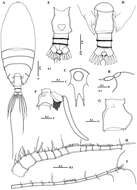
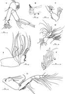
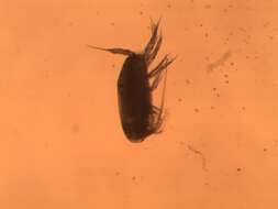
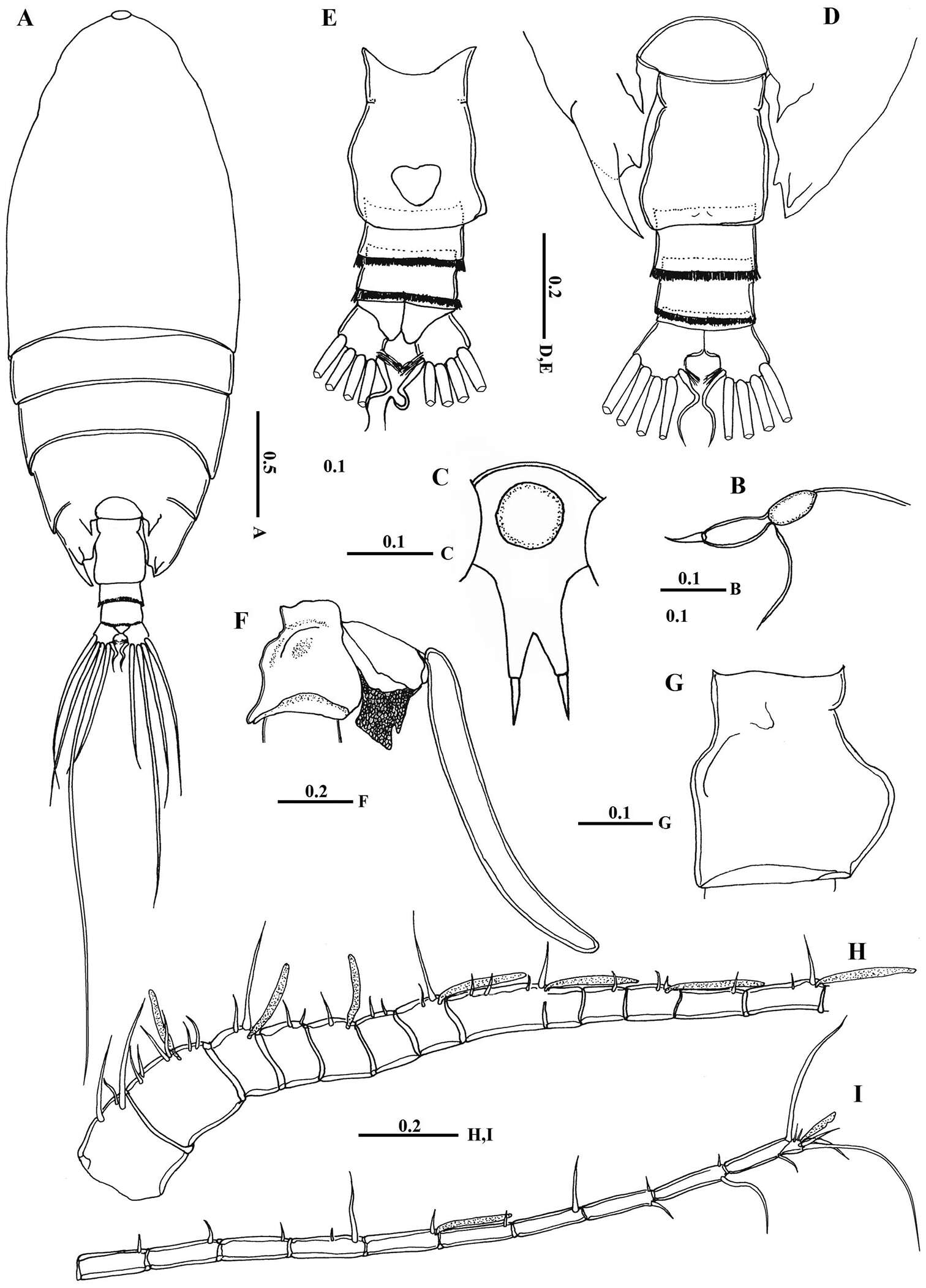
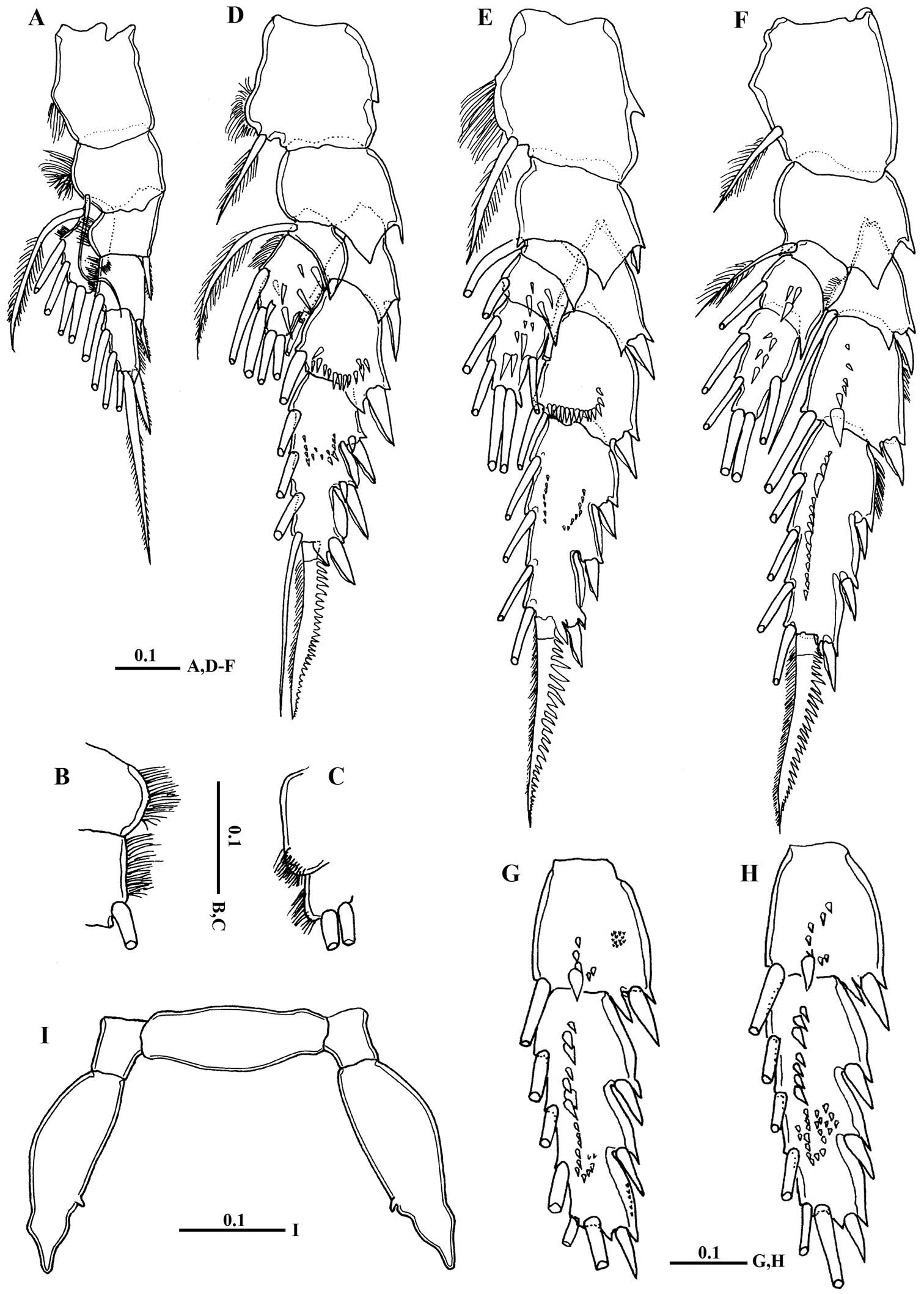
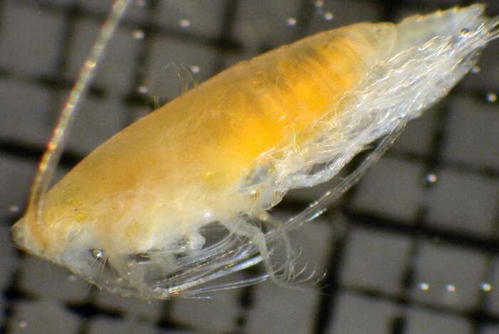
Figure 2.Macandrewella cochinensis female from the northern Red Sea. A habitus, dorsal view B rostrum, lateral view C rostrum, ventral view D posterior prosome and urosome, dorsal view E urosome, ventral view F genital double-somite with spermatophore, lateral view (right) G genital double-somite, lateral view (right) H–I antennules. All scale bars in mm.
-
Mohsen M. El-Sherbiny, Ali M. Al-Aidaroos
Zookeys
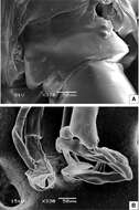
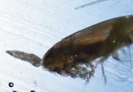
Figure 7.SEM micrographs of Macandrewella cochinensis male from the northern Red Sea. A genital somite, dorsal view B distal part of leg 5.
-
Scaphocalanus magnus (T. Scott, 1894)
-
Mohsen M. El-Sherbiny, Ali M. Al-Aidaroos
Zookeys
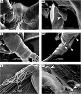
Figure 3.SEM micrographs of Macandrewella cochinensis female from the northern Red Sea. A rostrum and cuticular lens indicated by arrow, ventral view B serration of postero-dorsolateral process of prosomal end indicated by arrow, lateral view C urosome, anterodorsal protrusions and posterodorsal swelling on left side indicated by arrows, dorsal view D urosome, posterodorsal swelling on left side indicated by arrow, lateral view (left) E maxillary endopod F leg 5 indicated by arrow.
-
Mohsen M. El-Sherbiny, Ali M. Al-Aidaroos
Zookeys
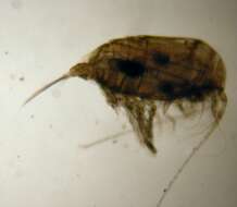
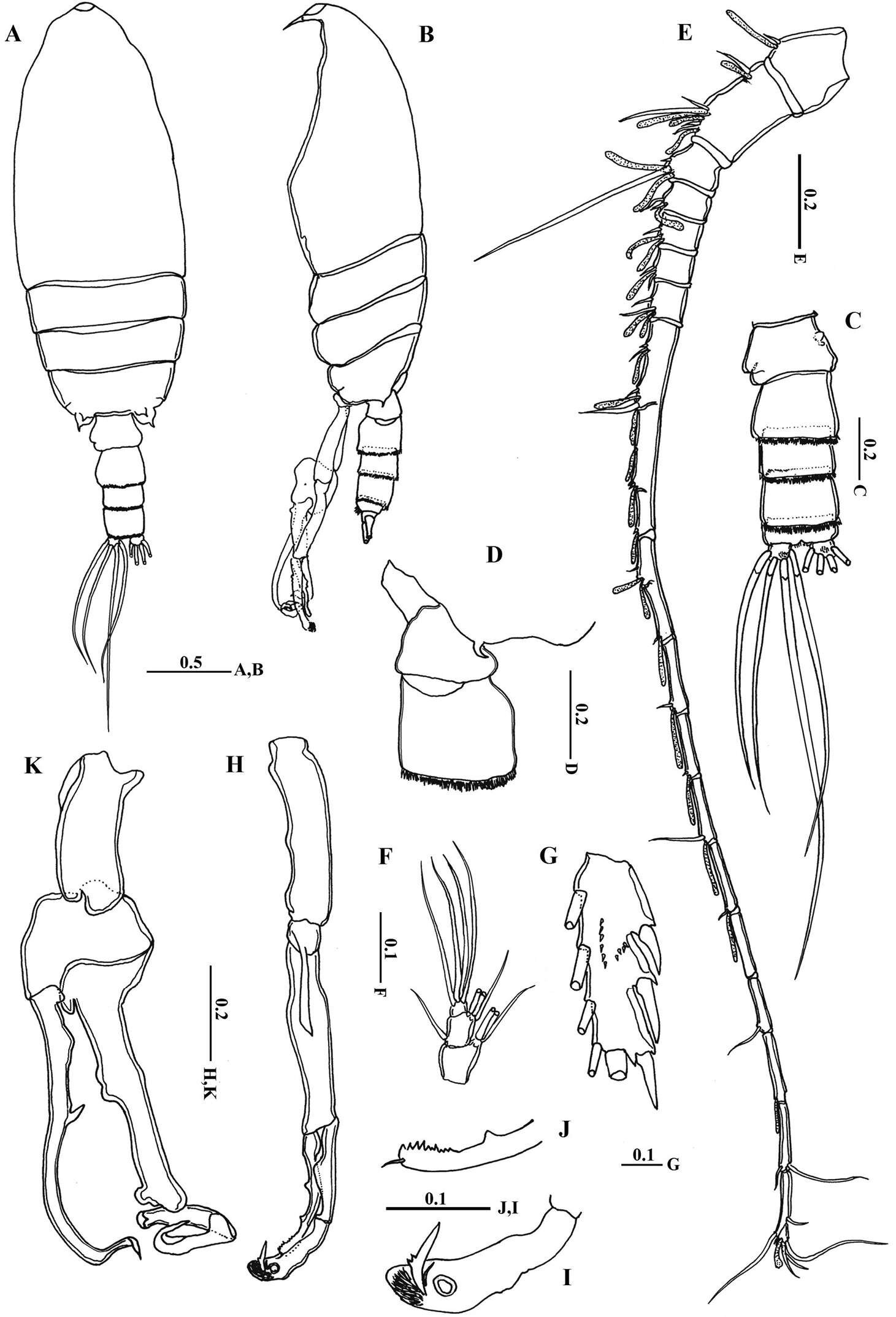
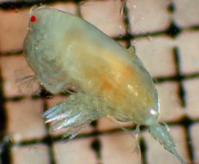
Figure 4.Macandrewella cochinensis female from the northern Red Sea. A antenna B mandibular gnathobase cutting edge C mandibular palp D maxillule E maxilla F maxilla endopod G maxilliped. All scale bars in mm.
-
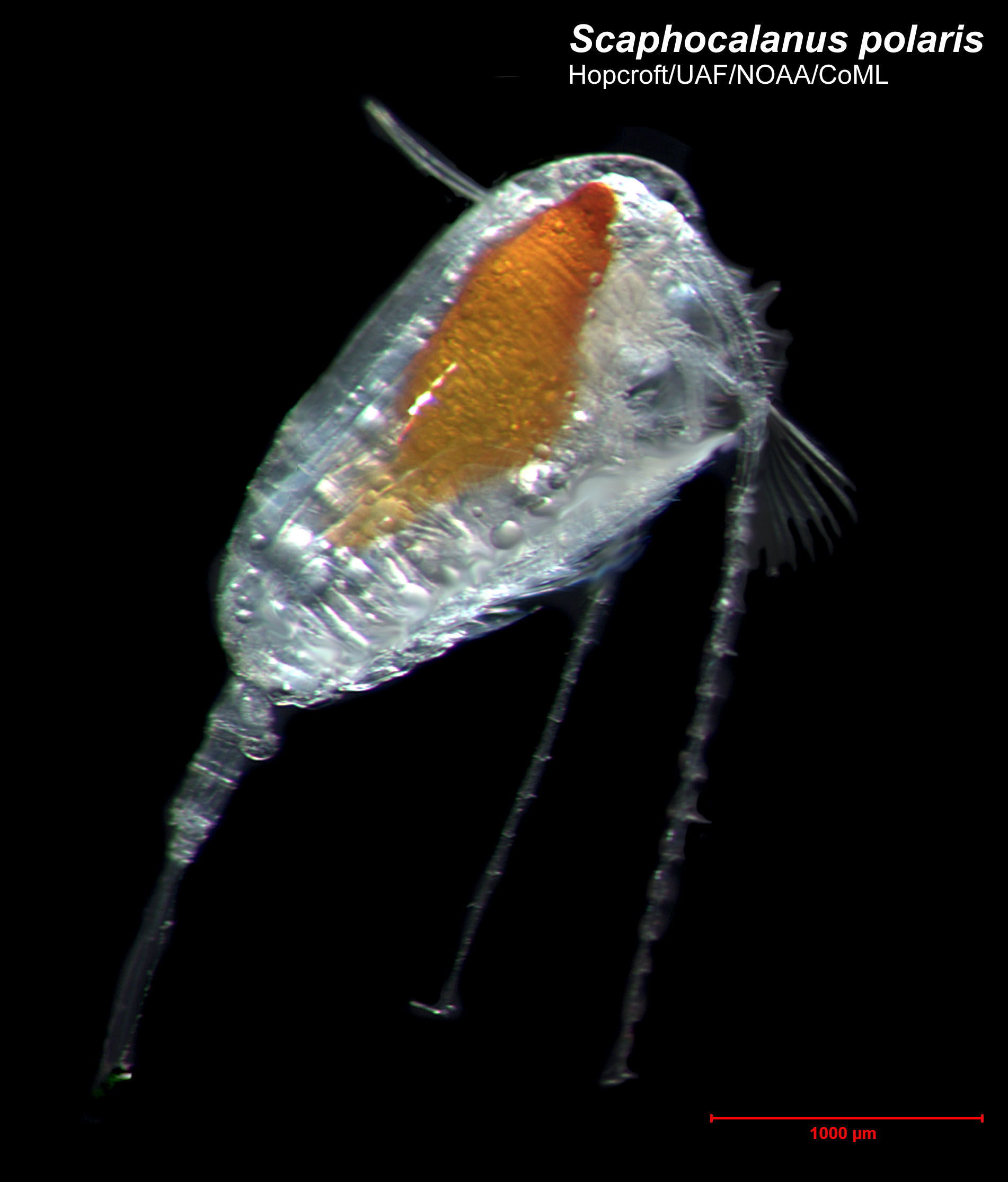
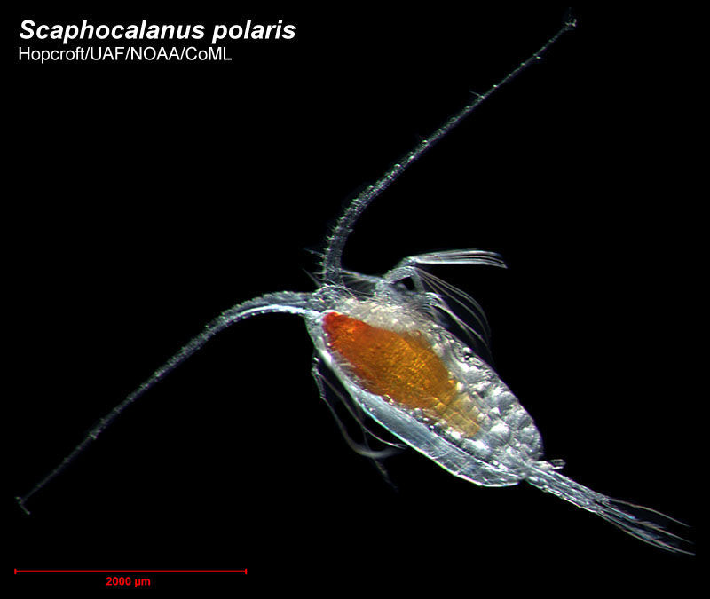
Scaphocalanus polaris (Brodsky, 1950 )
-
Mohsen M. El-Sherbiny, Ali M. Al-Aidaroos
Zookeys
Figure 5.Macandrewella cochinensis female from the northern Red Sea. A Leg 1, anterior surface B medial margin of first and second exopodal segments of Leg 1 C lateral distal margin of leg 1 endopod D leg 2, posterior surface E Leg 3, posterior surface F leg 4, posterior surface G–H second and third exopodal segments of leg 4, anterior surface I leg 5, anterior surface. All scale bars in mm.
-
Mohsen M. El-Sherbiny, Ali M. Al-Aidaroos
Zookeys
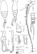
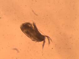
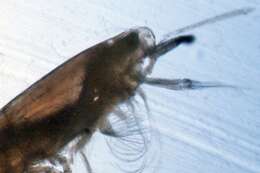
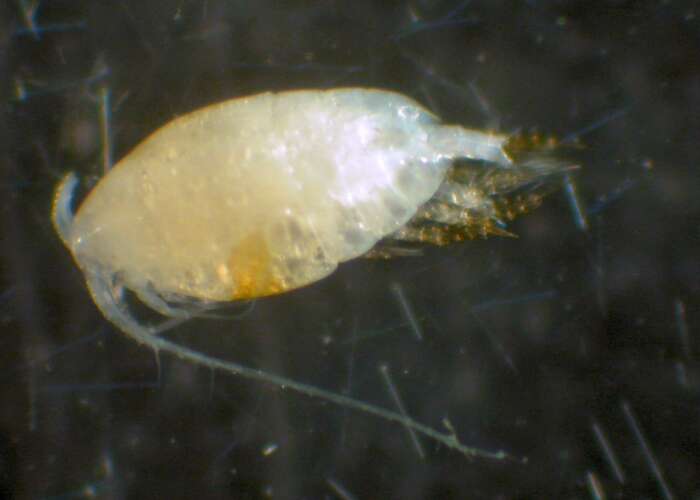
Figure 6.Macandrewella cochinensis male from the northern Red Sea. A habitus, dorsal view B habitus, lateral view C urosome, dorsal view D first and second urosomal segment, lateral view (right) E left antennule F maxilliped, terminal endopod segments G Exopod segment 3 of leg 2 H left leg 5 I terminal portion of left exopodal of leg 5 J terminal portion of left endopod of leg 5 K right leg 5. All scale bars in mm.
-
Scaphocalanus polaris (Brodsky, 1950 )
-
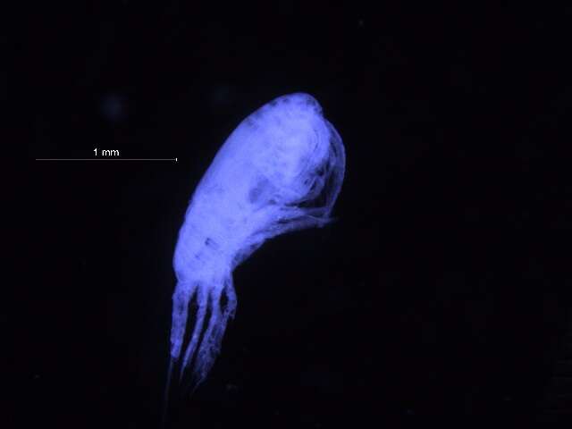
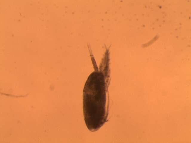
Lateral. Catalog no.: 145559. Specimen ID: 8576863. Field no.: COPCLAD436. Taxon rep.: Scaphocalanus brevicornis. Image quality: 1. Aspect ratio: 1.333.
-
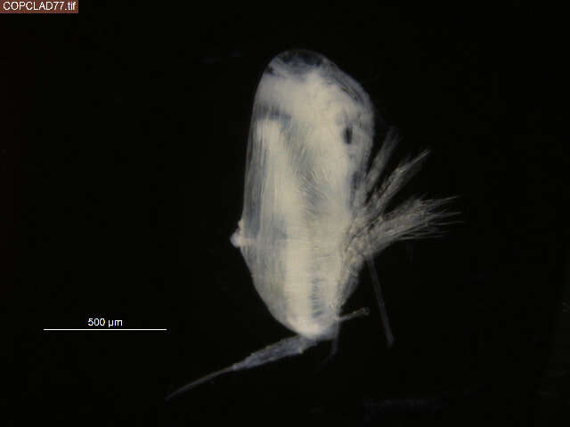
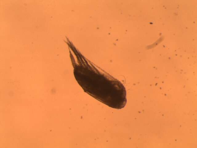
Lateral. Catalog no.: 145200. Specimen ID: 5666421. Field no.: COPCLAD77. Taxon rep.: Scolecithricella. Image quality: 4. Aspect ratio: 1.333.
-
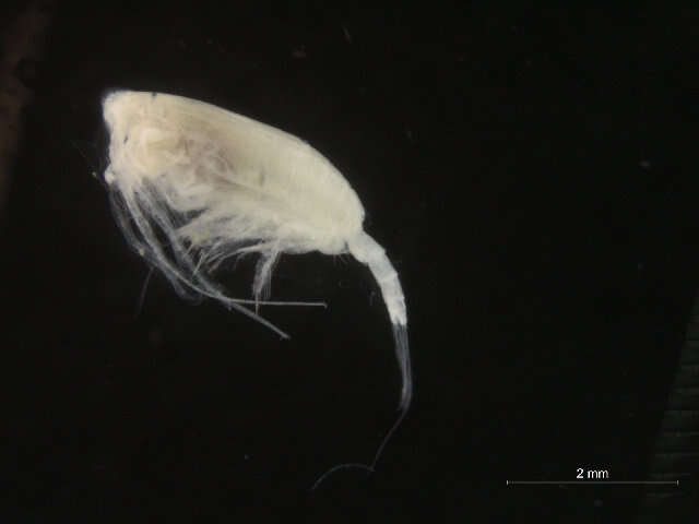
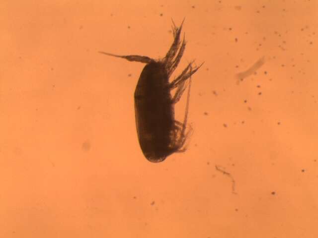
Lateral. Catalog no.: 145497. Specimen ID: 7568089. Field no.: COPCLAD374. Taxon rep.: Scaphocalanus. Image quality: 1. Aspect ratio: 1.333.
-
R. John Nelson. Fisheries and Oceans Canada. R. John Nelson. Year: 2019. Contact: moira.galbraith@dfo-mpo.gc.ca.
Barcode of Life Data Systems
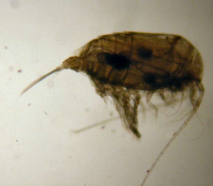
Dorsal. Specimen ID: 10925021. Field no.: IOS2018034-457. Taxon rep.: Mixtocalanus. Image quality: 1. Aspect ratio: 1.336.
-
Lateral..
-
Lateral..
-
Lateral..
-
Lateral..
-
female
-
female
-
female
-
female
-
-
-