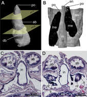
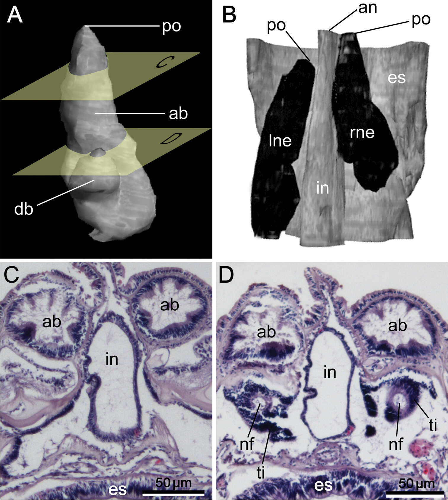
Figure 10.Reconstructed three-dimensional images and transverse sections of the nephridium of Phoronis emigi sp. n., based on NSMT-Te 721 (paratype). A Lateral view, showing the different lengths of the ascending and descending branches B dorsal view, showing the offset arrangement of the nephridia, with the nephridiopores at different levels along the body axis C transverse section through the ascending branch D transverse section through the tip of the descending branch, showing the nephridial funnels. Abbreviations: ab ascending branch; an anus; db descending branch; es esophagus; in intestine; lne left nephridium; nf nephridial funnel; p nephridiopore; rne right nephridium; ti funnel tissue. Planes C and D in panel A indicate the positions of the transverse sections in C and D.
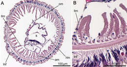
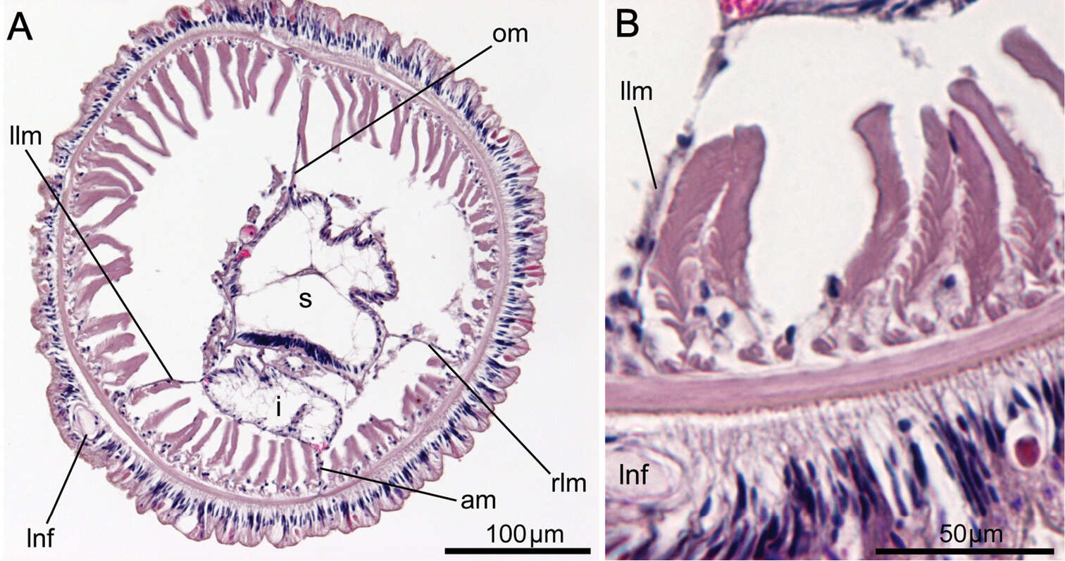
Figure 11.Phoronis emigi sp. n., NSMT-719 (paratype). A Transverse section through the posterior part of the body, showing four mesenteries and the position of the giant nerve fiber B enlargement of longitudinal muscles of the long feathery type. Abbreviations: am anal mesentery; lnf left giant nerve fiber; i intestine; llm left lateral mesentery; om oral mesentery; rlm right lateral mesentery; s stomach.