Diagnosis: Modal fin-ray counts of D-X,12 A-III,8 indicate Lutjanus mahogoni or L. synagris. Juveniles with the lateral line running through the middle of the lateral spot indicate L. mahogoni. (DNA)
Pretransitional mostly-unmarked stage, usually from 17-21 mm SL: Body: Pretransitional larvae develop a row of melanophores on the side of the body near the base of the dorsal fin. The row starts as small melanophores, often lined up along the anterior aspect of the pterygiophores below the soft dorsal fin. The row fills in under the soft dorsal fin and extends forward just below the spinous portion of the fin, first as a few spots beneath the third to fifth spines and the seventh to ninth, and then filling in up to the level of the third dorsal-fin spine. The two rows on each side of the dorsal fin merge into a line of melanophores lining the dorsal midline of the caudal peduncle (usually made up of a short row of deeper and larger larval melanophores overlain by a band of smaller melanophores). A similar line develops along the ventral midline of the caudal peduncle extending forward and ending just before a single large melanophore underlying the pterygiophores of the last anal-fin rays. There are a few deep melanophores at the end of the lateral midline of the caudal peduncle and a fine speckling of small melanophores around the central caudal peduncle. Head: Melanophores on the head consist of dense patches overlying the brain and on the surface braincase. There are small melanophores around the tips of the upper and lower jaws. The opercular area is covered in iridescence extending down to the pelvic-fin insertion. The inner cleithral surface of the gill cavity is speckled with large melanophores and there are internal melanophores lining the dorsal aspect of the swim bladder and peritoneum extending down to the vent and overlain by a silvery camouflage layer. Fin Spines: The dorsal and anal-fin spines are relatively stout, usually with some internal reticulations. The tip of the second dorsal-fin spine often curves slightly upward (the preopercular spine often curves slightly upward as well). The second dorsal-fin spine is usually longer than the third and typically the tip overlaps or extends beyond the tip of the third. The dorsal-fin spines then become progressively and evenly shorter such that the profile of the spinous tips forms a straight downward-sloping line. The anal-fin spines do not show anterior serrations (a rare individual has small remnant serrations). The second anal-fin spine is longer than the third, but the tips are closely-approximated or the third extends slightly farther back than the tip of the second when folded down. Fins: Melanophores are prominent along most of the length of the membrane just behind the second dorsal-fin spine and are concentrated on the membrane tag extending beyond the spine. Smaller melanophores speckle the outer third of all of the subsequent membranes of the spinous portion of the dorsal fin. On the caudal fin there are a few small melanophores at the base of some of the upper as well as most of the lower segmented rays. A row of melanophores develops along the anal-fin base, one at the base of each anal-fin-ray membrane, often including the membrane behind the third-spine. Pretransitional analogues: Pretransitional larvae of the two 12-dorsal-rayed snappers are best distinguished by the relative lengths of the second and third dorsal-fin spines: in L. mahogoni the second dorsal-fin spine is longer than the third spine with the tip often overlapping the tip of the third vs. shorter than the third spine in L. synagris. L. mahogoni can be separated from most of the 14-dorsal-rayed snappers by the dorsal-fin-ray count, as well as by having melanophores at the bases of the upper as well as the lower caudal-fin segmented rays at this early stage (sometimes shared by L. analis). The dorsal and anal-fin spines of larval L. mahogoni are also stouter than in L. analis, L. cyanopterus, and Ocyurus chrysurus. L. mahogoni larvae at this stage also do not have distinct anterior serrations persisting on the anal-fin spines as do L. griseus, L. apodus and L. jocu. Almost all pretransitional L. mahogoni captured over the reef already show at least a few melanophores at the lateral spot, unlike L. analis and the barred species.
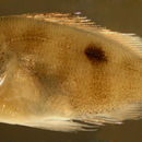
Transitional stage: Transitional L. mahogoni larvae develop a prominent lateral spot early, usually with the lateral line running through the middle of the spot (although some individuals clearly have the line running through the lower third). The spot is wider than the bar, distinctly expanding the outline of the bar. Larval L. mahogoni often have an upturned preopercular spine, but this character is not consistent later. Transitional analogues: The main difference between the two 12-rayed species is that the second dorsal-fin spine is longer than the third (sometimes about equal) for L. mahogoni (and L. analis) vs. shorter in L. synagris and this difference persists in juveniles up to 25 mm SL. The location of the lateral spot usually differs, although some individuals do overlap: the lateral line usually runs through the middle of the spot in L. mahogoni and through the lower third of the spot in L. synagris (variable in transitional L. analis). In addition, on L. synagris the bar forward of the lateral spot is not straight; it clearly curves away and brackets the spot. Transitional larvae of L. mahogoni always have a lateral spot, then they subsequently develop bars (vs. bars, then a spot on L. analis). Some transitional larvae and early recruits of L. analis after they develop the lateral spot can look remarkably similar to L. mahogoni. Other than the soft dorsal fin-ray counts, the species can be separated by some marking differences: the spot is larger and more elongated in L. mahogoni, expanding the bar from which it develops, while in L. analis the spot is only as wide as the bar from which it forms. Furthermore, transitional L. analis have three more typically distinct bars on the body behind the lateral spot while these bars are usually undeveloped on transitional L. mahogoni.
Juveniles: Juvenile L. mahogoni have an elongated lateral spot with the lateral line running through the middle of the spot. Juvenile analogues: Juveniles of L. mahogoni can be distinguished by the lateral-spot location, i.e. the lateral line through the middle of the spot in L. mahogoni and usually through the lower third in L. synagris and L. analis. Notably, the lateral spot is often elongated (in width) in juvenile L. mahogoni vs. rounded and within the bar in L. analis. The relative dorsal-spine-length differences persist in juveniles up to 25 mm SL. All have yellow stripes as juveniles, although the stripes become thinner and less conspicuous on juvenile L. mahogoni and thicker and more prominent on later juvenile L. synagris. The preopercular outline is not diagnostic in young stages, with L. mahogoni larvae and juveniles not showing the notch pattern that occurs later.
Description: Body wide and relatively thick with a sloping forehead and a large round eye and large terminal mouth. Dorsal-fin base long and anal-fin base short. Prominent dorsal, anal, and pelvic-fin spines and a large non-serrated preopercular spine.