-
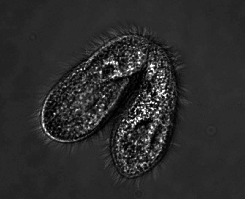
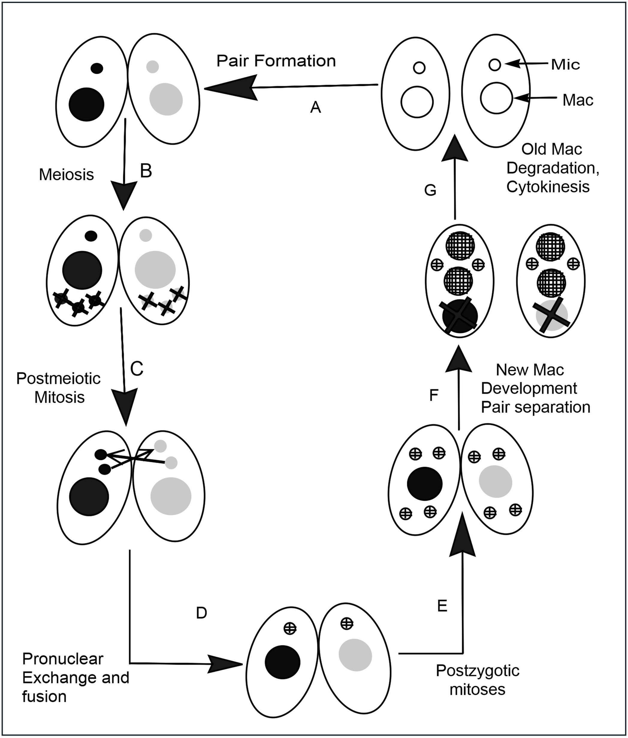
Description: English: Two cells of complementary mating types pair to exchange nuclei during sexual conjugation. Date: 8 March 2016. Source: Own work. Author:
Jmf368w.
-
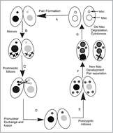
Description: English: Tetrahymena conjugation When nutrients are scarce, two individuals (A) pair with each other and begin sexual reproduction (conjugation). (B) The diploid micronucleus in each individual undergoes meiosis to form four haploid nuclei, and three of these are degraded. (C) The remaining haploid nucleus divides mitotically to form two pronuclei in each cell. (D) One of the two pronuclei in each cell is exchanged with the mating partner, and fusion leads to he formation of the diploid zygotic nucleus. (E) The zygotic nucleus divides twice mitotically to form four nuclei. (F) Two nuclei become micronuclei, and the other two differentiate to become macronuclei; the original parental macronucleus is degraded. (G) Cell division occurs and the nuclei are distributed to the daughter cells so that each progeny receives one micronucleus and one macronucleus. Date: 16 April 2014, 11:58:27. Source: Own work. Author:
Chaya5260.
-
-
-
-
-
-
-
-
Galende, Castilla y Len, Espaa
-
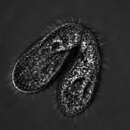
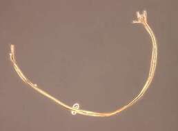
Tetrahymena (tet-ra-high-men-a), only one thread on the entire slide and it has to land on me.
-
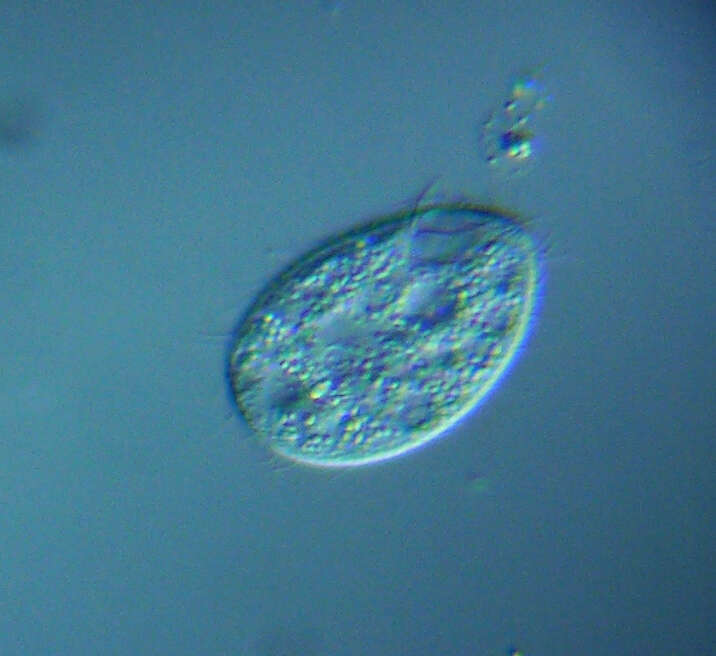
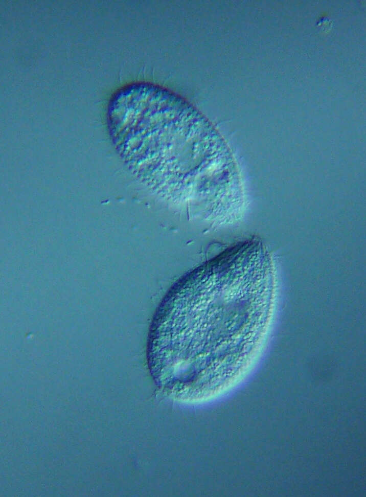
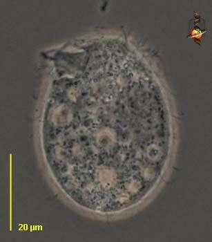
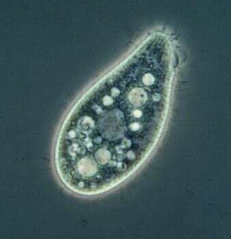
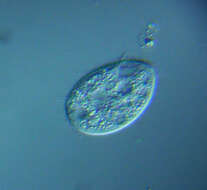
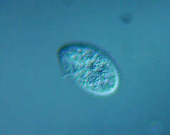
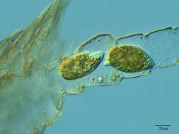
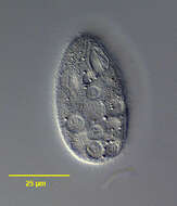
Tetrahymena (tet-ra-high-men-a) is an oligohymenophoran ciliate. There are cilia in about 20 kineties (rows) over the body and which are used for cell locomotion. There is also a group of three membranelles and an undulating membrane around the cytostome (upper left), and these are the buccal or oral cilia and are used in food capture. In nature often associated with damaged animals or dead tissue, may eat bacteria. Widely used in laboratory studies, and axenic (bacteria-free) cultures are maintained within high protein medium. This cell is slightly compressed. Phase contrast.
-
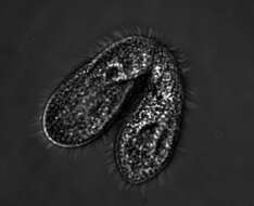
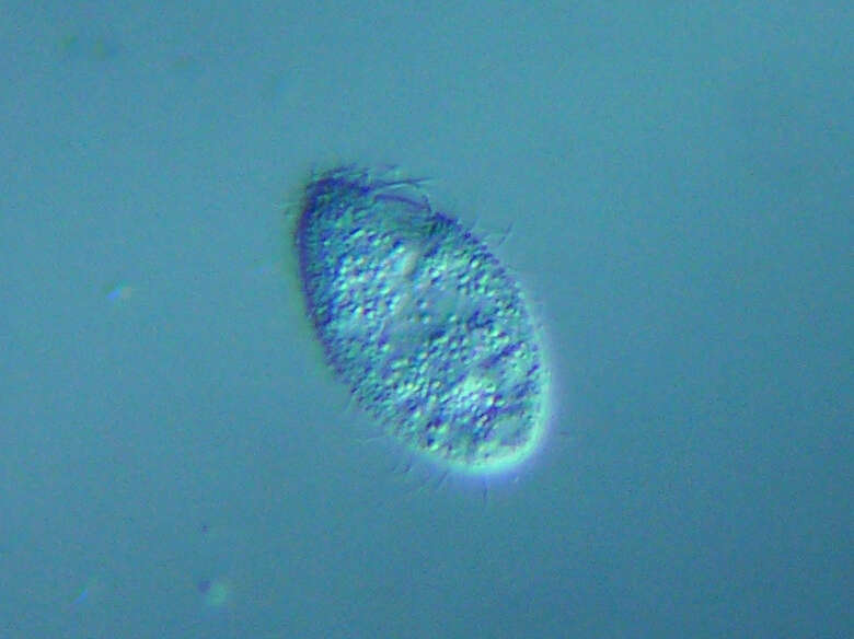
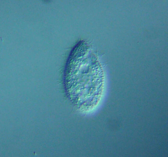
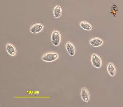
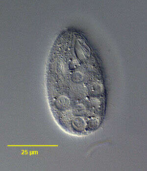
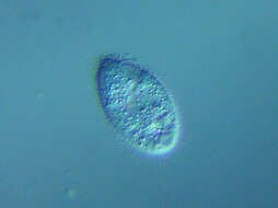
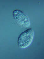
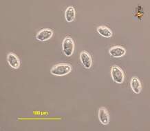
Tetrahymena (tet-ra-high-men-a) is an oligohymenophoran ciliate. There are cilia in about 20 kineties (rows) over the body and which are used for cell locomotion. There is also a group of three membranelles and an undulating membrane around the cytostome, and these are the buccal or oral cilia and are used in food capture. In nature often associated with damaged animals or dead tissue, may eat bacteria. Widely used in laboratory studies, and axenic (bacteria-free) cultures are maintained within high protein medium. cells pear-shaped. Phase contrast.
-
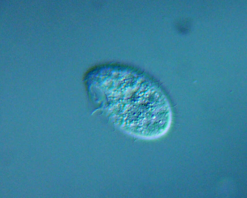
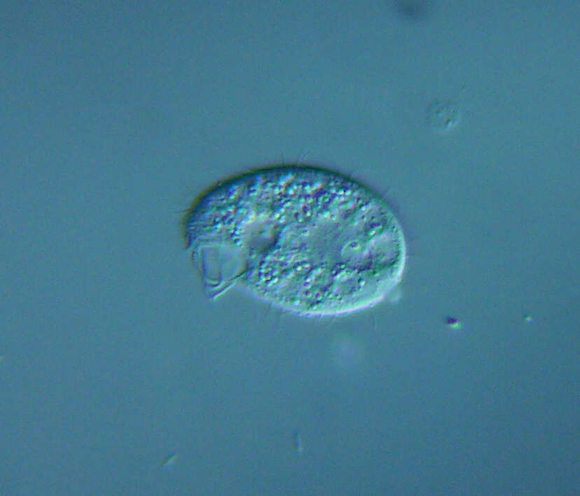
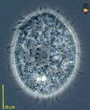
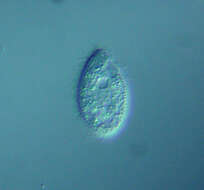
Tetrahymena (tet-ra-high-men-a) is an oligohymenophoran ciliate. There are cilia in about 20 kineties (rows) over the body and which are used for cell locomotion. There is also a group of three membranelles and an undulating membrane around the cytostome (upper right), and these are the buccal or oral cilia and are used in food capture. In nature often associated with damaged animals or dead tissue, may eat bacteria. Widely used in laboratory studies, and axenic (bacteria-free) cultures are maintained within high protein medium. Cells are pear-shaped. Differential interference contrast.
-
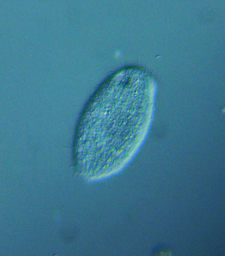
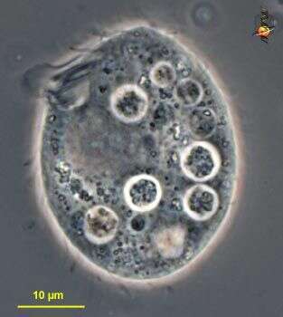
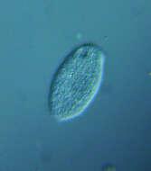
Tetrahymena (tet-ra-high-men-a) is an oligohymenophoran ciliate. There are cilia in about 20 kineties (rows) over the body and which are used for cell locomotion. There is also a group of three membranelles and an undulating membrane around the cytostome (upper left), and these are the buccal or oral cilia and are used in food capture. In nature often associated with damaged animals or dead tissue, may eat bacteria. Widely used in laboratory studies, and axenic (bacteria-free) cultures are maintained within high protein medium. Cells pear-shaped. Phase contrast.
-
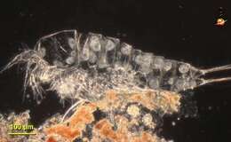
Tetrahymena (tet-ra-high-men-a) is an oligohymenophoran ciliate. Widely used in laboratory studies, and axenic (bacteria-free) cultures are maintained within high protein medium. In nature often associated with damaged animals or dead tissue. This occurrence within the cytoskeleton where they may feed on residual tissue (pieces left behind if the cytoskeleton has been shed) or on the body tissue (if the crustacea was damaged) is a good example of where this genus might be found in nature. Dark ground.
-
-
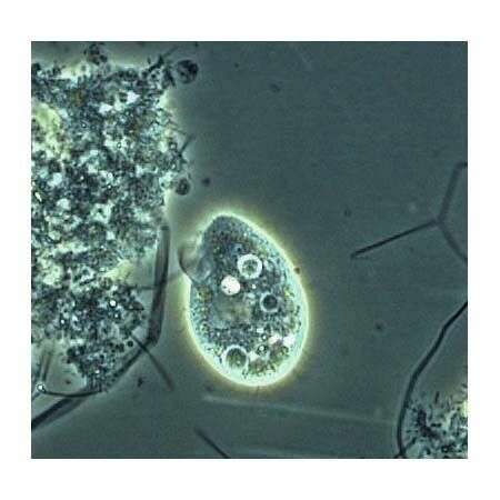
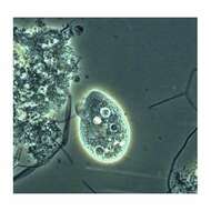
Tetrahymena: One of the most studied protozoans. This image was taken by Krishnakumar B. in a sample from an anaerobic bioreactor for organic rich wastewater treatment in Regional Research Laboratory-Trivandrum (CSIR-India).
-
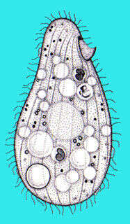
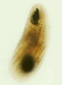
Portrait (ventral view) of the hymenostome ciliate,Tetrahymena pyriformis complex.
-
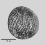
Right ventrolateral view of the silverline system (argyrome) of a ciliate from the Tetrahymena pyriformis complex.The oral aperture is to the viewer's upper right. Stained by the Klein-Foissner dry silver nitrate technic (see Foissner, W. Europ. J. Protistol., 27:313-330;1991). Brightfield, black and white.
-
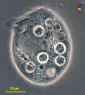
Cell from strain W, oral cilia are near the front of the cell (top of image), large clear area is the macronucleus, there is no micronucleus in this strain. Phase contrast image.
-
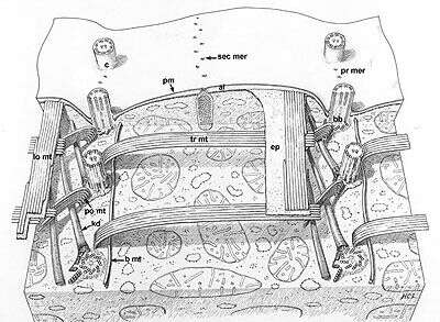
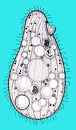
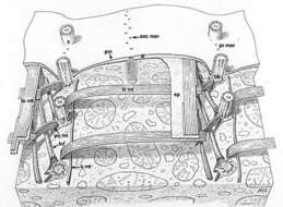
A classic drawing of the organization of this model ciliate. Segments of two kineties or rows of basal body (bb)/cilium (c) complexes. Kinetodesmal fibers (kd) arise from the left side of the proximal end of each basal body and angle up to the cell pellicle. Transverse microtubules (tr mt) extend to the right and postciliary microtubules (po mt) angle posteriorly and to the left of the kinety. One or two basal microtubules (b mt) extend along the right of the kinety at the proximal end of the basal bodies. A band of longitudinal microtubules (lo mt) lies between the epiplasm (ep) and the alveoli (al) to the left of the kinety. Mucocysts (mu) approach the plasma membrane (pm) through the septa separating alveoli along the primary (pr mer) and secondary meridians (sec mer). Drawn by H. C. Lyman.
This image is available in Richard Allen's collection.
-
The protargol stains the bases of the cilia and the nucleus. The image shows the kineties, the three membranelles and the single curved undulating membrane that make up the mouth ciliature, and the macronucleus (this strain lacks a micronucleus).