-
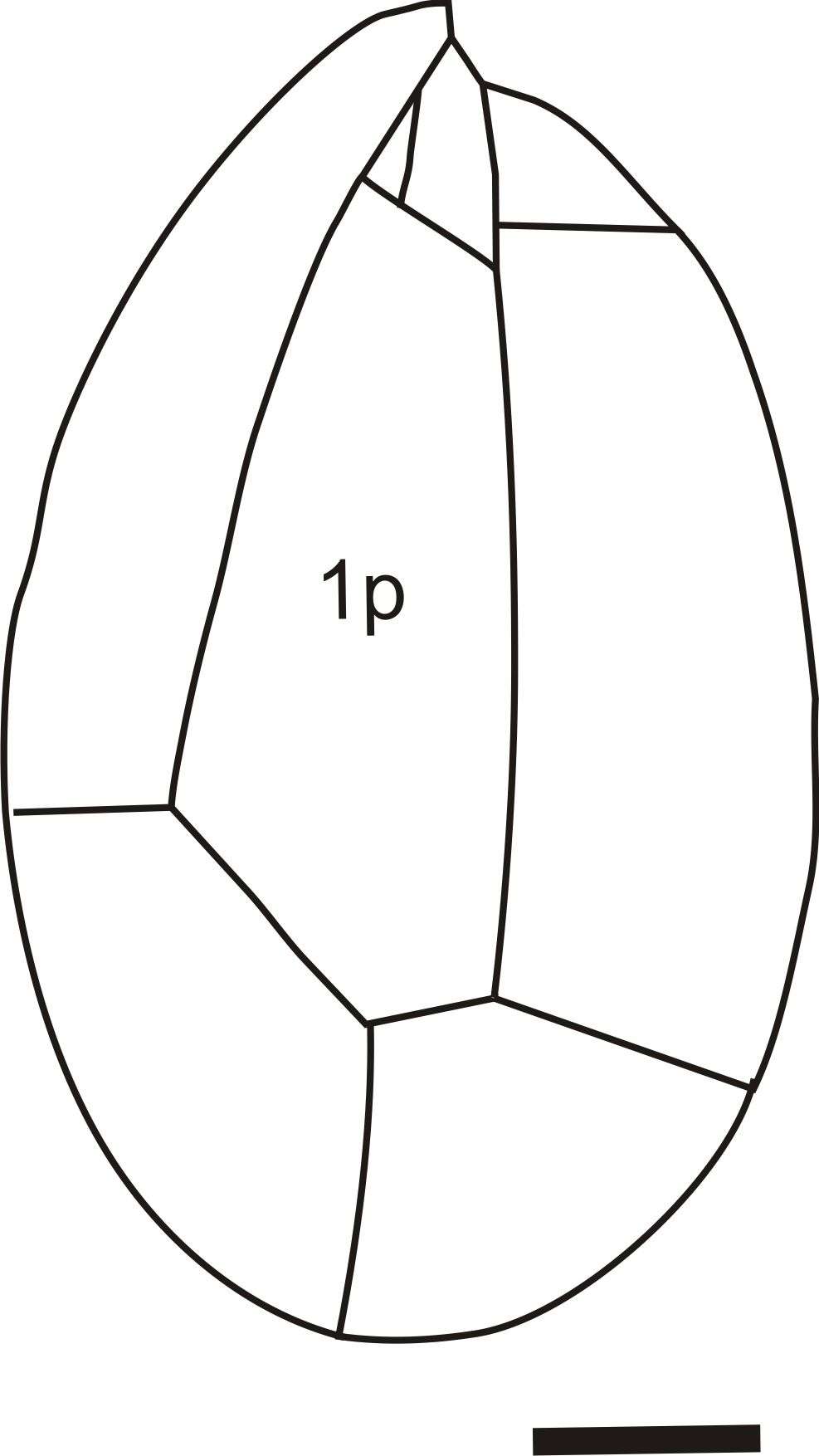
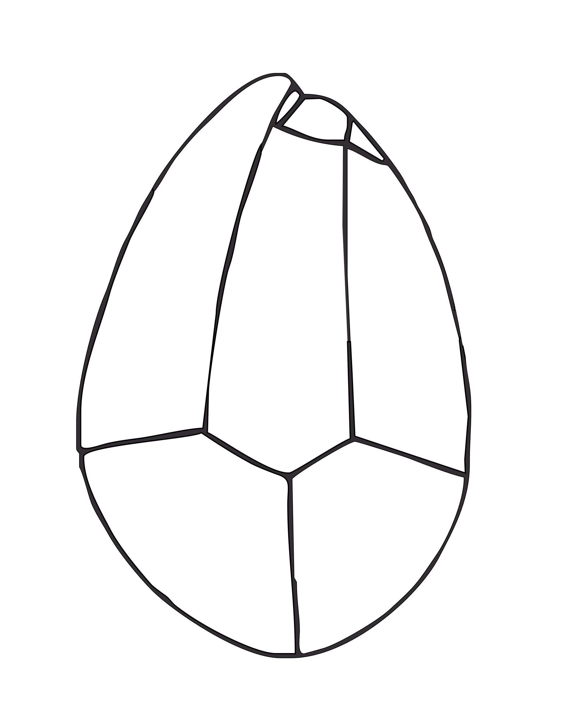
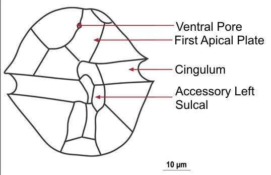
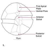
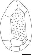
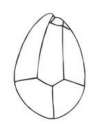
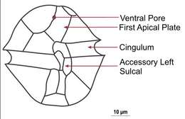
Fig 1: Alexandrium ostenfeldii Schematic drawing of a cell showing plate patterns on ventral side of cell
-
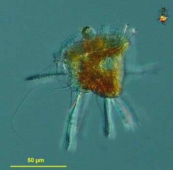
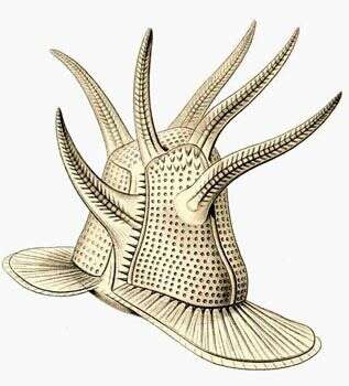
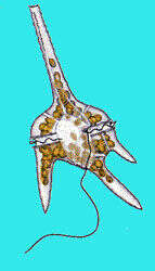
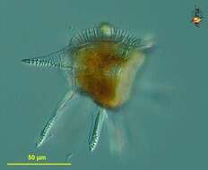
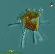
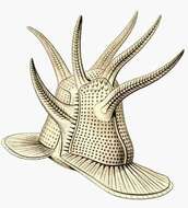
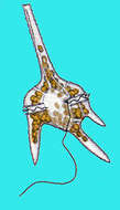
Ceratocorys (serr-at-owe-core-iss) horrida, a marine autotrophic dinoflagellate occurring in the plankton. Body armoured with projecting spines, some with wings. The girdle is edged with two projecting and ribbed flanges. Mostly from warm waters. Differential interference microscopy.
data on this strain.
-
San Martn de Castaeda, Castilla y Len, Espaa
-
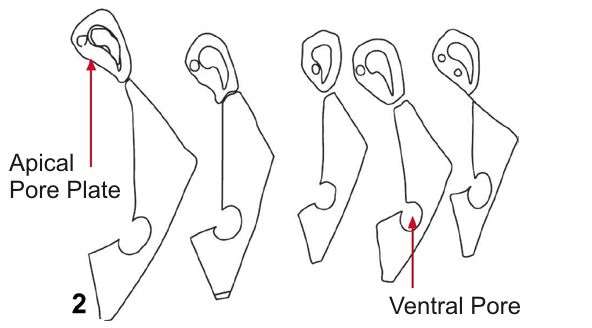
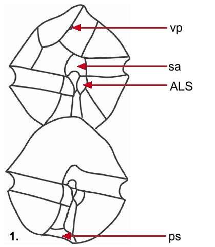
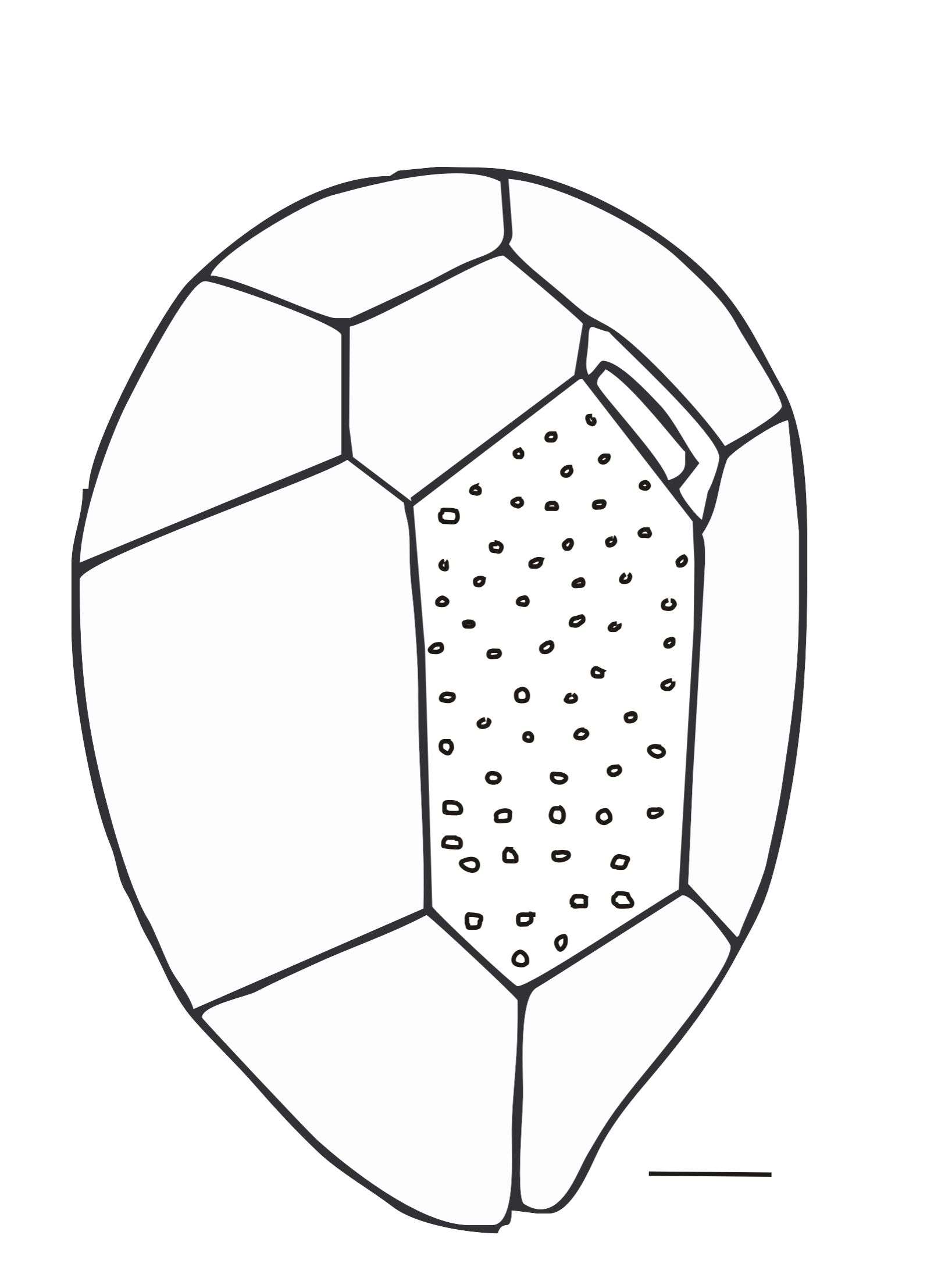
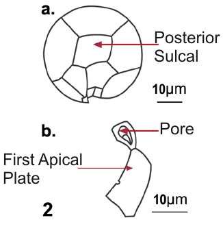
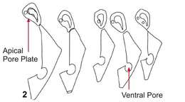
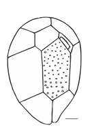
Fig 2: Alexandrium ostenfeldii Schematic drawing of a cell showing the morphological variation in APC and 1'
-
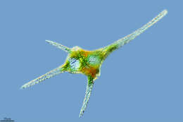
Ceratocorys (serr-at-owe-core-iss) horrida, a marine autotrophic dinoflagellate occurring in the plankton. Body armoured with projecting spines, some with wings. The girdle is edged with two projecting and ribbed flanges. Mostly from warm waters. Differential interference microscopy.
data on this strain.
-
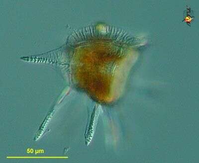
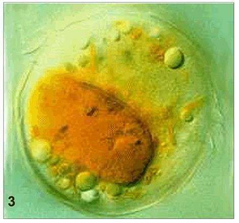
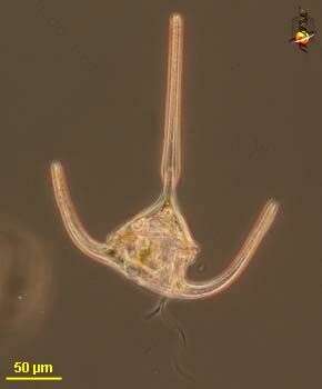
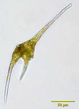
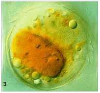
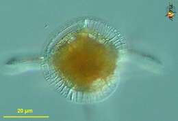
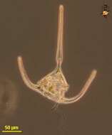
Fig 3: Alexandrium ostenfeldii whole cell with food vacuole
-
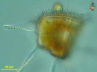
Ceratocorys (serr-at-owe-core-iss) horrida, a marine autotrophic dinoflagellate occurring in the plankton. Body armoured with projecting spines, some with wings. The girdle is edged with ridged flanges, one visible here at the top of the image. Mostly from warm waters. Differential interference microscopy.
data on this strain.
-
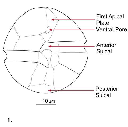
Fig 1: Schematic diagram of Ostreopsis ovata in hypothecal view. Scale bar = 10 ¦#181;m. Redrawn from Tomas et al. 1997
-
Ceratocorys (serr-at-owe-core-iss) horrida, a marine autotrophic dinoflagellate occurring in the plankton. This image shows the plastid, as well as the nucleus with the condensed chromosomes. Differential interference microscopy.
data on this strain.
-
Fig 2: Schematic diagram of Ostreopsis ovata in epithecal view. Scale bar = 10 ¦#181;m. Redrawn from Tomas et al. 1997
-
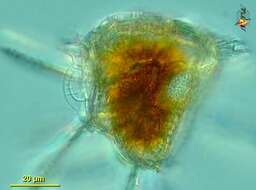
Ceratocorys (serr-at-owe-core-iss) horrida, a marine autotrophic dinoflagellate occurring in the plankton. Body armoured with projecting spines, some with wings. The girdle is edged with two projecting and ribbed flanges, shown in this view from the apex. Mostly from warm waters. Differential interference microscopy.
data on this strain.
-
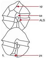
Fig 1: Schematic diagram of Alexandrium affine, showing ventral (top) and dorsal (bottom) views.
-
Ceratocorys horrida.
-
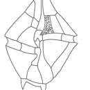
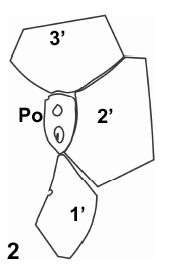
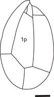
Fig 2: Schematic drawing of the Po and surrounding apical plates
-
-
Fig 1: Ostreopsis siamensis Schematic diagram (hypothecal view) redrawn from Tomas et al. 1997.
-
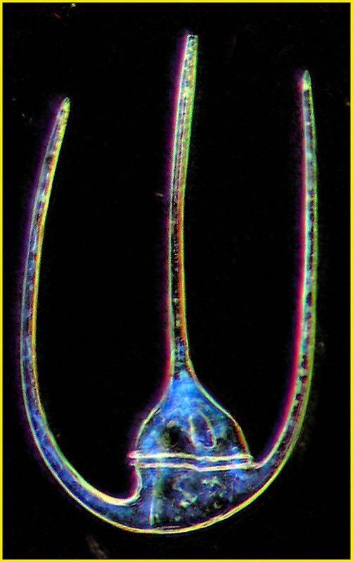
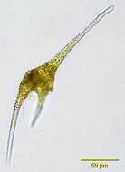
Ceratium (serr-at-ee-um) longipes, a representative of a large and distinctive genus of marine autotrophic dinoflagellates - made distinctive by having one anterior projection and two or as in this case three, posterior horns. Phase contrast microscopy.
-
Fig 2: Ostreopsis siamensis Schematic diagram (epithecal view) redrawn from Tomas et al. 1997.
-
Ceratium (serr-ate-ee-um), dinoflagellate, the chloroplasts of which are evident because they emit red light when illuminated with intense UV light. The UV light is filtered out so that only the red fluorescence is visible. This is a dinoflagellate. Fluorescence microscopy image by Dave Caron.
-
-
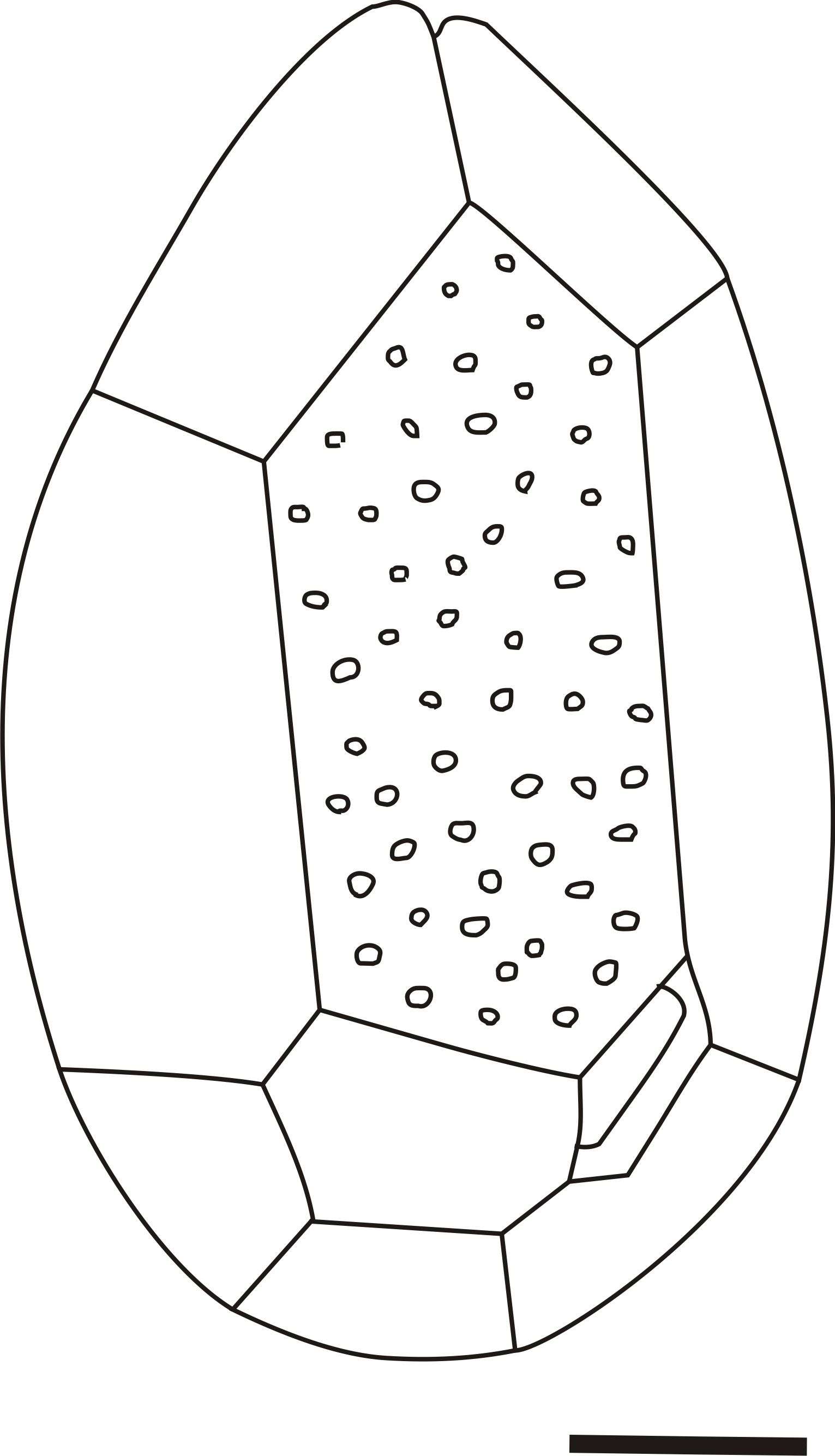
Fig 1: Alexandrium tamarense Schematic drawing of a cell showing plate patterns on ventral side of cell
-
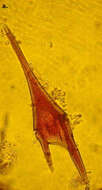
Bright field portrait of the dinoflagellate Ceratium furca (Ehrenberg) Claparéde and Lachmann 1858, trailing flagellum can be seenb. From a freshwater pond near Boise, Idaho.
-
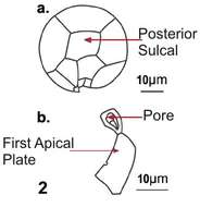
Fig 2: Alexandrium tamarense Schematic drawing of a cell in posterior view and b. apical pore coplex with 1'.
-
C. furca is a species with strongly developed apical horns, the left antapical horn is twice as long as the right one. The epithecal plates arereticulated to form ridges This species forms blooms in summer/ autumn in the North and Irish Sea.