-
-
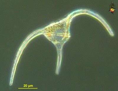
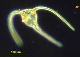
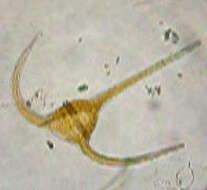
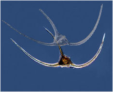
Ceratium bipes (serrate-ee-um try-poss), a common dinoflagellate encountered in marine waters, with both cones of the cell drawn into long horns, the circumferential flagellum lying in a groove in the expanded central region. With plastids. Dark ground illumination.
-
Ceratium bipes (serrate-ee-umtry-poss), a common dinoflagellate encountered in marine waters, with both cones of the cell drawn into long horns. Differential interference contrast
-
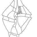
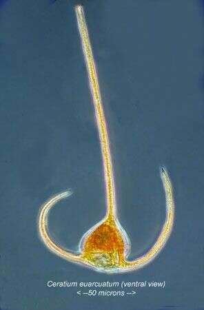
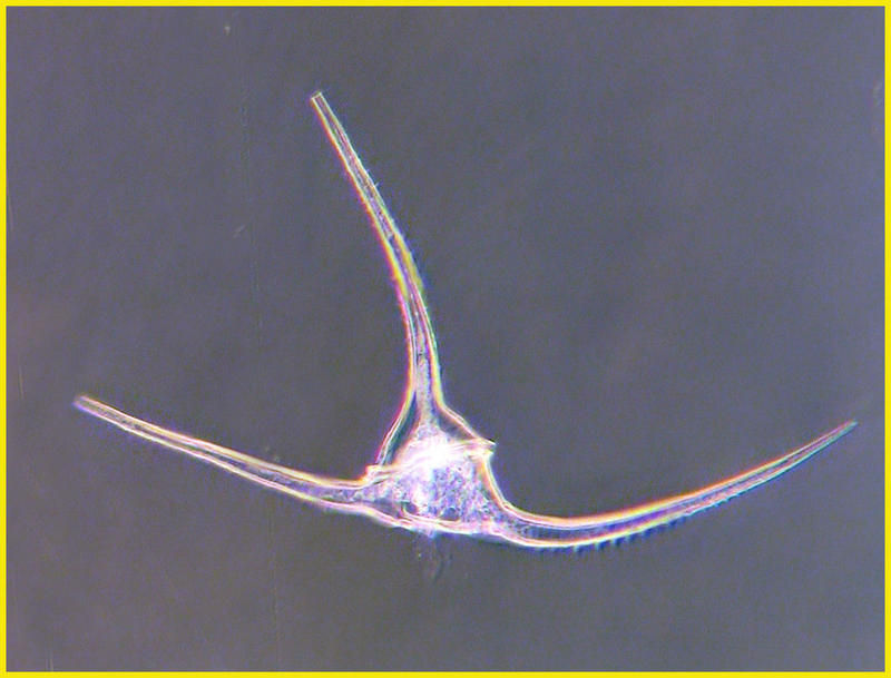
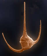
Ceratium tripos.
-
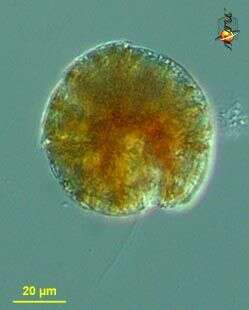
This specimen of Ceratium had a snack shown by the organge fluorescence of cryptophyte pigment- perhaps that of the ciliate Mesodinium rubrum
-
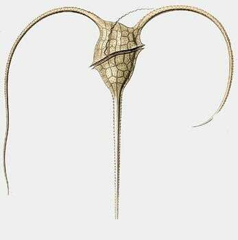
Its morphology is highly variable. It has a large dorsoventrally flattened cell body. The left antapical horn is more strongly developed than the right one. It is found in many areas of the Irish Sea but is never as abundant as C. furca.
-
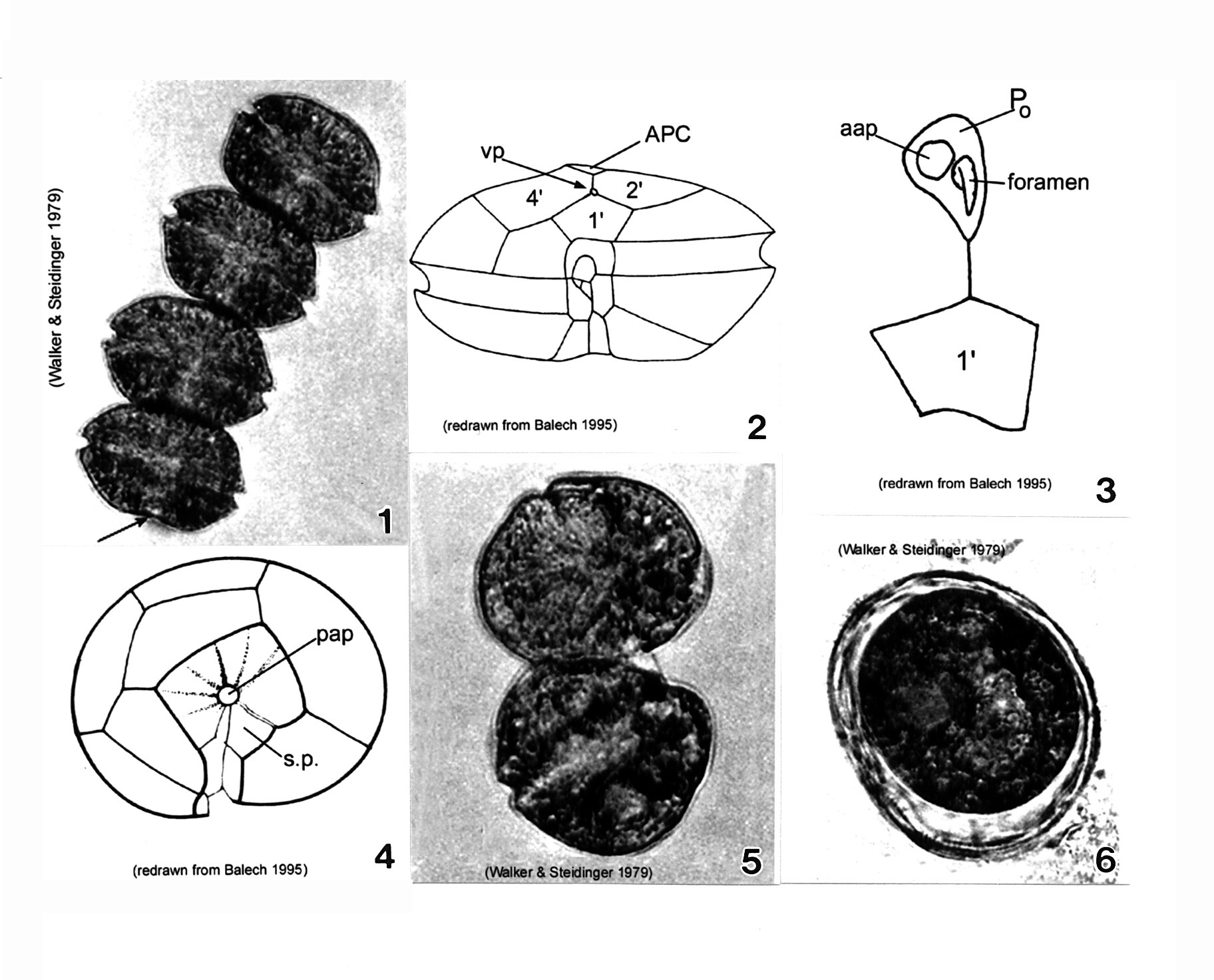
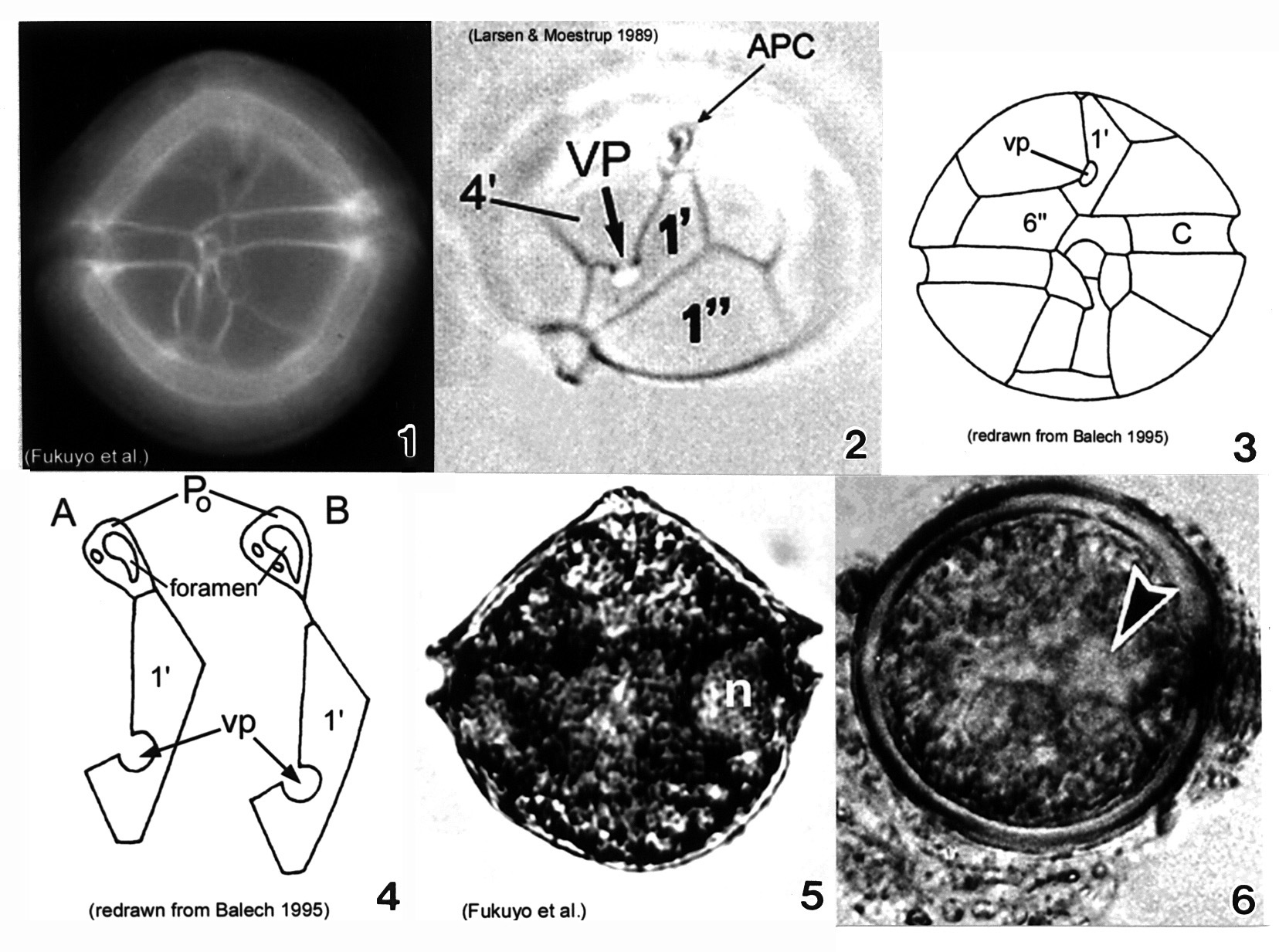
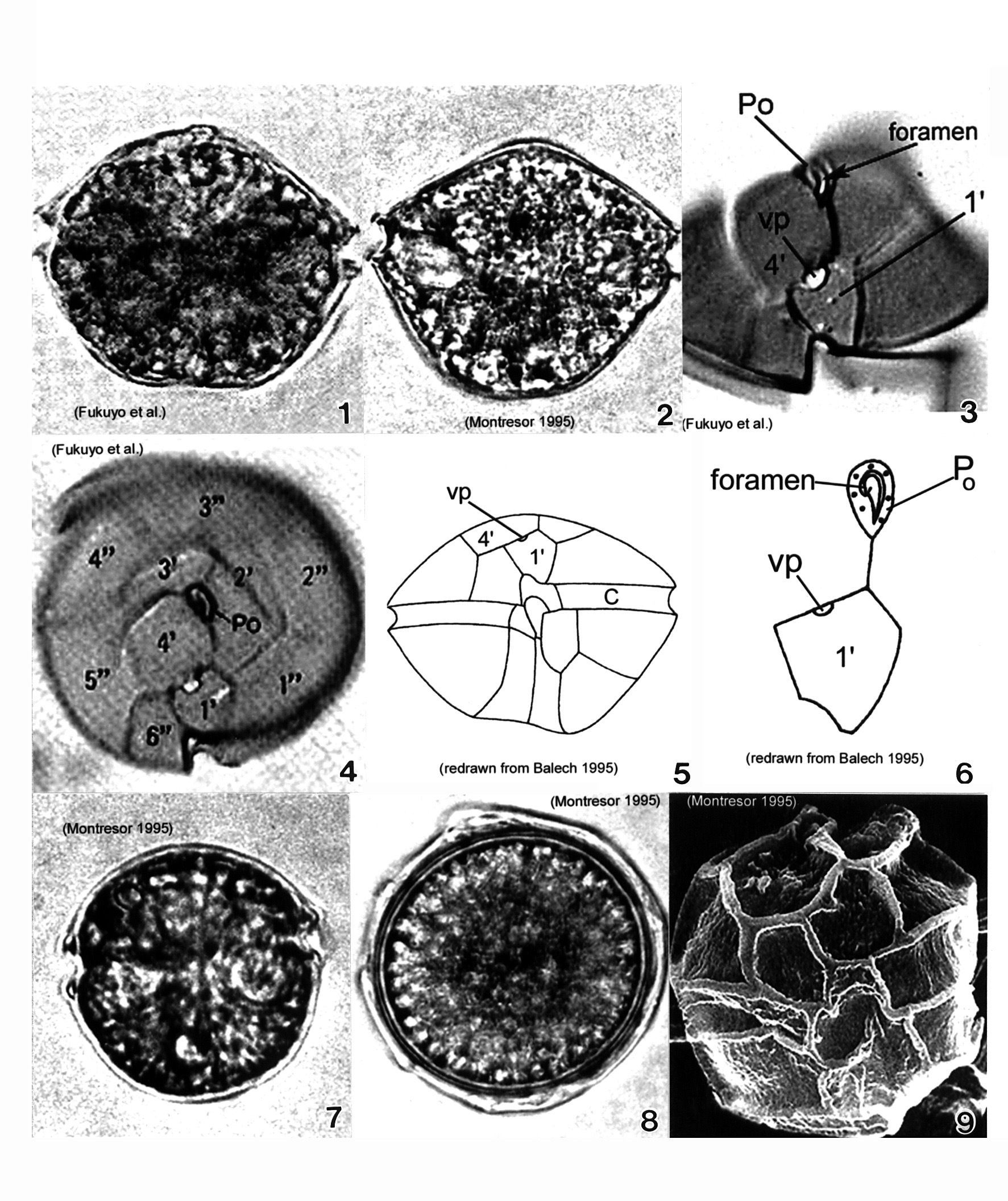
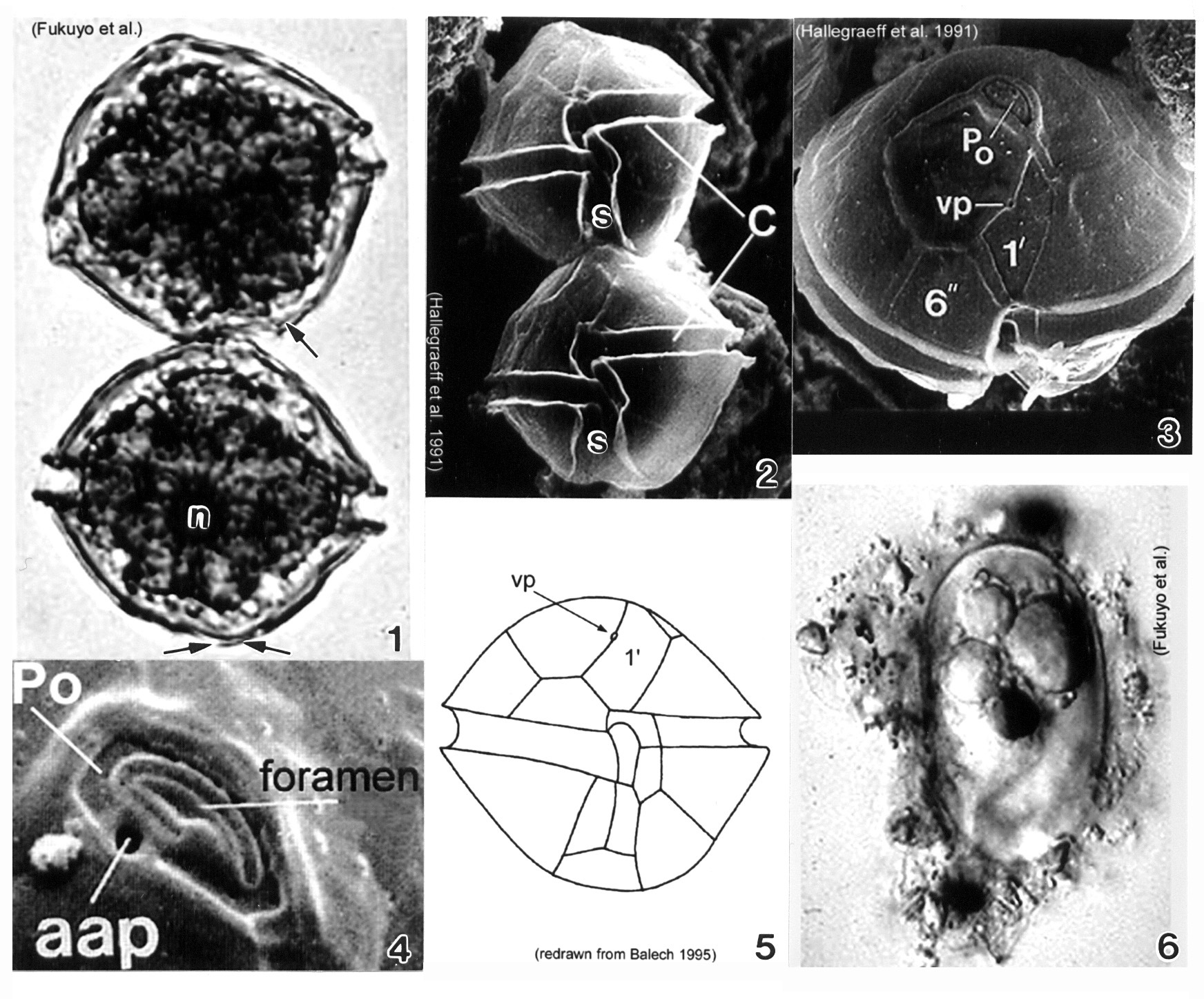
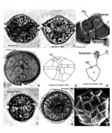
Plate 4. Alexandrium monilatum. Fig. 1. LM: four-cell chain. Cells large, wider than long, flattened anterio-posteriorly. Antapex slightly concave (arrow). Figs. 2-4. Line drawings. Fig. 2. Ventral pore (vp) depicted (Florida specimens) at anterior margin of 1' plate where it comes in contact with plates 2' and 4'. Cingulum (C) deeply excavated, wide, descending; displaced one time its width. Fig. 3. Apical pore plate (Po) does not come in contact with 1' plate. Anterior attachment pore (aap) large, round and dorsally situated in the APC. Foramen comma-shaped. Fig. 4. Antapical view: posterior sulcal plate (sp) large, rhomboid and concave with radial markings. Posterior attachment pore (pap) large and centrally located. Figs. 5-6. LM. Fig. 5. Two isogamous gametes fusing at oblique angles. Fig. 6. Mature resting cysts: dark and round, with a triple layered wall.
-
-
Lugol's-fixed specimen from the NW Med.
-
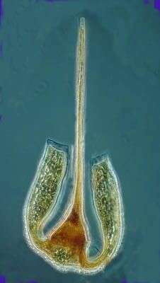
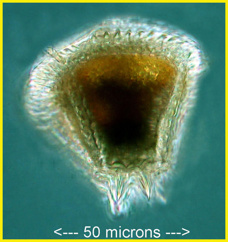
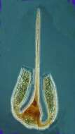
This species has a highly variable morphology. The cell body is small with long apical and antapical horns which are pointing towards the ventral side of the body. The horns are open ended. This species can be common in the North and Irish Sea in summer and autumn.
-
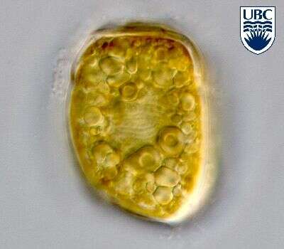
This species of Ceratium seems to be polymorphoric as the 'platy cornes' are variable in width & shape. This specimen was preserved with lugols & found in the NW Med in October.
-
Gambierdiscus (gam-beer-disk-us) toxicus, a toxic dinoflagellate with chloroplasts. With two flagella, but only one (the trailing flagellum) visible here. Small lobes of the plastid extend to the surface of the cell. Armoured, with fairly thick thecal plates. Flattened and this view is more or less polar. Mostly associated with sediments of warmer waters. Differential interference microscopy.
data on this strain.
-
Ceratium arcticum specimens from the Chukchi Sea.
-
Gambierdiscus (gam-beer-disk-us) toxicus, a toxic dinoflagellate with chloroplasts. With two flagella, but only one (the trailing flagellum) visible here. Armoured, with fairly thick thecal plates. Flattened and this view is more or less polar. Mostly from warmer waters. Differential interference microscopy.
data on this strain.
-
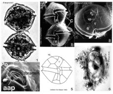
Plate 5. Alexandrium ostenfeldii. Figs. 1-3. LM. Fig. 1. Ventral view. Cell large and nearly spherical. Cingulum deeply excavated. Epitheca broad and convex-conical. Hypotheca hemispherical with an obliquely flattened antapex. Fig. 2. Epitheca: apical view. Ventral pore (vp) large and distinct. First apical plate (1') forms a 90 degree angle at the point where vp and 4' plate come in contact. Apical pore complex (APC) with comma-shaped foramen. Figs. 3-4. Line drawings. Fig. 3. Ventral view: 6'' plate wider than high. Cingulum (C) slightly excavated. Fig. 4. APC and 1' plate: a. Po in direct contact with 1'; b. Po in indirect contact with 1' via thin suture. Fig. 5. LM: vegetative cell. Small equatorial nucleus (n). Fig. 6. LM: temporary cyst large and spherical, covered in mucilage. Nucleus visible (arrowhead)(Mackenzie et al. 1996).
-
Gambierdiscus (gam-beer-disk-us) toxicus, a toxic dinoflagellate with chloroplasts (not shown in this image). Armoured, with fairly thick thecal plates. This image shows the plates and a large apical pore. Mostly from sediments of warmer waters. Differential interference microscopy.
data on this strain.
-
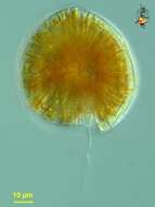
Amphidiniella (am-fee-din-ee-ella) sedentaria Horiguchi 1995. The image shows a cell in ventral view. The cingulum is near the anterior end of the cell and is slightly ascending.
-
-
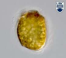
Adenoides spec., a so far undescribed taxon. Left lateral view, mid cell focus, note the granula reserve material in the cell.
-
-
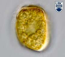
Left lateral view, mid cell focus. Note the centrally located nucleus and storage material (granules).
-
Plate 6. Alexandrium pseudogonyaulax. Figs. 1-4. LM. Fig. 1. Ventral view. Cell broadly pentagonal; wider than long. Epitheca short and dome-shaped. Hypotheca longer than epitheca. Cingulum shallow and barely displaced. Fig. 2. Dorsal view. Antapex obliquely concave. Fig. 3. Epitheca: ventral view. Apical pore plate (Po) with comma-shaped foramen. 1' plate pentagonal with large wide ventral pore (vp) on 4' plate margin. Fig. 4. Epitheca: apical view. 1' plate does not come in contact with Po. Po oval and longitudinal on apex. Figs. 5-6. Line drawings. Fig. 6. Po and 1' plate not in contact. Fig. 7. LM: isogamous gametes smaller and rounder than vegetative cells. Fig. 8. LM: round resting cyst. Fig. 9. SEM: paratabulate cyst.
-
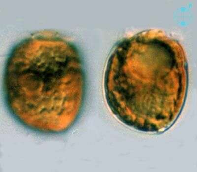
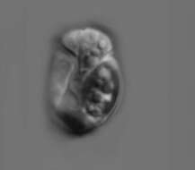
Adenoides (add-en-oi-dees) eludens (Herdman) Balech 1956. The image on the left shows the left lateral view of a cell, with yellow-brown plastids with a ring -like pyrenoid. The small epicone is almost not visible. The image on the right shows a mid-focus plane through a cell, with a pusule near the anterior end and the nucleus near the posterior end.
-
Plate 7. Alexandrium tamarense. Fig. 1. LM. Two cell chain: cells small to medium; slightly longer than wide, nearly spherical. Cingulum (C) deeply escavated and lipped. Left hypothcal lobe slightly larger than right. Nucleus (n) visible. Figs. 2-4. SEM. Fig. 2. Two cell chain: cingulum displaced 1X its width. Deep sulcus (s) widens posteriorly. Fig. 3. Epitheca: apical view. Apical pore plate (Po) rectangular; narrows ventrally. Po and first apical plate (1') in direct contact. Small ventral pore present on 1' plate. Fig. 4. Apical pore complex (APC): foramen large and fishhook shaped. Small round anterior attachment pore (aap) present (Hallegraeff 1991). Fig. 5. Line drawing. Fig. 6. LM. Oblong resting cyst with rounded ends, reddish lipid bodies; covered in mucilage.