Bythograea
vrijenhoeki
Juveniles or regressed adults with observations on two fixed and sectioned specimens from Bythograea vrijenhoeki collected on the American cruise PAR 5-388S, Dive 4089. On slide series 4089 Bv and series Carcino Bv (1). Worms small, 450-500 lm; found in mucous sheath on host. Ocelli not observed. Anterior proboscis chamber
pyriform, 15 lm long. Basis eosinophilic, robust, oblique section, 9 lm wide. Single stylet on basis, at least 10 lm long, with basal hub of 5 lm. Stylet to basis ratio not calculated. Two accessory stylet pouches anterolateral to stylet bulb. Anterior proboscis chamber not measured.Middle proboscis chamber 15 lm in diameter, glandular in
appearance. Posterior proboscis chamber glandular, 36-45 lm long by 17 lm wide, intensely basophilic, with weak fibrous coat. Proboscis sheath greatly reduced. Body
musculature with one layer of outer circular muscles, one layer of inner longitudinal muscles. Submuscular glands eosinophilic; elongate, slender, 10-20 lm long by 4-5 lm
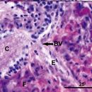
wide; numerous. In section, submuscular glands as a single row around the body, not arrayed as in O. davidi; interior to muscles. Frontal glands present in esophageal region
anterior to cerebrum; eosinophilic, as a diffuse field around esophagus. Gonads undeveloped.
American cruise PAR 5) Dive #4089: 23
March 2005, PAR-388S, Sebastian’s Steamer vent site,
37847.289S, 110854.519W
“Ovicides jonesi new species
Figs. 5 and 6
Material.—Juveniles or regressed adults with observations on two fixed and sectioned specimens from Bythograea vrijenhoeki collected on the American cruise PAR 5-38°S, Dive 4089. On slide series 4089 Bv and series Carcino Bv (1). Worms small, 450-500 µm; found in mucous sheath on host. Ocelli not observed. Anterior proboscis chamber
pyriform, 15 µm long. Basis eosinophilic, robust, oblique section, 9 µm wide. Single stylet on basis, at least 10 µm long, with basal hub of 5 µm. Stylet to basis ratio not
calculated. Two accessory stylet pouches anterolateral to stylet bulb. Anterior proboscis chamber not measured. Middle proboscis chamber 15 µm in diameter, glandular in
appearance. Posterior proboscis chamber glandular, 36-45 µm long by 17 µm wide, intensely basophilic, with weak fibrous coat. Proboscis sheath greatly reduced. Body
musculature with one layer of outer circular muscles, one layer of inner longitudinal muscles. Submuscular glands eosinophilic; elongate, slender, 10-20 µm long by 4-5 µm
wide; numerous. In section, submuscular glands as a single row around the body, not arrayed as in O. davidi; interior to muscles. Frontal glands present in esophageal region
anterior to cerebrum; eosinophilic, as a diffuse field around
esophagus. Gonads undeveloped.
Material.—Juveniles and regressed adults with observations on nine fixed and sectioned specimens from Bythograea laubieri collected on the American cruise PAR 5, Dive
4089. On slide series Carcino B.l. (1) (Accession number USNM 1097951). Worms small, 500-1000 lm long by 160-180 lm wide; found in mucous sheath on host. Ocelli
absent. Basis eosinophilic, robust, 27 µm long by 8-10 µm wide. Single stylet on basis, 9-10 µm long, with basal hub of 3-5 µm wide. Stylet to basis ratio 0.333-0.370. Two
accessory stylet pouches anterolateral to stylet bulb, 15 µm long by 11 µm wide, with developing stylets. Stylet bulb 45 µm long by 20 µm wide. Anterior proboscis chamber pyriform, 12 µm long. Middle proboscis chamber 18-25 µm in diameter, weakly eosinophilic, surrounded by muscles. Posterior proboscis chamber glandular, 40-50 µm long by 16-20 µm wide, intensely basophilic, with thin fibrous coat. Proboscis sheath greatly reduced. Body musculature with one layer of outer circular muscles, one layer of inner longitudinal muscles. Submuscular glands eosinophilic; elongate, slender, 8-12 µm long by 4-10 µm wide; numerous. In section, submuscular glands as a single row around the body, not arrayed as in O. davidi; interior to muscles. Cephalic glands in esophageal region anterior to cerebrum; developed as paired frontal organs; weakly basophilic in most worms, occasionally eosinophilic. Frontal organs in several worms weakly basophilic, with vesicular appearance. Gonads undeveloped. Two worms with regressed oöcytes.
Type Host and Site of Infestation.—On the sterna, pleopods and axillae of the pereiopods of Bythograea laubieri and B. vrijenhoeki.
Type Locality.—(American cruise PAR 5) Dive #4089: 23n March 2005, PAR-38°S, Sebastian’s Steamer vent site, 37°47.28’S, 110°54.51’W, 2204 m.
Holotype.—Regressed adult (Accession number USNM 1097951) on slide series Carcino B.l. (1) (worm 1, slides 1 through 3) deposited in the National Museum of Natural History, Smithsonian Institution, Washington, D.C., USA.
Paratypes.—Juveniles and regressed adults (Accession number USNM 1097952) on slide series Carcino B.l. (1) (worms 2-9, slides 2 and 3) in the National Museum of Natural History, Smithsonian Institution, Washington, D.C., USA. Juveniles or regressed adults (Accession number MNHN-NMRT 2) on slide series Carcino B.v. B.v. (2), slides 3 through 7, in the Muséum National d’Histoire Naturelle, Paris.
Etymology.—The species is named in honor of Dr. William ‘‘Joe’’ Jones of MBARI, whose hard work and careful attention to details made the oceanographic missions in the South Pacific successful and productive.
Remarks.—We place the worms from B. vrijenhoeki and B. laubieri into the genus Ovicides based on their carcinonemertid characters and the presence of the accessory stylet pouches. The worms from B. vrijenhoeki were smaller than those from B. laubieri and showed a few minor differences in morphology (somewhat larger submuscular glands, diffuse and eosinophilic frontal organs), but were otherwise quite similar. Therefore, we consider them to be the same species, O. jonesi. This worm has cephalic glands organized as a presumptive frontal organ in the esophageal region. In some of the worms, the organs are well organized and have a vesiculated appearance. These organs are better organized than the diffuse frontal organs present in O. davidi. Ovicides jonesi shares features with both O. jasoni (blind, single band of submuscular glands, similar sized stylet) and O. davidi (large basis, large posterior proboscis chamber), but it can be separated from each by the opposite characters (Tables 1 and 2). The frontal organ in O. jonesi is unusual in that it is well organized in some specimens, or only weakly developed in other specimens of the same species from the same host. This difference is difficult to explain, but it may be due to differences in maturity or metabolic state (juveniles vs. regressed adults that have fed previously). Ovicides jonesi is only the third species in the family known to possess a frontal organ, after O. davidi (see above) and C. australiensis (see Campbell et al., 1989). In O. jonesi, the frontal organ lies immediately anterior to the cerebrum and can be quite large, 15-30 lm in diameter. The frontal organ of Carcinonemertes australiensis is also large and well organized, whereas that of O. davidi is diffusely organized.”
(Shields and Segonzac, 2007; 686-690)