About
Education
Discuss
TraitBank
Sign In
Sign Up
Language
Deutsch
English
Español
français
italiano
Nederlands
Piemontèis
Português do Brasil
suomi
Türkçe
čeština
Ελληνικά
македонски
Українська
العربية
简体中文
繁體中文
names in breadcrumbs
vernacular
scientific
About
Education
Discuss
TraitBank
Sign In
Sign Up
en
Deutsch
English
Español
français
italiano
Nederlands
Piemontèis
Português do Brasil
suomi
Türkçe
čeština
Ελληνικά
македонски
Українська
العربية
简体中文
繁體中文
names in breadcrumbs
vernacular
scientific
Creatures
»
…
»
Animal
»
Cnidarians
»
Anemones And Corals
»
…
»
Sea Anemones
»
…
Creatures
»
Cellular Organisms
»
Eukaryotes
»
Opisthokonts
»
Animal
»
Cnidarians
»
Anemones And Corals
»
Hexacorallians
»
Sea Anemones
»
Enthemonae
»
Actinioidea
«
Actiniidae Rafinesque 1815
collect
overview
data
media
articles
names
No copyright
any license
CC-BY
CC-BY-NC
CC-BY-NC-SA
CC-BY-SA
No copyright
type
any type
image
map
sound
video
provider
any provider
Arnold Arboretum Photo Gallery
Barcode of Life Data Systems
Barry Armstead Photography, ALA
BioImages, the virtual fieldguide, UK
Botanical Illustrations
Bugs for Bugs, Atlas of Living Australia
CalPhotos
CephBase
Eastfield College SEM Lab
eMammal
Femorale
Fishbase
Flickr BHL
Flickr Group
Harvard Museum of Comparative Zoology
iNaturalist
Moorea Biocode
Mycokeys
NMNH Collection
Phytokeys
PlanetScott
SailinSteve
SI Wild
SINA images
TreatmentBank
Turbellarian Taxonomic Database
USDA PLANTS images
Wikimedia Commons
Wolf Spiders of Australia, ALA
World Register of Marine Species
1
2
3
4
5
…
Last »
cc-publicdomain
trusted
cc-publicdomain
trusted
cc-publicdomain
trusted
cc-publicdomain
trusted
cc-publicdomain
trusted
cc-publicdomain
trusted
cc-publicdomain
trusted
cc-publicdomain
trusted
cc-publicdomain
trusted
cc-publicdomain
trusted
cc-publicdomain
trusted
cc-publicdomain
trusted
cc-publicdomain
trusted
cc-publicdomain
trusted
cc-publicdomain
trusted
cc-publicdomain
trusted
cc-publicdomain
trusted
cc-publicdomain
trusted
cc-publicdomain
trusted
cc-publicdomain
trusted
cc-publicdomain
trusted
cc-publicdomain
trusted
cc-publicdomain
trusted
cc-publicdomain
trusted
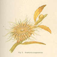
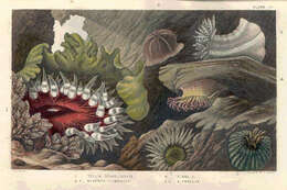
Anemonia sargassensis. 1914. Sea anemones; Anemonia.
cc-publicdomain
Freshwater and Marine Image Bank U Washington
Anemonia sargassensis.
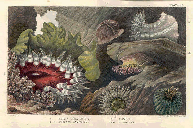
1. Tealia Crassicornis, 2. 3. Bunodes Cemmacea, 4. S. Ballii; 5. 6. S. Thallia. 1860. Sea anemones; Corals.
cc-publicdomain
Freshwater and Marine Image Bank U Washington
1. Tealia Crassicornis, 2. 3. Bunodes Cemmacea, 4. S. Ballii; 5. 6. S. Thallia.
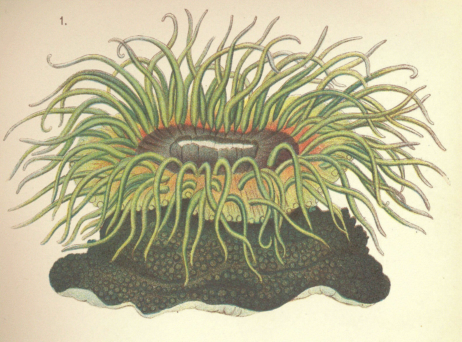
Phymactis veratra. 1890. Sea anemones.
cc-publicdomain
Freshwater and Marine Image Bank U Washington
Phymactis veratra.
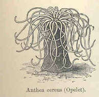
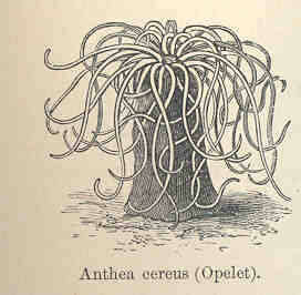
Anthea cereus (Opelet). 1892. Corals.
cc-publicdomain
Freshwater and Marine Image Bank U Washington
Anthea cereus (Opelet).
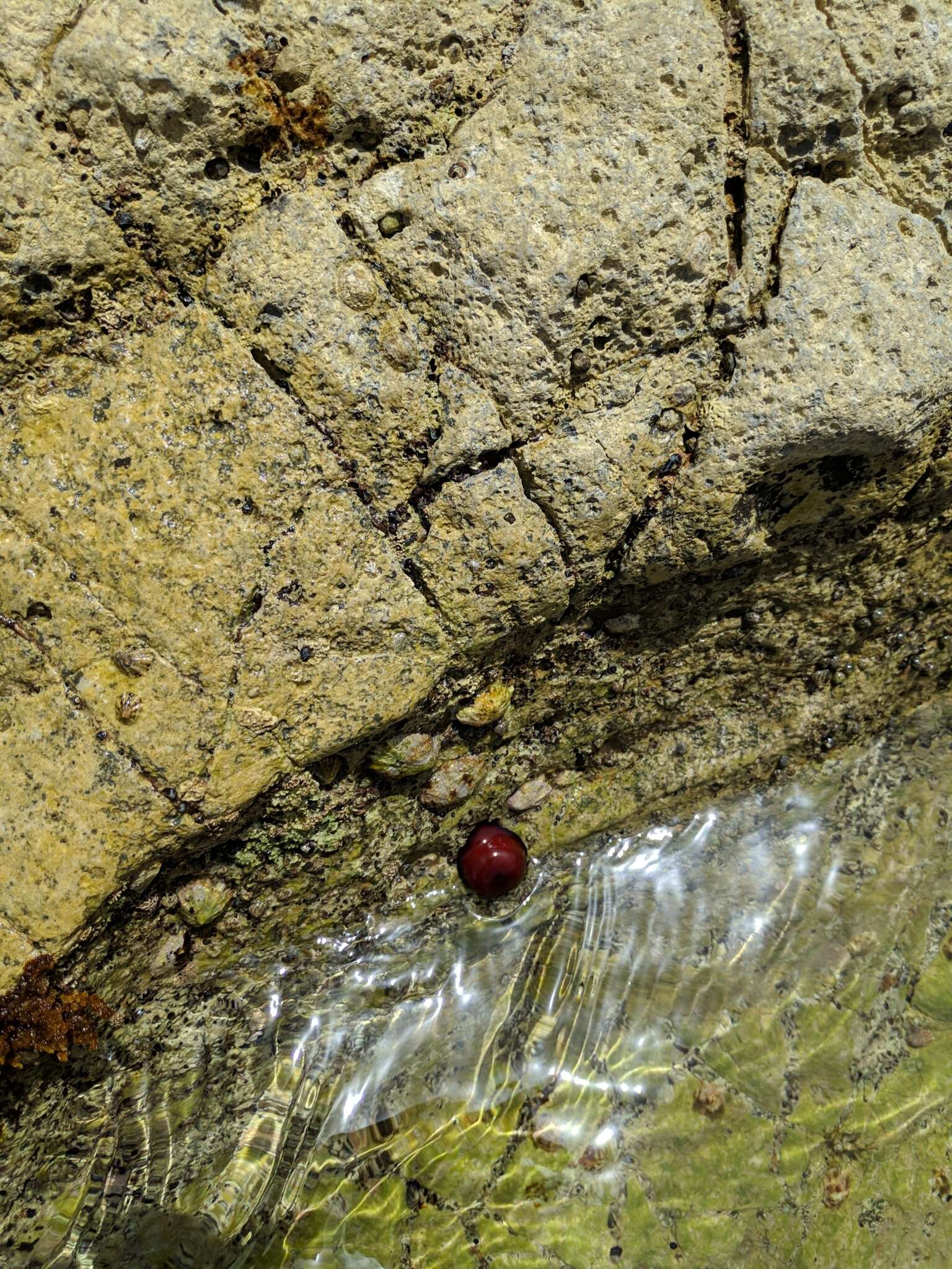
"
cc-publicdomain
Pieter Huybrechts
iNaturalist
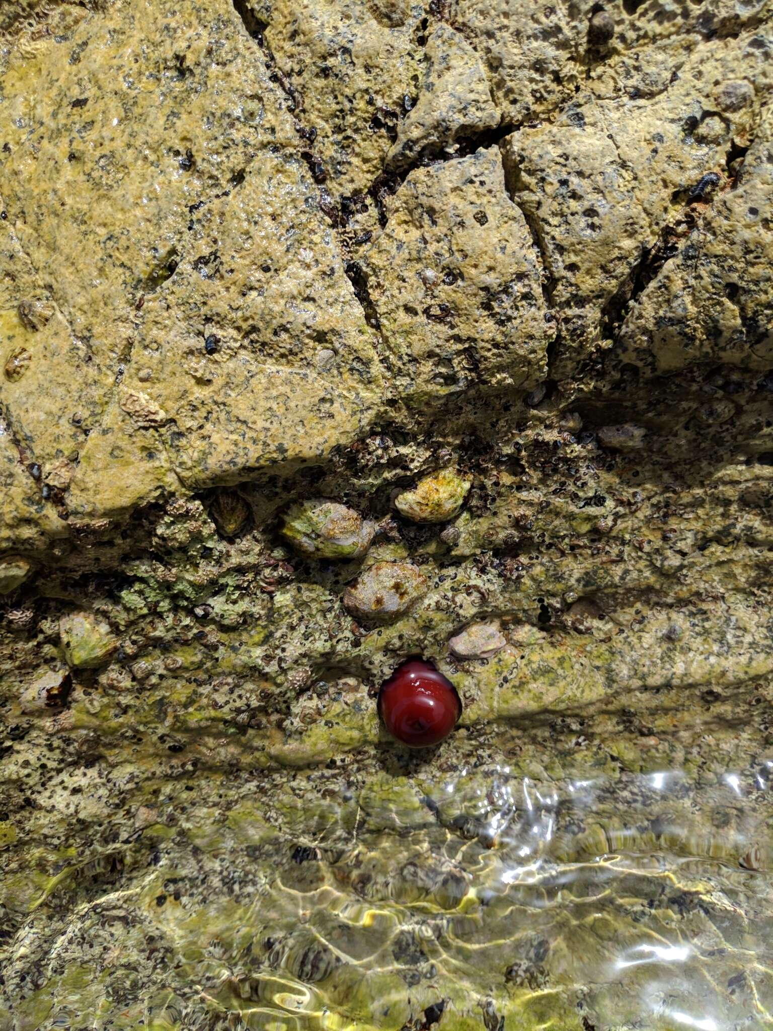
"
cc-publicdomain
Pieter Huybrechts
iNaturalist
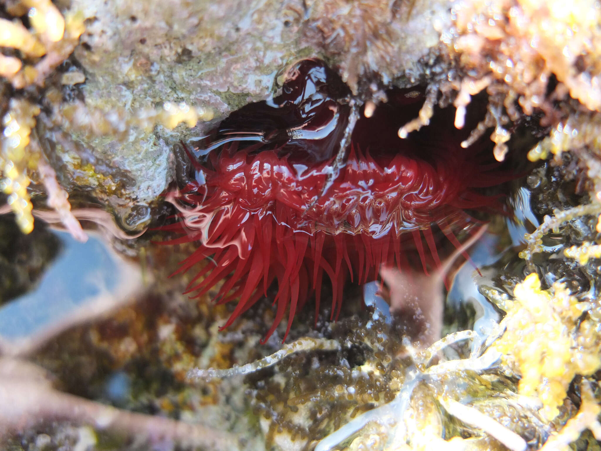
"
cc-publicdomain
Paul Braun
iNaturalist
"
cc-publicdomain
Светлана Царахова
iNaturalist
"
cc-publicdomain
Светлана Царахова
iNaturalist
"
cc-publicdomain
Anna N Chapman
iNaturalist
"
cc-publicdomain
Scott Loarie
iNaturalist
"
cc-publicdomain
Andrew Deacon
iNaturalist
"
cc-publicdomain
natalie
iNaturalist
"
cc-publicdomain
natalie
iNaturalist
"
cc-publicdomain
Gabe Schp
iNaturalist
"
cc-publicdomain
Dan Horowitz
iNaturalist
"
cc-publicdomain
Sunita Singh
iNaturalist
"
cc-publicdomain
peter-f
iNaturalist
"
cc-publicdomain
ANDRÉ SIMÕES
iNaturalist
"
cc-publicdomain
Alex Heyman
iNaturalist
"
cc-publicdomain
Alex Heyman
iNaturalist
"
cc-publicdomain
Alex Heyman
iNaturalist
"
cc-publicdomain
Alex Heyman
iNaturalist
"
cc-publicdomain
Alex Heyman
iNaturalist
1
2
3
4
5
…
Last »