-
O. sinensis is closely related to O. mobiliensis and O. regia but the processes are nearly parallel to the cell axis and the processes are close to the processes. The valve face between the processes is flat or concave.
-
Phasde contrast micrograph of living diatom - cracked.
-
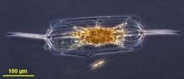
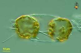
Eucampia (you-camp-ee-a) zoodiacus is a filament forming diatom (stramenochrome). Adjacent cells are attached by two interlocking apical elevations. Differential interference microscopy.
data on this strain.
-
Chain forming diatom, which produces wing like extensions one valve end, which link adjacent cells. This is a cosmopolitan species.
-
Biddulphia pulchella.
-
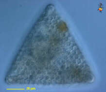
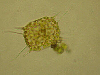
Biddulphia ?. A triangular version? Collected by ATOL special protist hunters 1st ocean taw in Woods Hole during the Protistology Workshop at MBL, October-November 2005. Isolation and art by Adrian Reyes-Prieto.
-
Eucampia (you-camp-ee-a) zoodiacus is a filament forming centric diatom (stramenochrome). Adjacent cells are attached by two interlocking apical elevations. Detail showing peripheral disc-shaped plastids and central nucleus. Differential interference microscopy.
data on this strain.
-
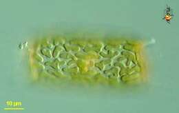
Odontella (owe-don-tell-a) mobiliensis, a centric diatom. The frustule or shell is formed of two valves joined by girdle bands. Many small peripheral chloroplasts and a central nucleus. Differential interference microscopy.
data on this strain.
-
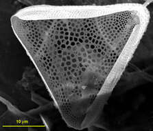
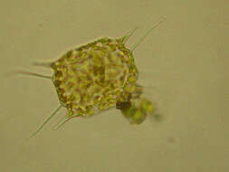
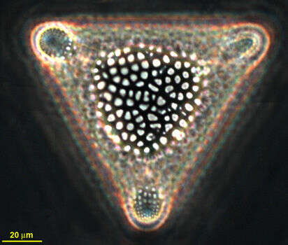
Biddulphia sp? (Triangular guy). Collected by ATOL special protist hunters 1st ocean taw. Woods Hole Massachusetts for the Protistology Workshop at MBL. October-November 2005. Isolation and art by Adrian Reyes-Prieto, SEM by Charles O'Kelly and Shauna Murray.
-
Triceratium moronense.
-
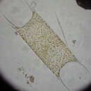
Odontella (owe-don-tell-a) mobiliensis, a centric diatom. The frustule or shell is formed of two valves joined by girdle bands. With horns (spines) emerging from the apical margins of the valves and more spines (referred to as apical processes) arising closer to the centre of the valves. Many small peripheral chloroplasts and a central nucleus. Two daughter cells located within frustule of parental cell. Differential interference microscopy.
data on this strain.
-
Biddulphia sp? (Triangular guy). Collected by ATOL special protist hunters 1st ocean taw. Woods Hole Massachusetts for the Protistology Workshop at MBL. October-November 2005. Isolation and art by Adrian Reyes-Prieto, SEM by Charles O'Kelly and Shauna Murray.
-
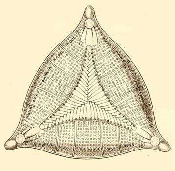
Triceratium pentacrinus.
-
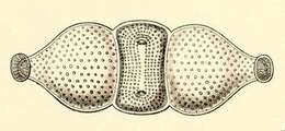
Cells are single or united into short chains by the long spines extending from the elevated central part of the valve face. The processes are slender and point diagonally outward. This species can be confused with O. regia.
-
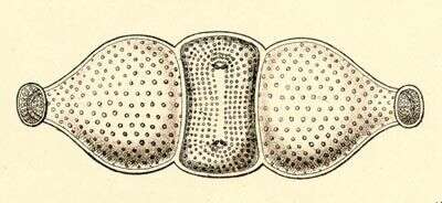
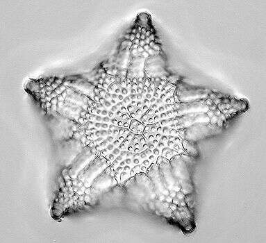
Trigonium (try-go-knee-um) or Hydrocera (high-dro-see-ra) is a marine diatom. It is a centric diatom in which a three pointed asymmetry has been imposed. Test only. Differential interference contrast,
-
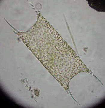
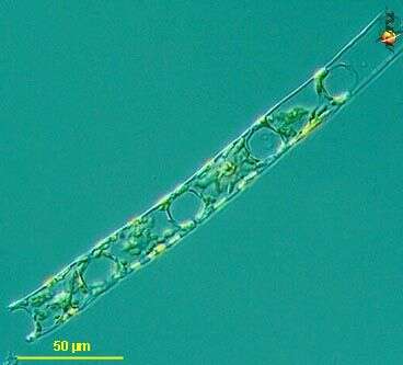
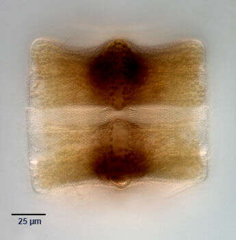
Portrait (girdle view) of two frustules of the centric marine diatom Triceratium pentacrinus (Ehrenberg). Collected from a commercial saltwater aquarium in Boise, Idaho, october 2004. DIC.
-
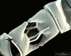
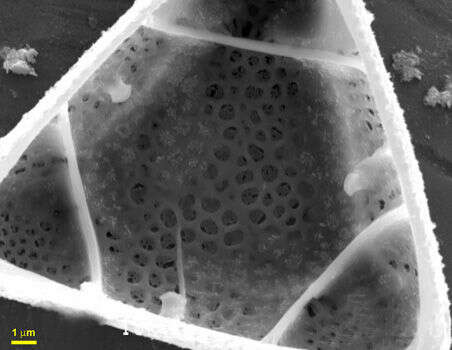
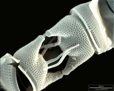
Scale bar indicates 10 µm. Sample from North Sea near Heligoland (spring diatom bloom). Use of SEM equipment courtesy of Lab Dr. Karl-Heinz Schäffner, Solingen, Germany.
-
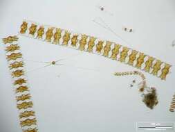
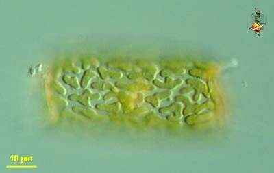
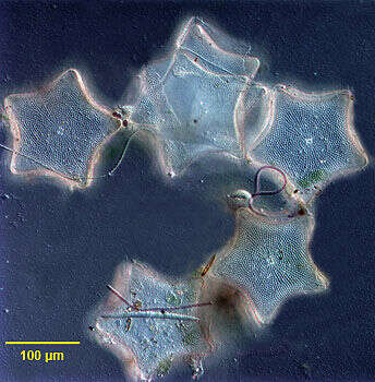
Group portrait (valve view)of the centric marine diatom, Triceratium pentacrinus (Ehrenberg). Collected from a commercial saltwater aquarium in Boise, Idaho October 2004. DIC.
-
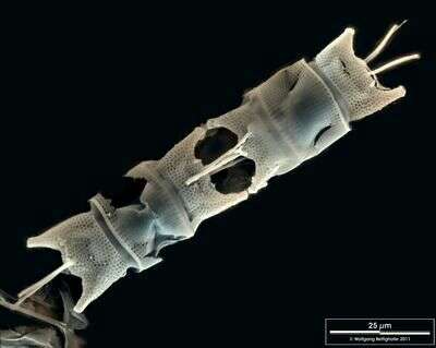
Scale bar indicates 25 µm. Sample from North Sea near Heligoland (spring diatom bloom). Use of SEM equipment courtesy of Lab Dr. Karl-Heinz Schäffner, Solingen, Germany.
-
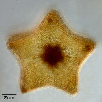
Portrait (valve view) of the centric marine diatom, Triceratium pentacrinus (Ehrenberg). Collected from a commercial saltwater aquarium in Boise, Idaho, October 2004. DIC.
-
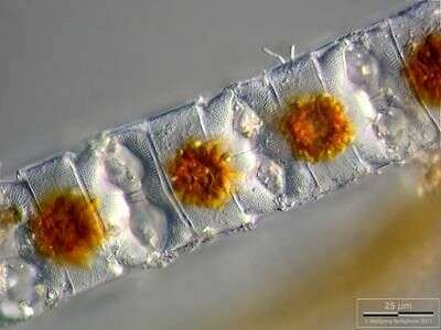
Depth-of-focus image exhibiting structure of the frustules. Chloroplasts are concentrated in cell centers due to long lasting exposition with microscope's illumination. Scale bar indicates 25 µm. The image was built up using several photomicrographic frames with manual stacking technique. Sample from North Sea near Heligoland (spring diatom bloom). Images were taken using Zeiss Universal with Olympus C7070 CCD camera.
-
-
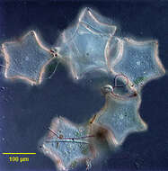
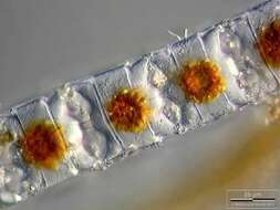
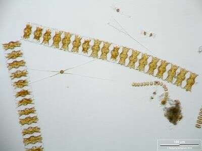
Long chains of Odontella aurita accompanied by Chaetoceros danicus and Thalassiosira nordenskjoeldii. Scale bar indicates 100 µm. The image was built up using several photomicrographic frames with manual stacking technique. Sample from North Sea near Heligoland (spring diatom bloom). Images were taken using Zeiss Universal with Olympus C7070 CCD camera.
-
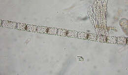
E. zodiacus forms long spiralling chains. It has blunt processes connecting adjacent cells. This species can be abundant in autumn