-
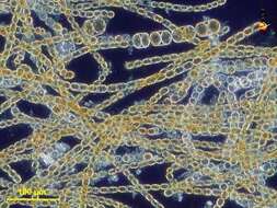
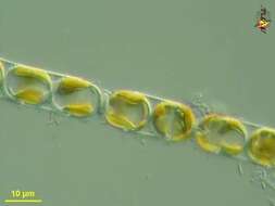
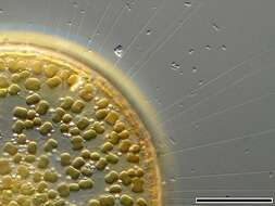
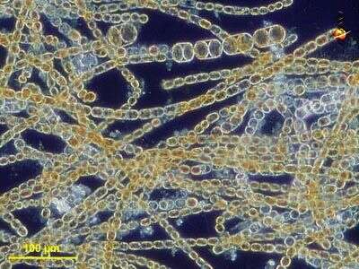
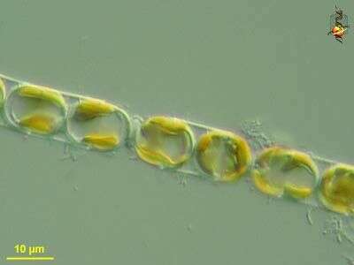
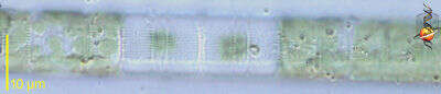
Melosira (mellow-sire-a) nummuloides, filament forming centric diatom, with multiple small plastids within the cell. Dark ground illumination. Leptosiropsis (leapt-owe-sire-op-sis) torulosa, green alga with organic wall that is produced in layers. Phase contrast microscopy.
data on this strain.
-
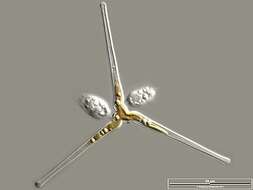
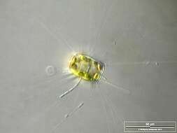
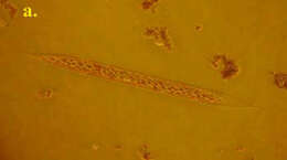
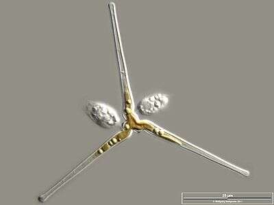
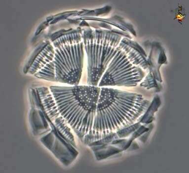
The oblique view exhibits short silicous spines, the so called occluded processes. Some chitinous spines protruding from the fultoportulae (also called strutted processes) along the dotted valve margin are also visible. Scale bar indicates 50 µm. The image was built up using several photomicrographic frames with manual stacking technique. Sample from North Sea near Heligoland (spring diatom bloom). Images were taken using Zeiss Universal with Olympus C7070 CCD camera.
-
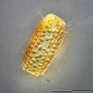
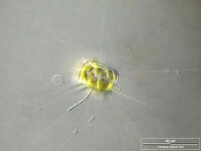
Cell accompanied by epibiotic flagellates. Scale bar indicates 50 µm. Sample from the Federsee near Lake Constance. The image was built up using several photomicrographic frames with manual stacking technique. Images were taken using Zeiss Universal with Olympus C7070 CCD camera.Image under Creative Commons License V 3.0 (CC BY-NC-SA).
-
-
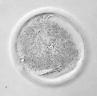
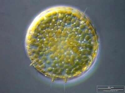
Cyclotella (sike-low-tell-a). Centric diatom, frustule only, seen from valve view, frustule broken from cover-slip pressure to show the brittle nature of the frustule. The pattern of pores in the frustule is used in identification. Phase contrast.
-
Melosira (mellow-sire-a) nummuloides, filament forming centric diatom, with multiple small plastids within the cell clearly shown in this micrograph. Differential interference microscopy.
data on this strain.
-
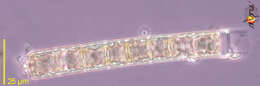
Silicious processes (the labiate and the occluded ones) are visible. Scale bar indicates 25 µm. The image was built up using several photomicrographic frames with manual stacking technique. Sample from North Sea near Heligoland (spring diatom bloom). Images were taken using Zeiss Universal with Olympus C7070 CCD camera.
-
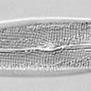
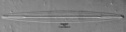
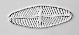
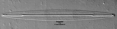
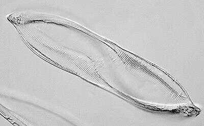
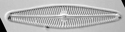
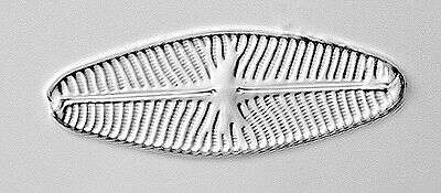
This image of Amphipleura pellucida was taken using an Olympus SPlanapo 100X/1.40, Zeiss 1.40 achro plan condenser, DIC, Wratten 47 deep blue filter, The Imaging Source 1024X768 digital camera, mosaic of 4 images (every image is an average of 32 frames in order to reduce noise). Software Panorama Maker 3.0 and Photoshop.
-
The thin, barely visible floating extensions are made of chitin. Furthermore, filamentous bacteria colonies are attached. Scale bar indicates 50 µm. Sample from the Lake Constance (vicinity of Bodman). The image was built up using several photomicrographic frames with manual stacking technique. Images were taken using Zeiss Universal with Olympus C7070 CCD camera.Image under Creative Commons License V 3.0 (CC BY-NC-SA).
-
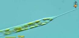
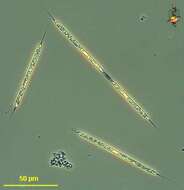
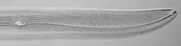
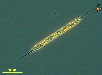
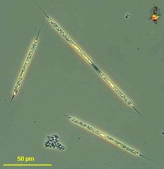
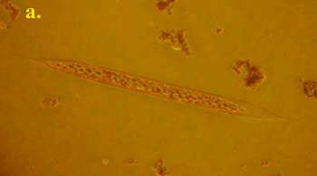
Rhizosolenia (rye-so-so-lean-ee-a) setigera, one of the common genera of marine phytoplantkonic diatoms, a centric diatom in which the valves, at the ends of the cells, are conical and give rise to spines. Much of the long cylindrical body is enclosed with hoop-shaped girdle bands. Phase contrast microscopy.
data on this strain.
-
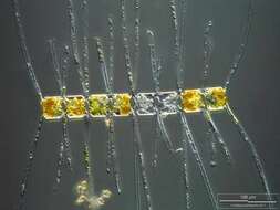
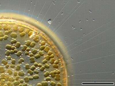
The image shows numerous chitinous spines which minimize their sedimentation speed. Scale bar indicates 25 µm. The image was built up using several photomicrographic frames with manual stacking technique. Sample from North Sea near Heligoland (spring diatom bloom). Images were taken using Zeiss Universal with Olympus C7070 CCD camera.Image under Creative Commons License V 3.0 (CC BY-NC-SA).
-
-
Optical tranversal section, showing the nucleus. The thin, barely visible floating extensions are made of chitin. Furthermore, filamentous bacteria colonies are attached. Scale bar indicates 50 µm. Sample from the Lake Constance (vicinity of Bodman). The image was built up using several photomicrographic frames with manual stacking technique. Images were taken using Zeiss Universal with Olympus C7070 CCD camera.Image under Creative Commons License V 3.0 (CC BY-NC-SA).
-
Rhizosolenia (rye-so-so-lean-ee-a) setigera, one of the common genera of marine phytoplantkonic diatoms, a centric diatom in which the valves, at the ends of the cells, are conical and give rise to spines. Much of the long cylindrical body is enclosed with hoop-shaped girdle bands. This image shows the plastids and the valve region of the cell. Differential interference microscopy.
data on this strain.
-
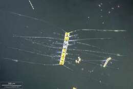
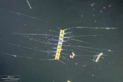
Note that setae also contain protoplasm and even chloroplasts. Scale bar indicates 200 µm. Sample from North Sea near Heligoland (spring diatom bloom). The image was built up using several photomicrographic frames with manual stacking and stitching technique. Images were taken using Zeiss Universal with Olympus C7070 CCD camera.
-
-
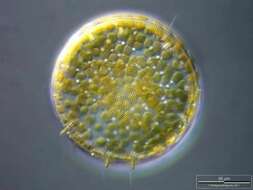
Melosira (mell-o-sigh-ra) is a centric diatom. The cells are like old-style hat boxes, or old-style pill boxes, or like petri-dishes. In Melosira, many cells are joined end to end to create a filament. The less substantial rings on the lower image are where the two halves of the frustule are joined together by the girdle bands, the more visible connections are where two cells are joined together. Each cell has a radial symmetry. As with other diatoms, plastids have chlorophylls a and c and so have a yellow brown colour. The lower picture reveals the individual disc-shaped plastids. Phase contrast.
-
Rhizosolenia (rye-so-so-lean-ee-a) setigera, one of the common genera of marine phytoplantkonic diatoms, a centric diatom in which the valves, at the ends of the cells, are conical and give rise to spines. Much of the long cylindrical body is enclosed with hoop-shaped girdle bands. Phase contrast microscopy.
data on this strain.
-
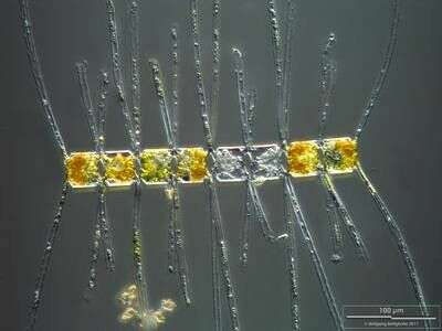
Note that setae also contain protoplasm and even chloroplasts. Scale bar indicates 100 µm. Sample from North Sea near Heligoland (spring diatom bloom). The image was built up using several photomicrographic frames with manual stacking technique. Images were taken using Zeiss Universal with Olympus C7070 CCD camera.
-
-
Melosira (mell-o-sire-a) is a centric diatom. The cells are like old-style hat boxes, or old-style pill boxes, or like petri-dishes. In Melosira, many cells are joined end to end to create a filament. The less substantial rings on the lower image are where the two halves of the frustule are joined together by the girdle bands, the more visible connections are where two cells are joined together. Each cell has a radial symmetry. As with other diatoms, plastids have chlorophylls a and c and so have a yellow brown colour. Differential interference contrast.
-
Cells are conical with long processes, otaria are absent. Diameter: 5-50 microns. R. setigera is a cold water species, which can occur throughout the year.
-
-