-
-
Note that the delicate spines are chitinous. Focus on valve surface. Scale bar indicates 50 µm. The image was built up using several photomicrographic frames with manual stacking technique. Sample from North Sea near Heligoland (spring diatom bloom). Images were taken using Zeiss Universal with Olympus C7070 CCD camera.
-
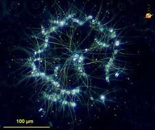
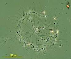
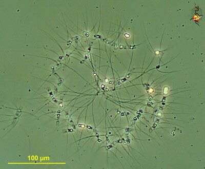
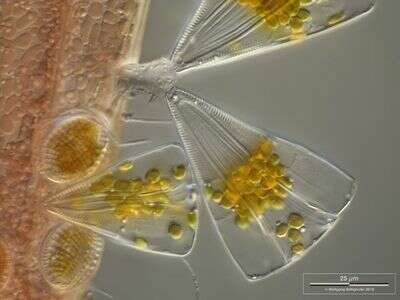
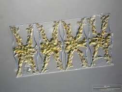
Chaetoceros socialis (key-toss-err-oss sew-see-ah-liss), a centric diatom in with long spines. In this species dozens or hundreds of cells are linked loosely together by their spines. Common in marine ecosystems. Dark ground.
-
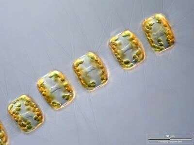
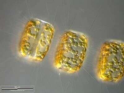
Chain of Porosira glacialis. Note that the delicate spines are chitinous. Focus on frustule surface. Scale bar indicates 50 µm. The image was built up using several photomicrographic frames with manual stacking technique. Sample from North Sea near Heligoland (spring diatom bloom). Images were taken using Zeiss Universal with Olympus C7070 CCD camera.
-
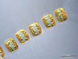
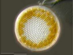
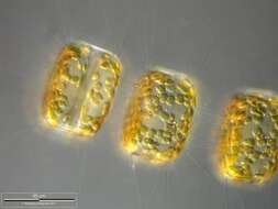
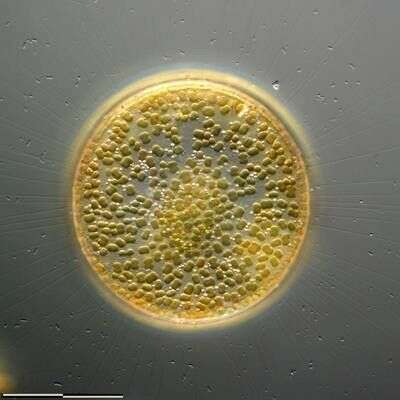
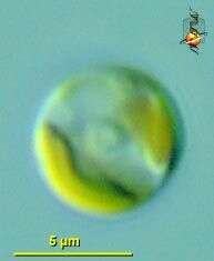
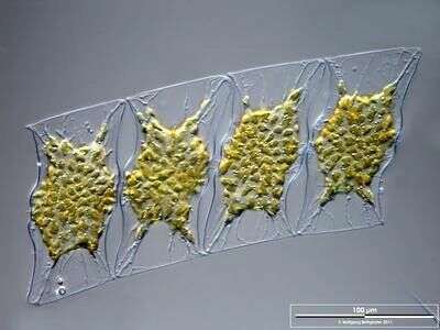
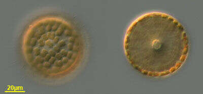
Pinguiococcus (ping-wee-cock-us) pyrenoidosus ) pyrenoidosus (pinguiophyte), a small marine phytoplankter, with a deeply bilobed golden chloroplast and a large central pyrenoid (basis for the specific epithet). Name not formally published at the this time. Differential interference microscopy.
-
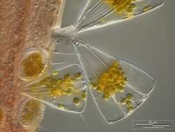
Image shows chloroplasts, small sparkling droplets of storage matter, and the fine connection tubes between the cells. Scale bar indicates 100 µm. Sample from North Sea near Heligoland (spring diatom bloom). Images were taken using Zeiss Universal with Olympus C7070 CCD camera.
-
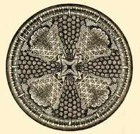
Actinoptychus heliopelta.
-
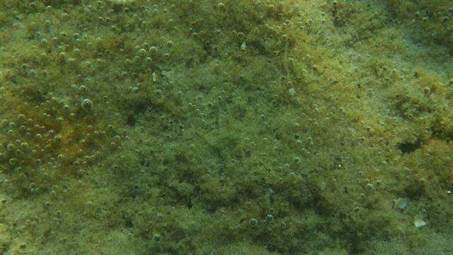
Fjellerup Strand
-
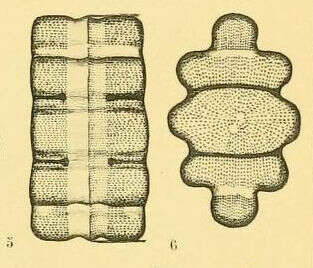
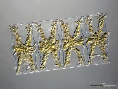
Terpsinoe americana by H. and M. PeragalloDiatomées marines de France et des districts maritimes voisins, par H. et M. Peragallo (b. 1851 and 1853, respectively). Publication info: Grez-sur-Loing,M. J. Tempère,1897-1908. Plate 90
-
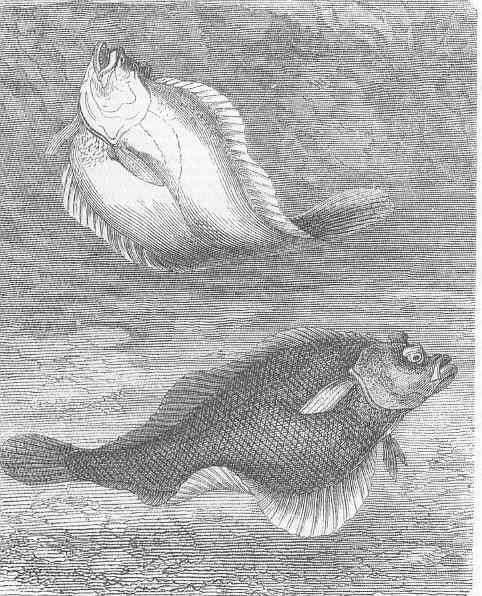
Dab (platessa Imanda).
-
-
Marginal silica processes are visible. Scale bar indicates 25 µm. The image was built up using several photomicrographic frames with manual stacking technique. Sample from North Sea near Heligoland (spring diatom bloom). Images were taken using Zeiss Universal with Olympus C7070 CCD camera.
-
Chaetoceros socialis (key-toss-err-oss sew-see-ah-liss), a centric diatom in with long spines. In this species dozens or hundreds of cells are linked loosely together by their spines. Common in marine ecosystems. Phase contrast.
-
Chain of Porosira glacialis. Note that the delicate spines are chitinous. Focus on cell center showing cytoplasmic accumulation around the nucleus. Scale bar indicates 50 µm. The image was built up using several photomicrographic frames with manual stacking technique. Sample from North Sea near Heligoland (spring diatom bloom). Images were taken using Zeiss Universal with Olympus C7070 CCD camera.
-
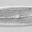
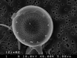
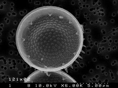
Inside view of lacunate valve. Note the position of the fultoportula on a valve face stria, taking the place of an areola. Specimen from single cell culture from Cleveland Harbor, Cleveland, Ohio, by E. Theriot.
-
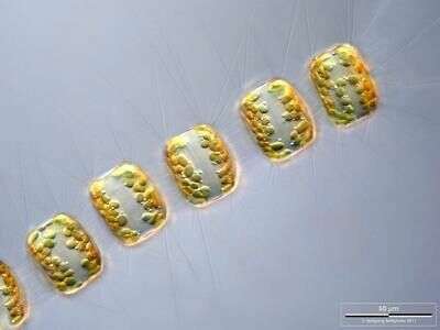
Image shows chloroplasts, small sparkling droplets of storage polysaccharide chrysolaminarin used as carbohydrate food reserve, and the fine connection tubes between the cells. The image was built up using several photomicrographic frames with manual stacking technique. Scale bar indicates 50 µm. Sample from North Sea near Heligoland (spring diatom bloom). Images were taken using Zeiss Universal with Olympus C7070 CCD camera.
-
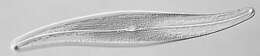
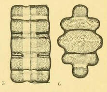
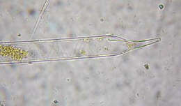
Valves are cylindrical terminating in a proboscis. Processes are absent. This is a temprate, coastal species.
-
Fjellerup Strand
-
Terpsinoe americana by Henri CoupinFrom: Album général des diatomées marines, d'eau douce ou fossiles : album représentant tous les genres de diatomées et leurs principales espèces / par Henri Coupin (b. 1868).Plate 313: frustrules (600x magnification). sea water
-
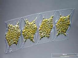
Licmophora juergensii living together with other araphid diatoms, peritrich ciliates and filamentous cyanobacteria on a red alga. They use jelly stalks for fixation on the substratum. Collected from Bodden, the brackish waters lying between the isles of Hiddensee and Ruegen (German Baltic Sea). This image was taken using Zeiss Universal with Olympus C7070 CCD camera.
-
Cells at two focal levels. Material from a plankton tow off Martha's Vineyard, Massachusetts. Image by Jeff Cole.
-
This species consists of very small cells which are united into curved chains. Cells have three short setae and one long one which causes the formation of larger secondary colonies by linking up in the centre of the colony with the long setae of other chains
-
Closeup of Porosira glacialis chain. Note that the delicate spines are chitinous. Focus on frustule surface. Scale bar indicates 50 µm. The image was built up using several photomicrographic frames with manual stacking technique. Sample from North Sea near Heligoland (spring diatom bloom). Images were taken using Zeiss Universal with Olympus C7070 CCD camera.
-
Scanning electron micrograph of interior of scutate valve. Note position of fultoportula in central area of valve face. Specimen from culture of cell isolated from Cleveland Harbor, Cleveland, OH, USA, by E. Theriot.