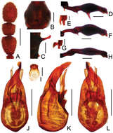
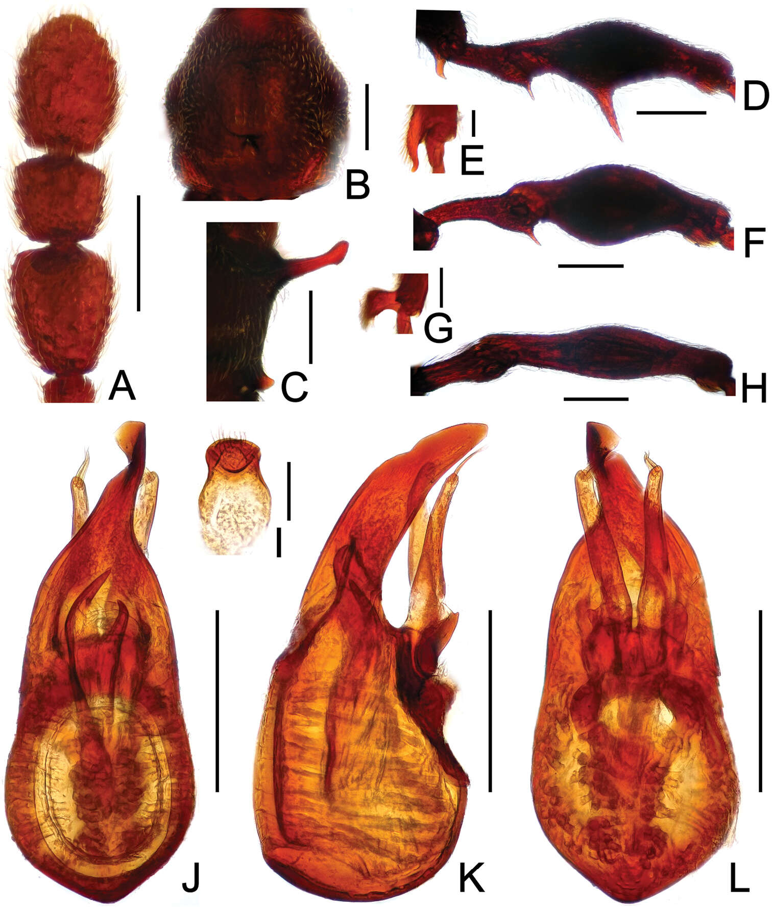
Figure 17.Diagnostic features of Pselaphodes tibialis. A antenna B pronotum C median meteventral process, in lateral view D procoxa, protrochanter and profemur E apical portion of protibia F mesotrochanter and mesofemur G apical portion of mesotibia H metatrochanter and metafemur I sternite IX J aedeagus, in dorsal view K same, in lateral view L same, in ventral view. Scales (mm): A, B, C, D, F, H, J, K, L = 0.2; I = 0.1; E, G = 0.05.