-
-
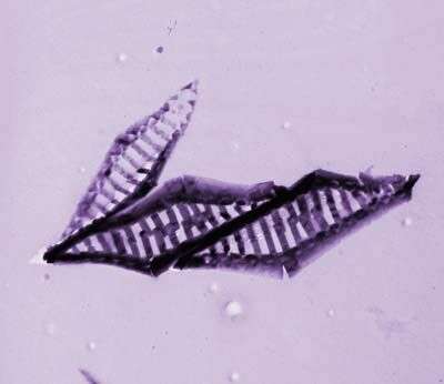
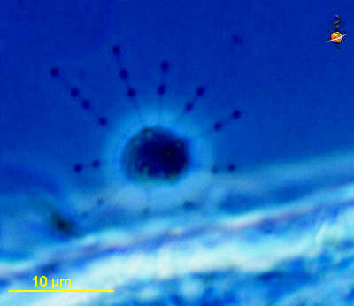
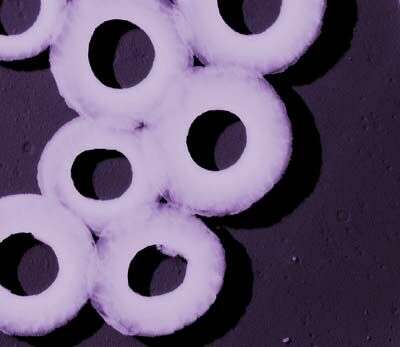
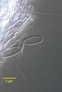
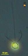
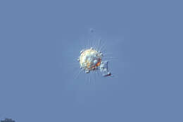
This image was made from samples taken during a scientific cruise in the Pacific. Water was filtered to concentrate the organisms that were present, then dried onto a thin sheet of plastic and then shadowed with a fine layer of metal to provide contrast. The preparation was then observed with an electron-microscope. This technique has been used to document the diversity of marine microbes, especially, protists in the oceans.
-
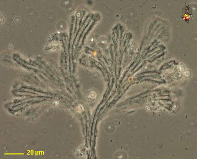
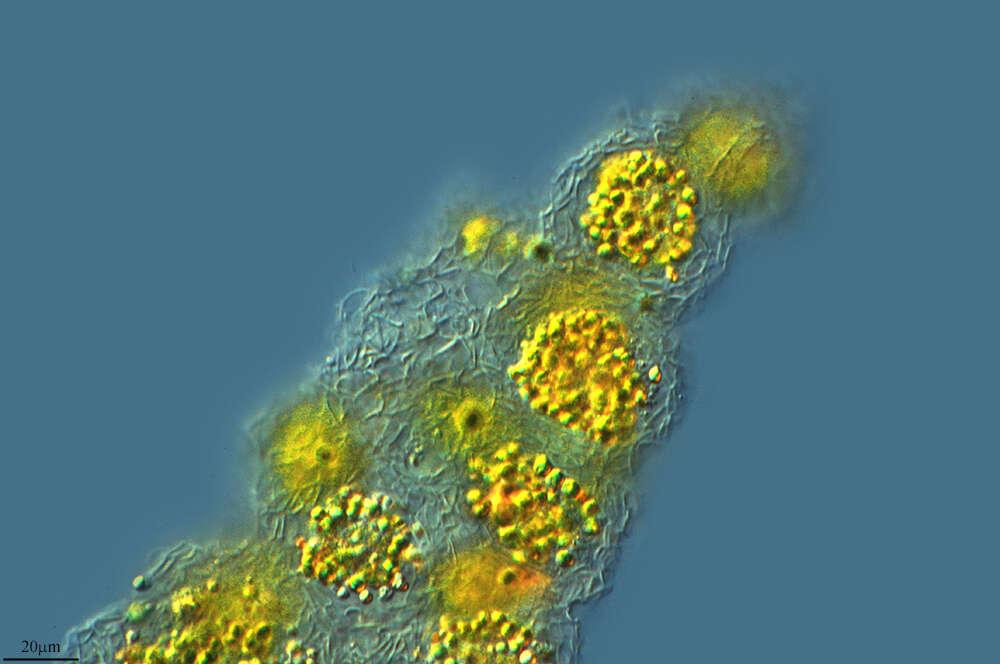
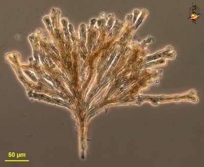
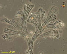
Rhipidodendron (rip-ee-doe-dend-ron, is a colonial spongomonad flagellate, in which the cells are located at the end of a branching (aborescent) colony. The matrix of the colony is formed from adhering small globules of mucilage. The branches are flat, with several channels in each blade. One cell is located at the end of each channel (many of the cells were dislodged from this preparation). Phase contrast.
-
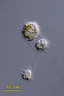
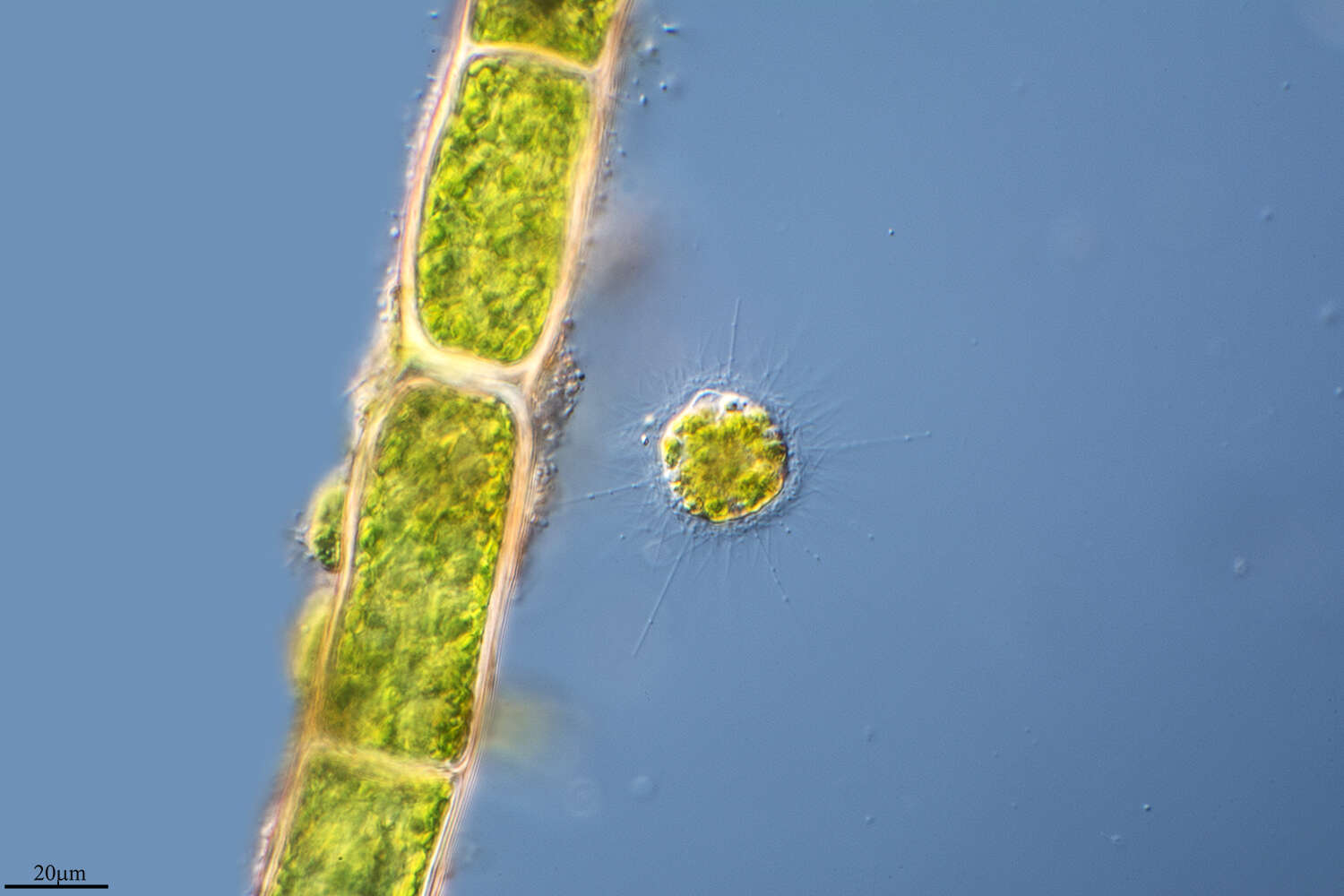
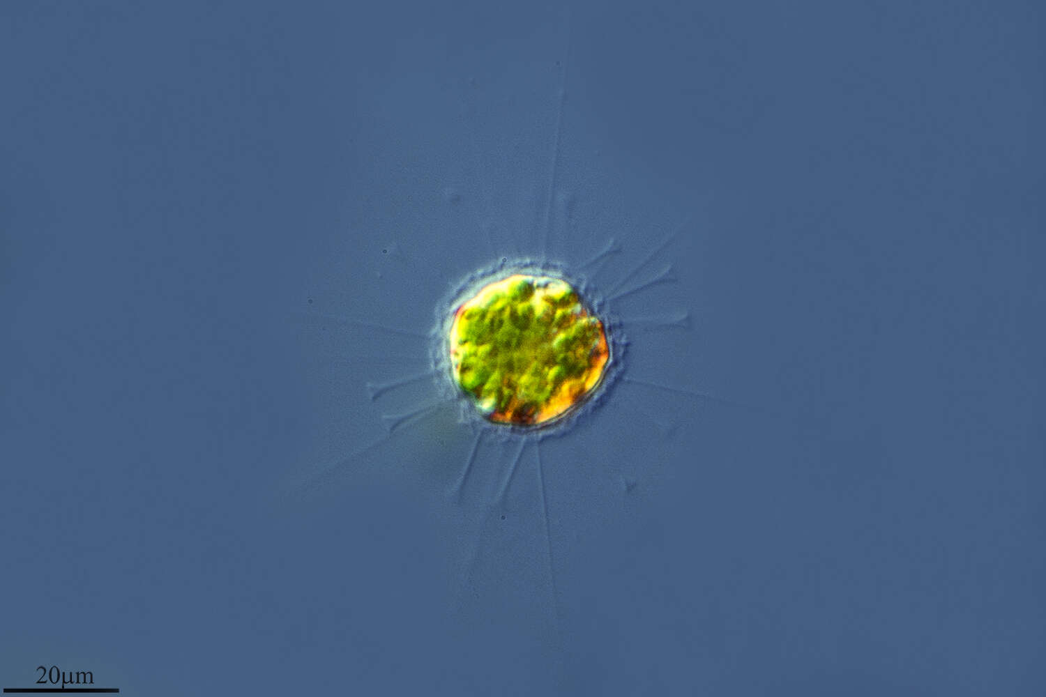
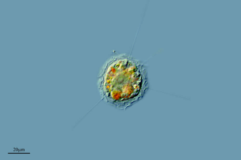
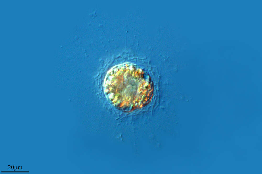
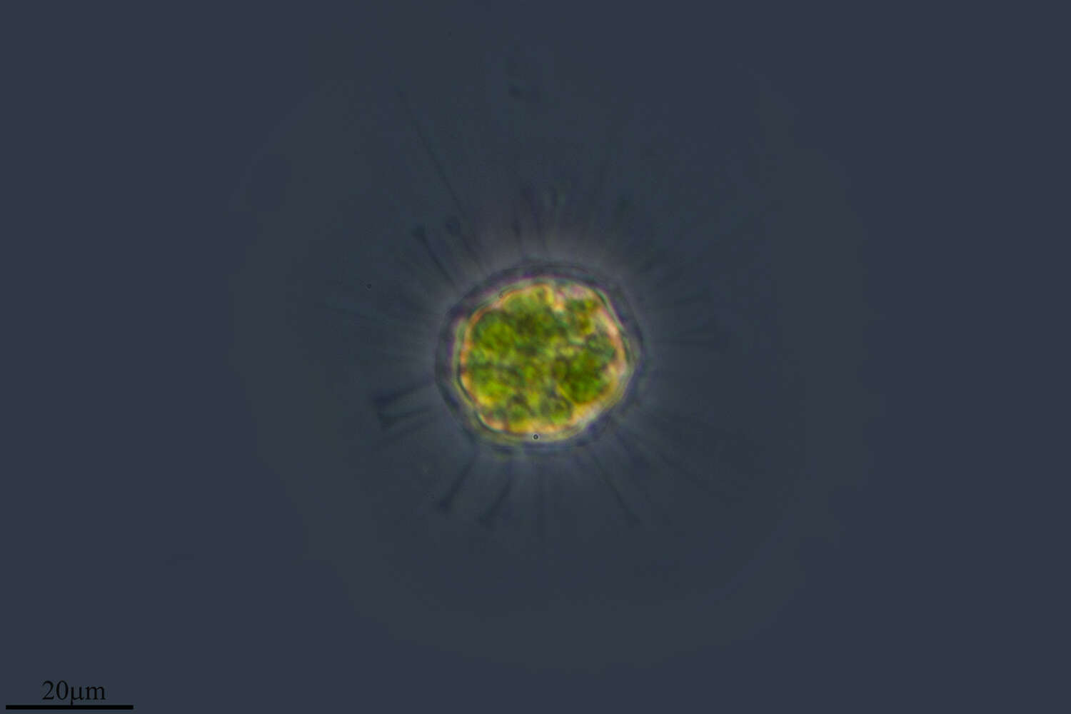
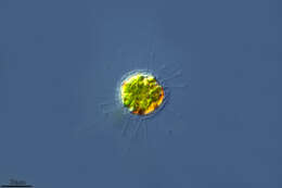
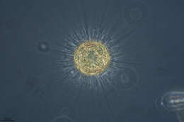
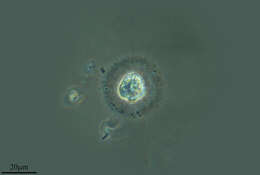
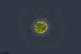
Raphidiophrys (raff-fid-ee-off-riss) ambigua is a solitary species in this genus of centrohelid heliozoa. It forms three kinds of spindle shaped spicules that accumulate around the pseudopodia. The first form are spindle shaped and pointed at both ends, the second form are medium sized, broadly spindle shaped and rounded at the ends, and the final form are very small (5 - 6 mm long) and elliptically shaped. The spicules are embedded in a gelatinous envelope of the cell. Cytoplasm without symbiotic algae but often coloured greenish, brownish or yellowish by food vacuoles. Three closely arranged specimen of Raphidiophrys ambigua, possibly shortly after cell division. Note the yellow brownish food vacuoles that are characteristic of this species. From a pond near Konstanz, Germany.Differential interference contrast.
-
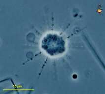
Detail of the siliceous scales of the centroheliozoan, Polyplacocystis symmetrica (Penard, 1904) Mikrjukov, 1996. An axopodium with a bead-like extrusome is visible below the scale to the viewer's left. Collected from a freshwater aquaculture pond near Boise, Idaho. November 2003. DIC.
-
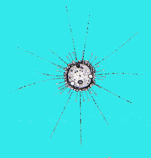
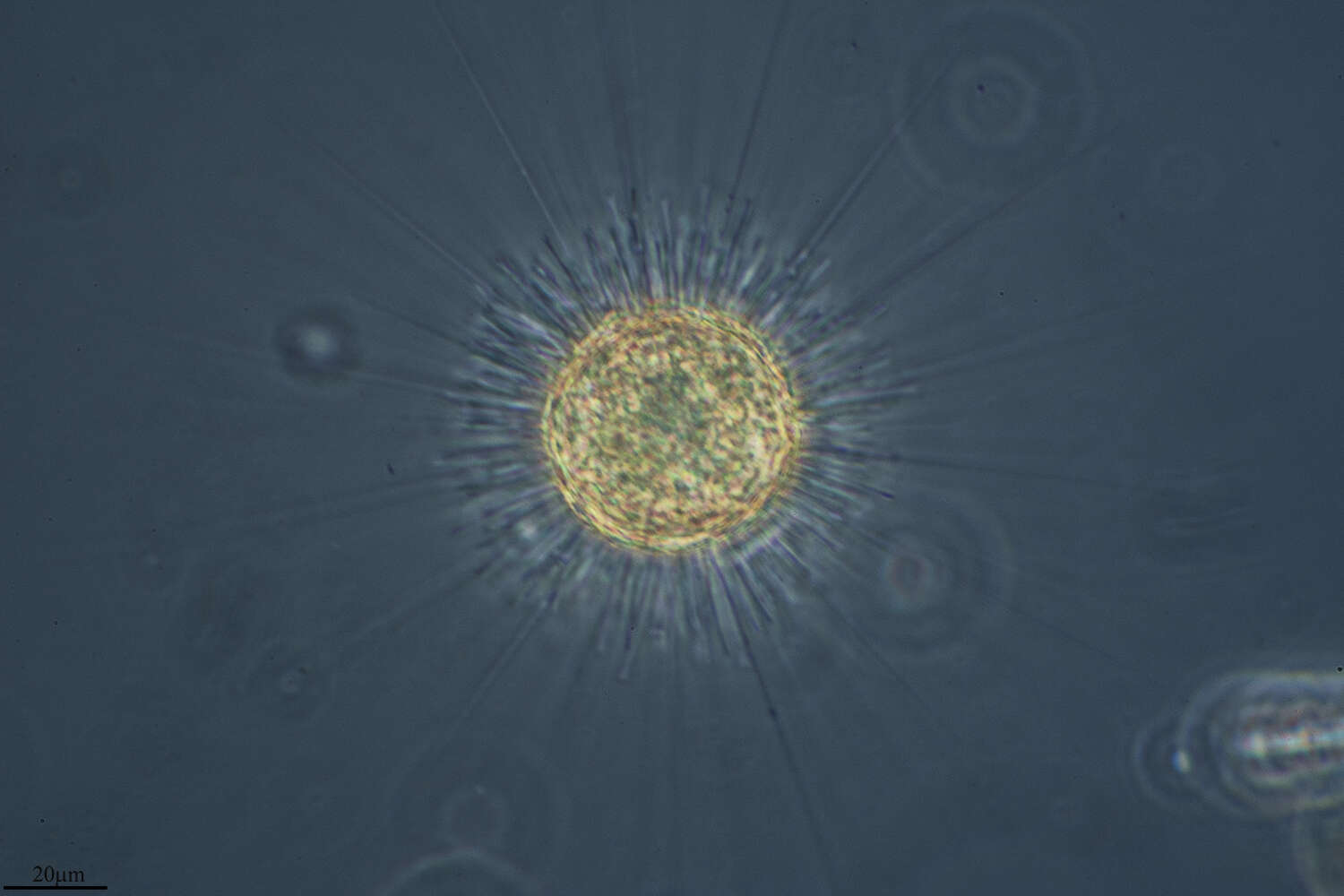
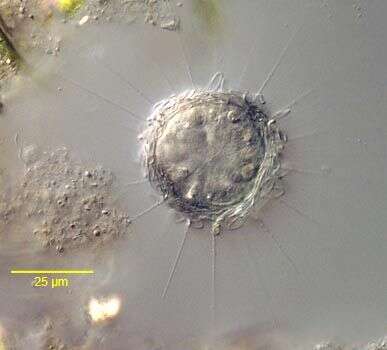
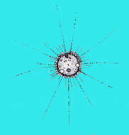
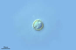
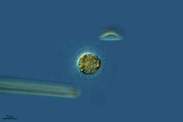
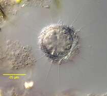
Oxnerella (ox-ner-ell-a) is a centrohelid heliozoon - the most speciose group of heliozoa. As with other heliozoa, it has radiating arms which intercept swimming prey which are captured by the extrusomes (the lumps on the arms) and then ingested. This genus includes spicules with fine spicules. Those spicules may be so delicate as not to be readily visible (if at all) by light microscopy. Heterophrys is similar but has delicate organic spicules. These may be very hard to see by light microscopy, and this can easily lead to misidentification. Phase contrast.
-
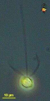
Chrysochromulina (cry-so-crumb-you-line-a) ericina a single-celled haptophyte, with two similar flagella, long anterior haptonema and a golden colour from two yellow-brown chloroplasts. Small scales lie on the surface of the cell but these are not evident in this image. Phase contrast microscopy.
data on this strain.
-
Madrid, Madrid, Spain
-
Mahide, Castille and Leon, Spain
-
Ribadelago, Castille and Leon, Spain
-
Muelas del Pan, Castille and Leon, Spain
-
Rozas de Puerto Real, Comunidad de Madrid, Espaa
-
Arboli, Catalonia, Spain
-
Ribadelago de Franco, Castille and Leon, Spain
-
Ribadelago, Castille and Leon, Spain
-
Ribadelago de Franco, Castille and Leon, Spain
-
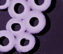
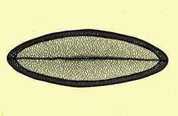
This image was made from samples taken during a scientific cruise in the Pacific. Water was filtered to concentrate the organisms that were present, then dried onto a thin sheet of plastic and then shadowed with a fine layer of metal to provide contrast. The preparation was then observed with an electron-microscope. This technique has been used to document the diversity of marine microbes, especially, protists in the oceans. According to Jeremy Young, this is a fragment of a coccosphere of Umbilicosphaera sibogae.
-
Rhipidodendron (rip-ee-doe-dend-ron, is a colonial spongomonad flagellate, in which the cells are located at the end of a branching (aborescent) colony. The matrix of the colony is formed from adhering small globules of mucilage. The branches are flat, with several channels in each blade. One cell is located at the end of each channel (many of the cells were dislodged from this preparation). Phase contrast.
-
Single whole plate scale viewed by transmission electron microscopy. The siliceous scales are formed within the cell and then form a loose layer or periplast around the outside of the cell.
-
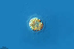
Portrait of the centroheliozoan, Polyplacocystis symmetrica (Penard, 1904) Mikrjukov, 1996. Collected from a freshwater aquaculture pond near Boise, Idaho. November 2003. DIC.
-
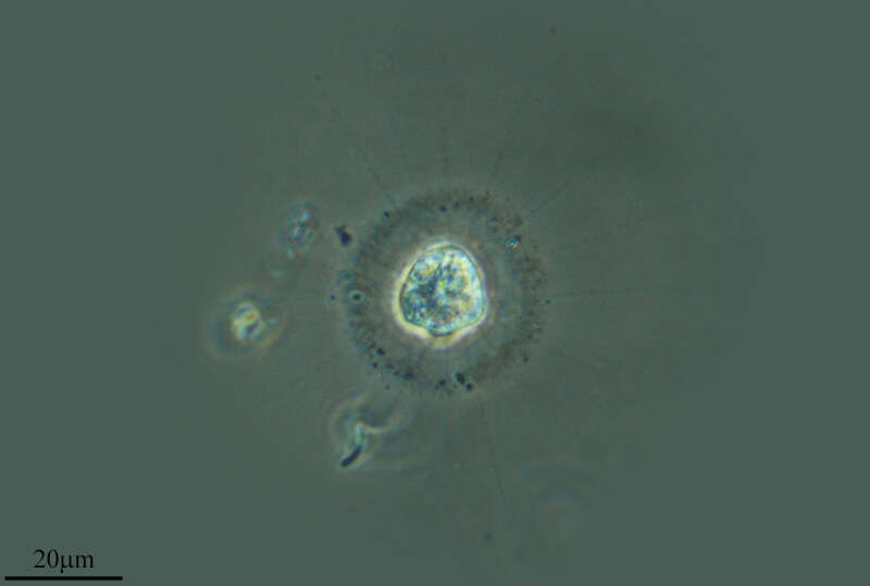
Oxnerella (ox-nerr-ell-a) is a centrohelid heliozoon, distinguished from the other centrohelids because there are no spicules or other materials around the outside of the cell. As with other centroheliozoa, the axopods are thin, parallel sides, and the extrusomes seem relatively large. Phase contrast micrograph.
-
Mahide, Castille and Leon, Spain
-
Villar del Pedroso, Extremadura, Spain
-
Lumbreras, La Rioja, Spain