-
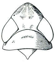
Stirellus labiatus, head, pronotum, and mesonotum, dorsally (Oman, P. W. 1949, The Nearctic leafhoppers (Homoptera: Cicadellidae). A generic classification and check list. Memoirs of the Entomological Society of Washington. 3:1-253.)
-
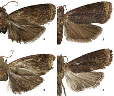
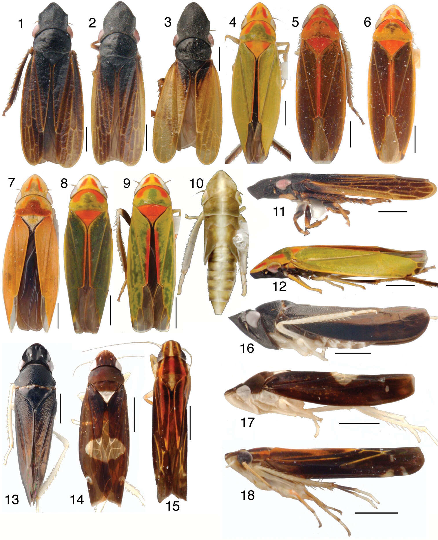
Figures 1–18.Mileewinae, scale bars = 1 mm 1–10 Tungurahualini, dorsal habitus 1 Tungurahuala basilisca, male from Colombia 2 Tungurahuala acuminata, male 3 same, female 4 Ilyapa bifida, male 5 Ilyapa loca, male 6 Ilyapa longispina, male 7 Ilyapa ochrescens, male 8 Ilyapa recurvata, male 9 Ilyapa viridis, male 10 same, 5th instar nymph 11–12 Tungurahualini, lateral habitus 11 Tungurahuala acuminata, male 12 Ilyapa viridis, male 13–18 Other Mileewinae 13 Makilingia sp., male from Thailand, dorsal habitus 14 same, Mileewa margheritae, male 15 same, Tinteromus sp., male from Colombia (full length of antenna not shown) 16 Makilingia sp., lateral habitus 17 same, Mileewa margheritae 18 same, Tinteromus sp.
-
Carolina Cuezzo, David A. Nickle
Zookeys
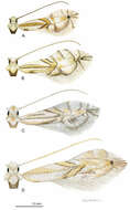
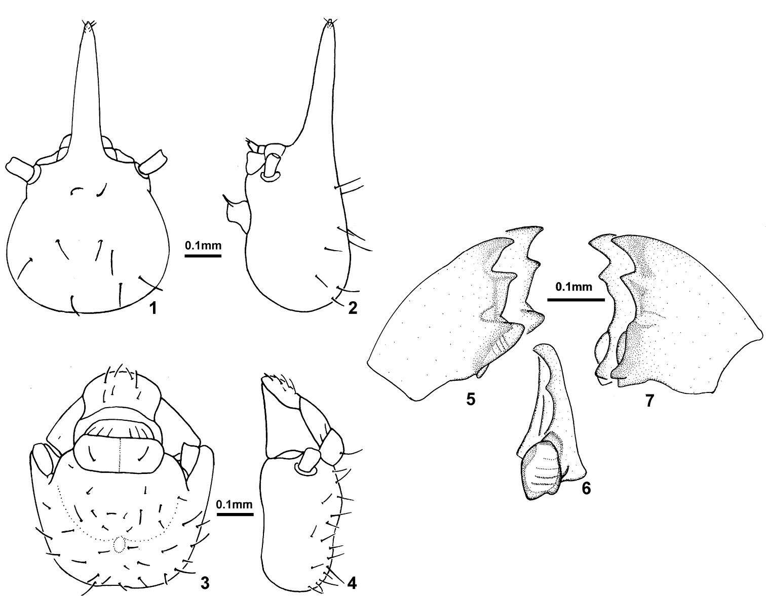
Figures 1–7.Sinqasapatermes sachae 1 soldier head in dorsal view 2 soldier head in profile 3 worker head in dorsal view 4 worker head in profile 5 worker left mandible in dorsal view 6 worker right mandible, showing molar plate in frontal view 7 worker right mandible in dorsal view.
-
Juli Pujade-Villar, Paul Hanson, Claudia A. Medina, Miguel Torres, George Melika
Zookeys
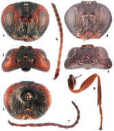
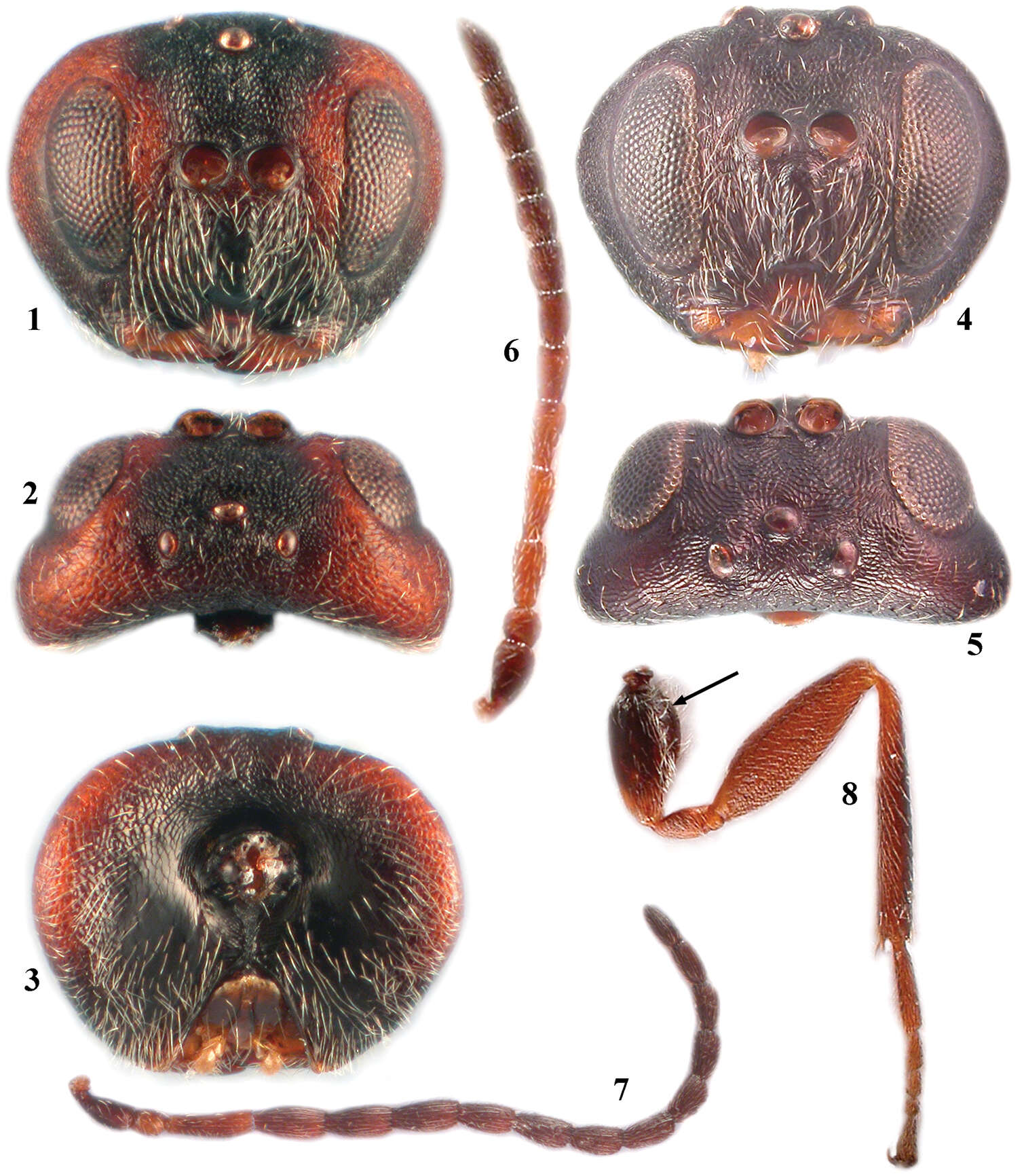
Figures 1–8.Zapatella grahami 1 head, female (anterior view) 2 head, female (dorsal view) 3 head, female (posterior view) 4 head, male (anterior view) 5 head, male (dorsal view) 6 antenna, female 7 antenna, male 8 hind leg, female (arrow indicates the dense white setae on dorsoposterior surface of coxa).
-
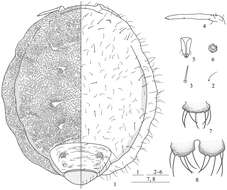
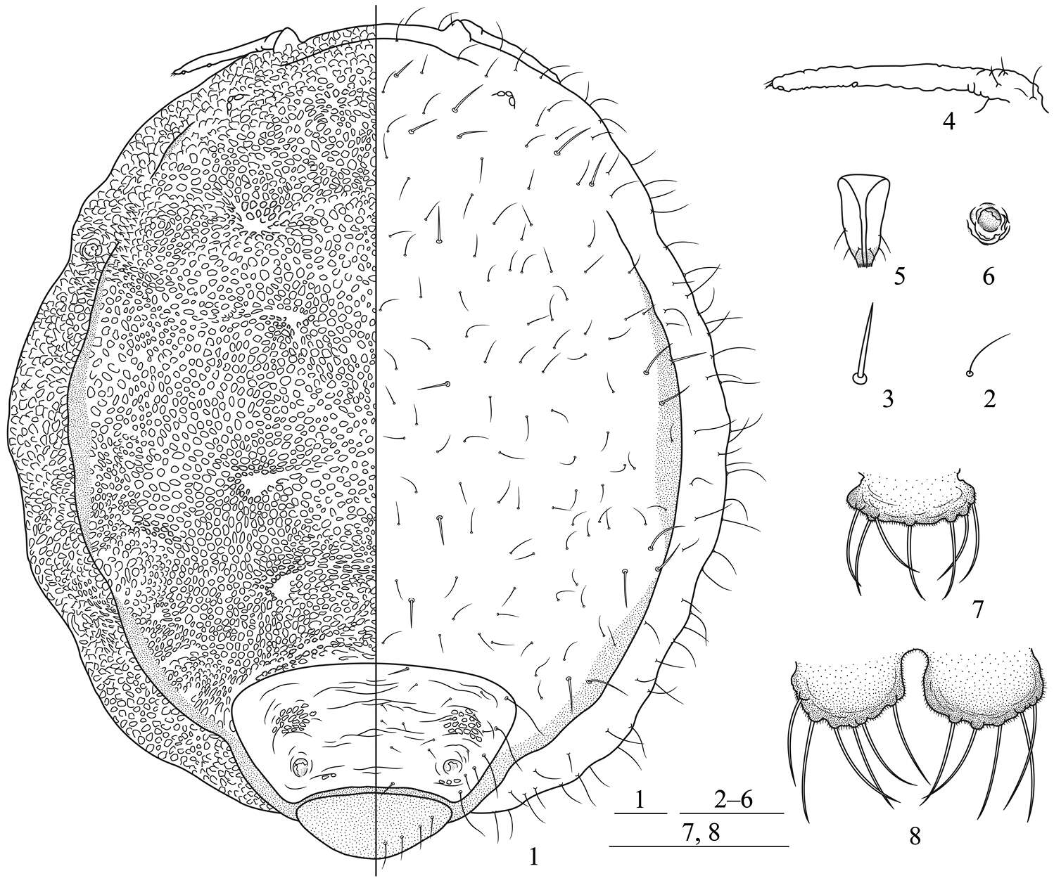
Figures 1–8.Neonipponaphis pustulosis sp. n. Apterous viviparous female: 1 dorsal view of body, with pustules in left and chaetotaxy in right 2 fine and pointed scattered dorsal seta 3 long, thick, and stiff dorsal seta 4 antenna 5 ultimate rostral segment 6 siphunculus 7 cauda 8 anal plate. Scale bars = 0.10 mm.
-
Boonsatien Boonsoong, Dietrich Braasch
Zookeys
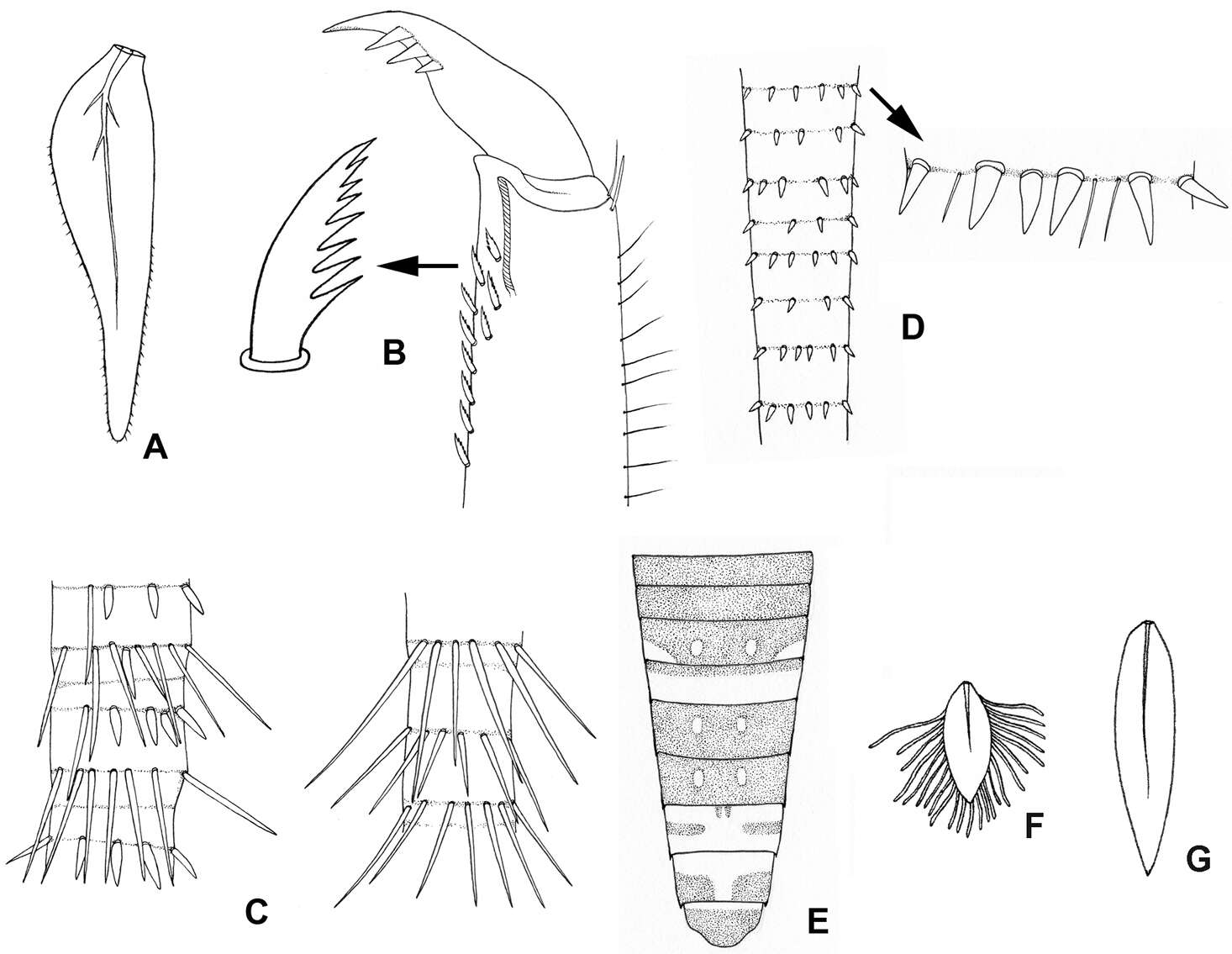
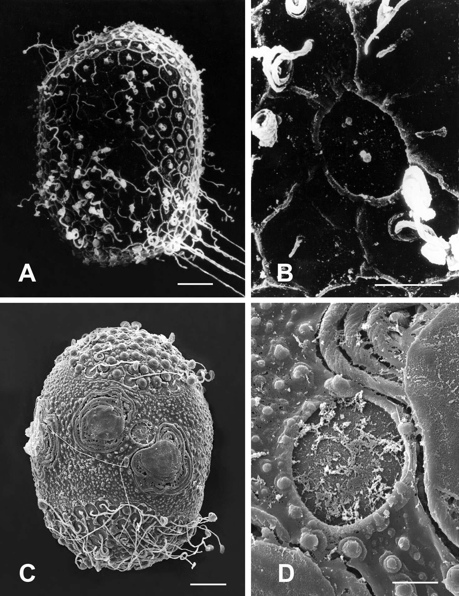
Figure 4.A–B Lamella of gills 7 (A) and setae on inner surface of hind tarsi (B) of Asionurus primus Braasch & Soldán, 1986 C bristles on cerci of Rhithrogeniella tonkinensisSoldán & Braasch, 1986 D bristles on cerci of Asionurus namnaoensis Braasch & Boonsoong, 2010 E dorsal view of abdomen of Asionurus rubromaculatus You, Wu, Gui & Hsu, 1981 F lamella of gills 1 of Asionurus gilliesiana Braasch, 1990 G lamella of gills 7 of Asionurus rainulfiana Braasch, 1990.
-
Jia Long, Ren Guo-Dong, Yu You-Zhi
Zookeys
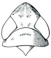
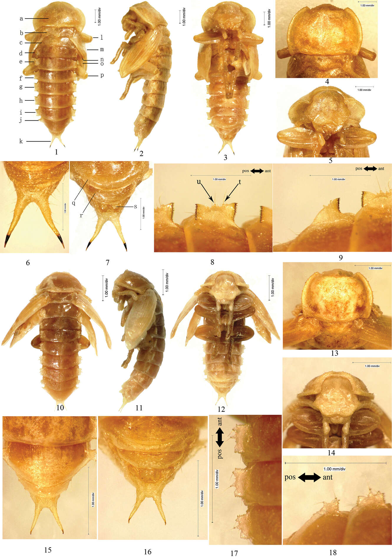
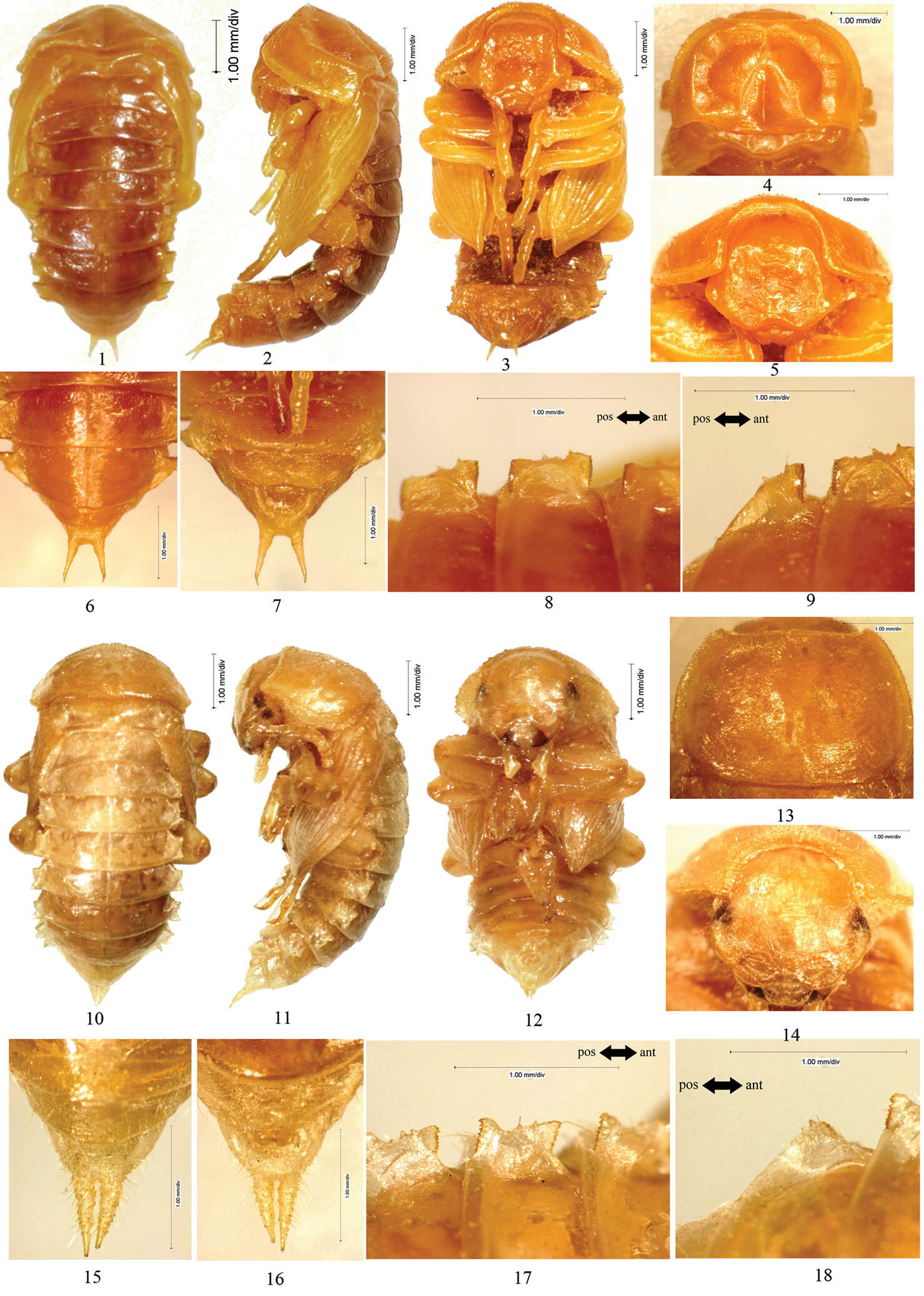
Figure 1.1–9 Scleropatrum horridum horridum Reitter, 1898 1 Pupal habitus in dorsal view 2 Pupal habitus in lateral view 3 Pupal habitus in ventral view 4 Pronotum 5 Head 6 Urogomphi in dorsal view 7 Urogomphi in ventral view 8 Lateral process of abdominal tergite V 9 Lateral process of abdominal tergite VII 10–18 Gonocephalum reticulatum Motschulsky, 1854 10 Pupal habitus in dorsal view 11 Pupal habitus in lateral view 12 Pupal habitus in ventral view 13 Pronotum 14 Head 15 Urogomphi in dorsal view 16 Urogomphi in ventral view 17 Lateral process of abdominal tergite V 18 Lateral process of abdominal tergite VII. a Pronotum b Mesonotum c Metanotum d Abdominal segment I e Abdominal segment II f Abdominal segment III g Abdominal segment IV h Abdominal segment V i Abdominal segment VI j Abdominal segment VII k Urogomphi l Profoot m Elytral sheath n Mesofoot o Metathoracic wing sheath p Metafoot q Abdominal sternite VII r Abdominal sternite VIII s Gonotheca t large setose tubercle I u large setose tubercle II.
-
Felipe N. Soto-Adames, Steven J. Taylor
Zookeys
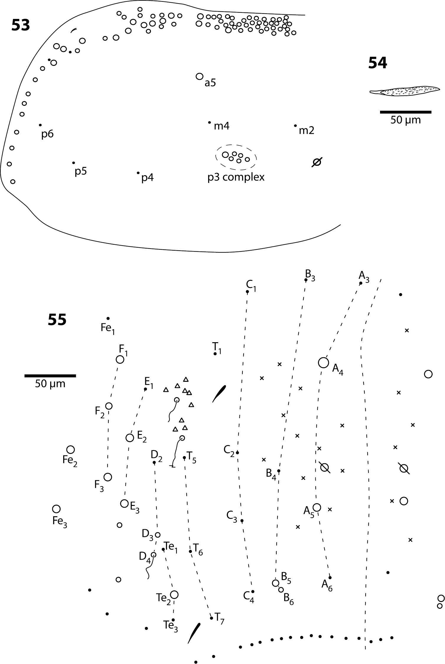
Figures 53–55.Trogolaphysa jataca 53 Mesothorax chaetotaxy 54 Second abdominal segment seta p5 55 Complete chaetotaxy of fourth abdominal segment.
-
Donald M. Windsor, Guillaume J. Dury, Fernando A. Frieiro-Costa, Susanne Lanckowsky, Jacques M. Pasteels
Zookeys
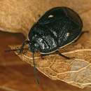
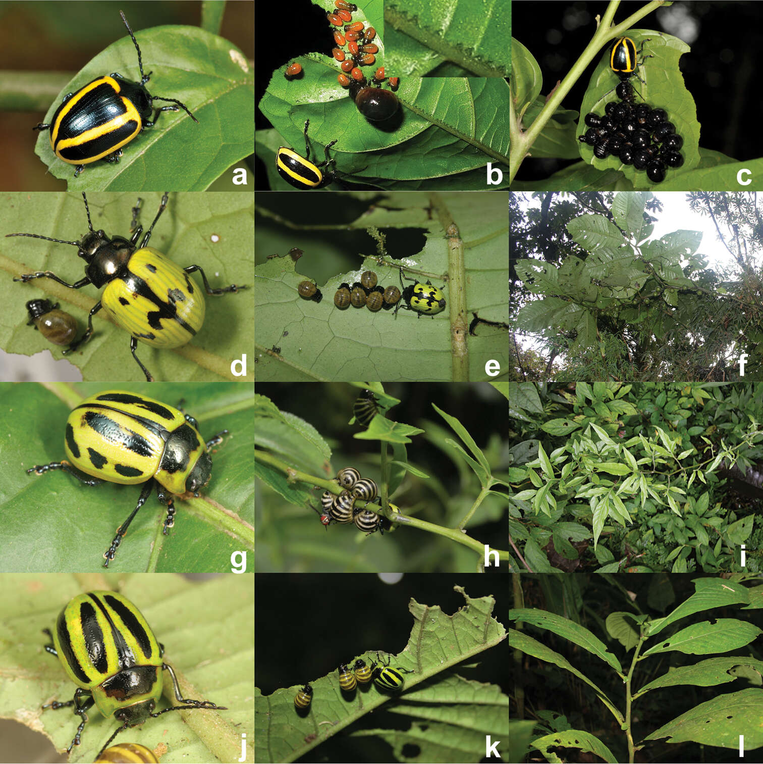
Figure 3.Maternal care providing Proseicela species, a Proseicela vittata adult (Photo by D.W.) b Proseicela vittata female and larvae from two cohorts. Insert shows detail of vein pinching along approximately 1cm of the primary vein (Photo by D.W.) c Proseicela vittata female with late stage larvae (Photo by D.W.) d Proseicela bicruciata adult female, (photo by G.D.) e Proseicela bicruciata female tending larvae (photo by G.D.) f Proseicela bicruciata food plant, Solanum abitaguense (photo by G.D.) g Proseicela spectabilis adult (photo by G.D.) h Proseicela spectabilis with nearly full-grown larval brood and tachinid parasitoid (photo by G.D.) i. Proseicela spectabilis host plant, Solanum sp. (photo by G.D.) j Proseicela sp. n. adult female (photo by G.D.) k the same female tending three feeding larvae feeding on Cuatresia sp. (Solanaceae) (photo by G.D.) l wider view of the host plant (photo by G.D.).
-
Lizhi Huo, Xingmin Wang, Xiaosheng Chen, Shunxiang Ren
Zookeys
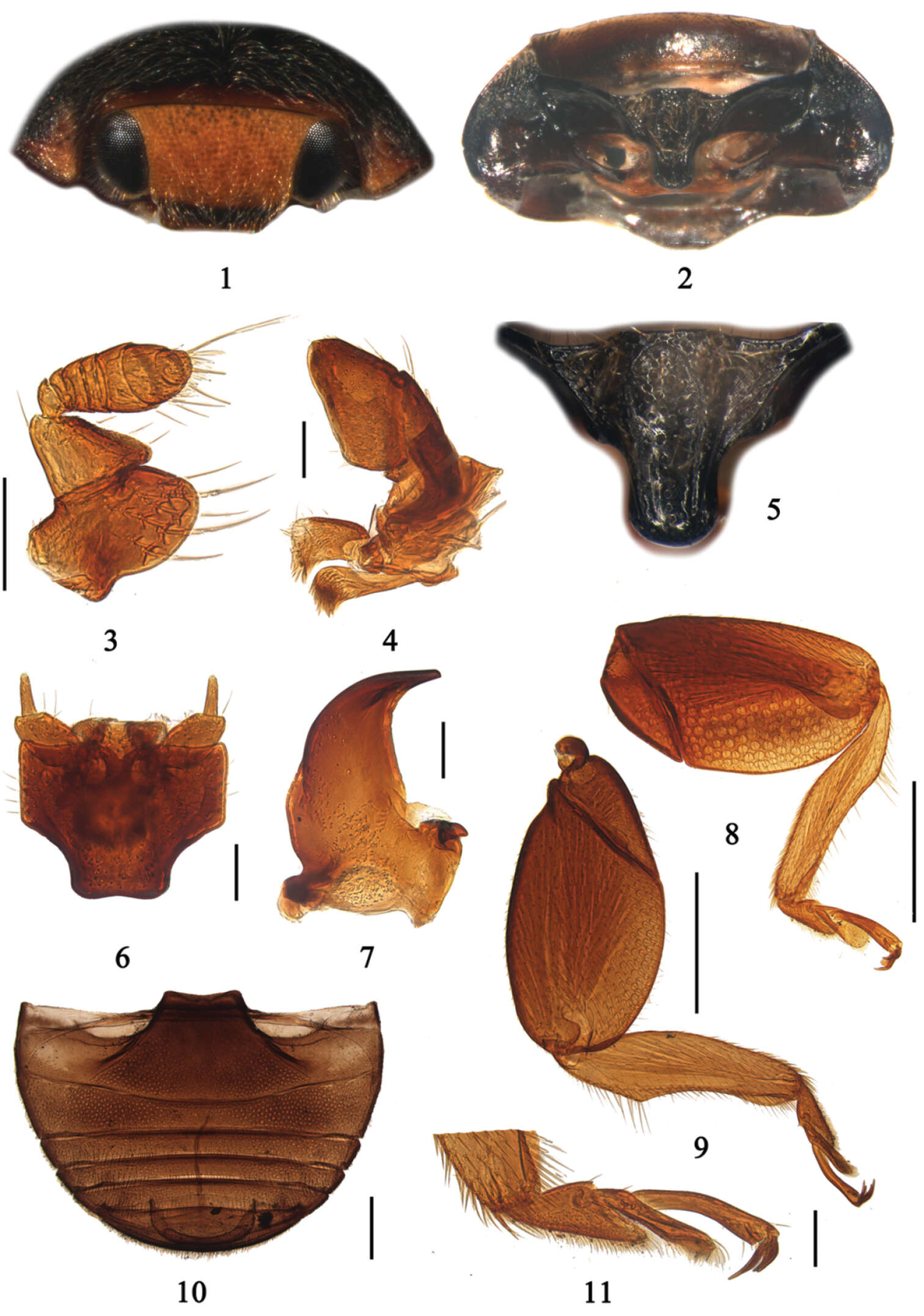
Figures 1–11.Aspidimerus matsumurai Sasaji, 1986. 1 head, frontal view 2 prothorax, ventral view 3 antenna 4 maxilla 5 prosternal process 6 labium 7 mandible 8 front leg 9 hind leg 10 abdomen 11 tarsi. Scale bars: Figures 1–7, 11: 0.1mm; Figures 8–10: 0.5mm.
-
Na-sen Wei, Paolo Rosa, Zai-fu Xu
Zookeys
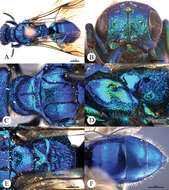
Plate 9.Cleptes seoulensis Tsuneki, 1959, male from Anhui. A Habitus dorsal B Head anterior C Pronotum and mesoscutum dorsal D Mesopleuron and metapleuron lateral E Mesoscutellum, metanotum and propodeum dorsal F Metasoma dorsal. Scale bars in mm.
-
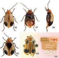
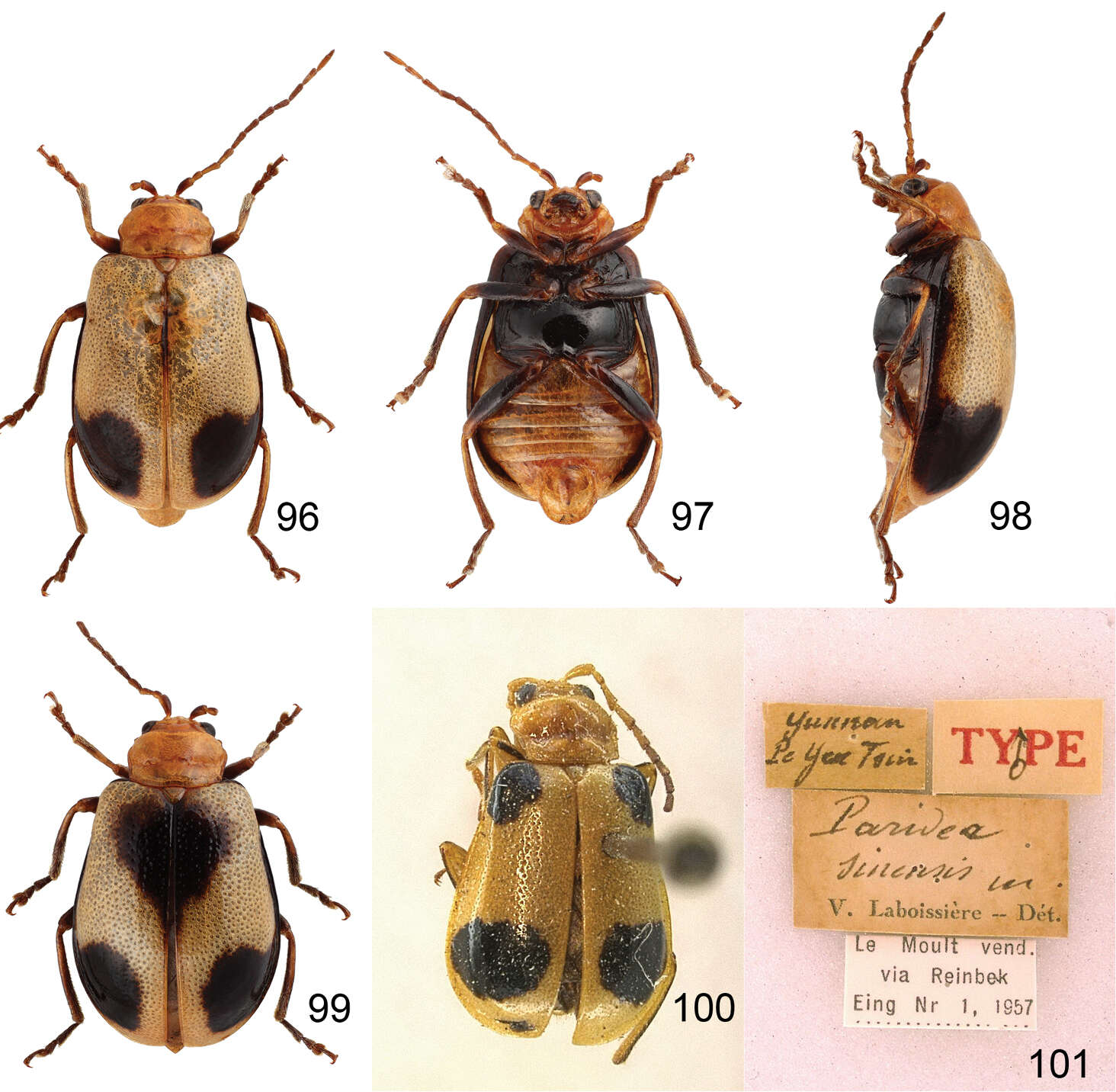
Figures 96–101.Paridea species. 96 Paridea (Semacia) angulicollis, male, dorsal view 97 ditto, ventral view 98 ditto, lateral view 99 Paridea (Semacia) angulicollis, female, dorsal view 100 Paridea (Paridea) sinensis, lectotype, dorsal view 101 Paridea (Paridea) sinensis, lectotype, labels.
-
Francisco Hita Garcia, Brian L. Fisher
Zookeys
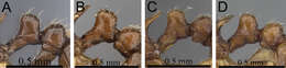
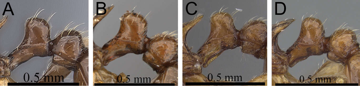
Figure 2.Petiole and postpetiole in profile. A Tetramorium alperti (CASENT0042547)B Tetramorium enkidu (CASENT0045673)C Tetramorium naganum (CASENT0280584)D Tetramorium gilgamesh (CASENT0247312).
-
Figures 1–4.Rhithrogeniella ornata Ulmer, 1939. 1 Genitalia of the male imago (holotype) in ventral view 2 Foreleg of a male subimago (paratype) 3 Hindleg of a male subimago (paratype) 4 Penis lobes of a male subimago (paratype): plain line, cuticular structures of the subimago; dotted line, outline of the imago penis lobes.
-
Alicia E. Timm, John W. Brown
Zookeys
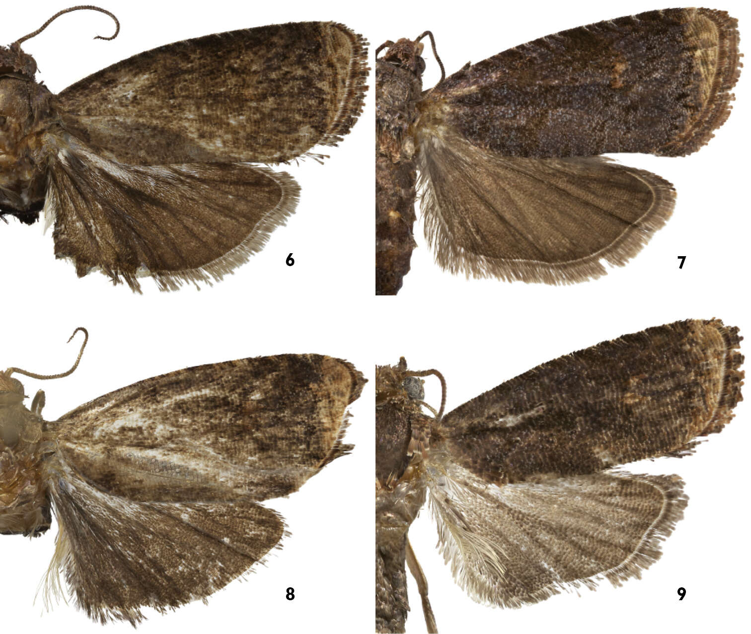
Figures 6–9.Upper suface of wings of Thaumatovalva. 6 Thaumatovalva deprinsorum 7 Thaumatovalva albolineana 8 Thaumatotibia spinai [image enhanced using best parts of both forewings] 9 Thaumatovalva limbata.
-
Donald R. Davis, David L. Wagner
Zookeys
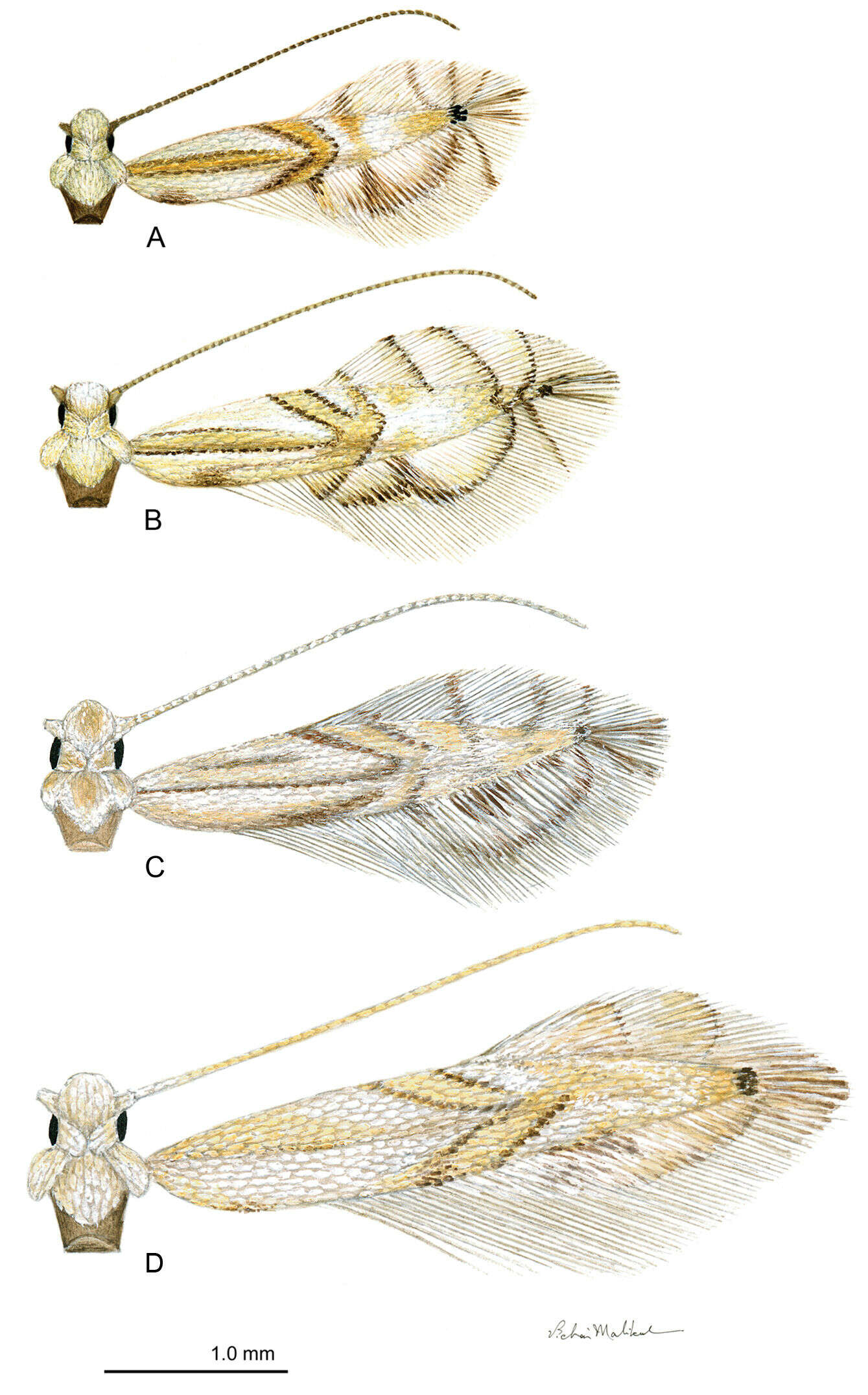
Figure 2.Phyllocnistis adults. A Phyllocnistis hyperpersea sp. n. (2.1 mm) B Phyllocnistis subpersea sp. n. (2.5 mm) C Phyllocnistis longipalpa sp. n. (2.6 mm) D Phyllocnistis perseafolia sp. n. (3.0 mm). (Drawn approximately to scale; forewing length in parentheses.)
-
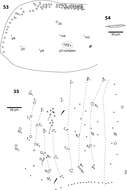
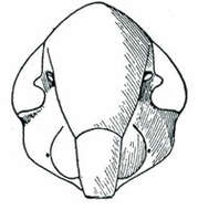
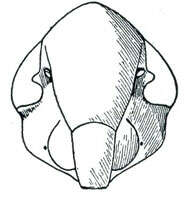
Stirellus labiatus, face (Oman, P. W. 1949, The Nearctic leafhoppers (Homoptera: Cicadellidae). A generic classification and check list. Memoirs of the Entomological Society of Washington. 3:1-253.)
-
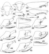
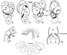
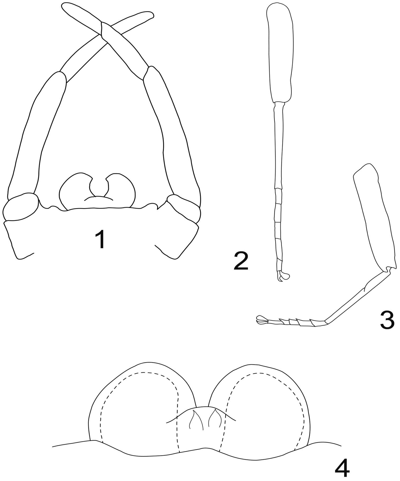
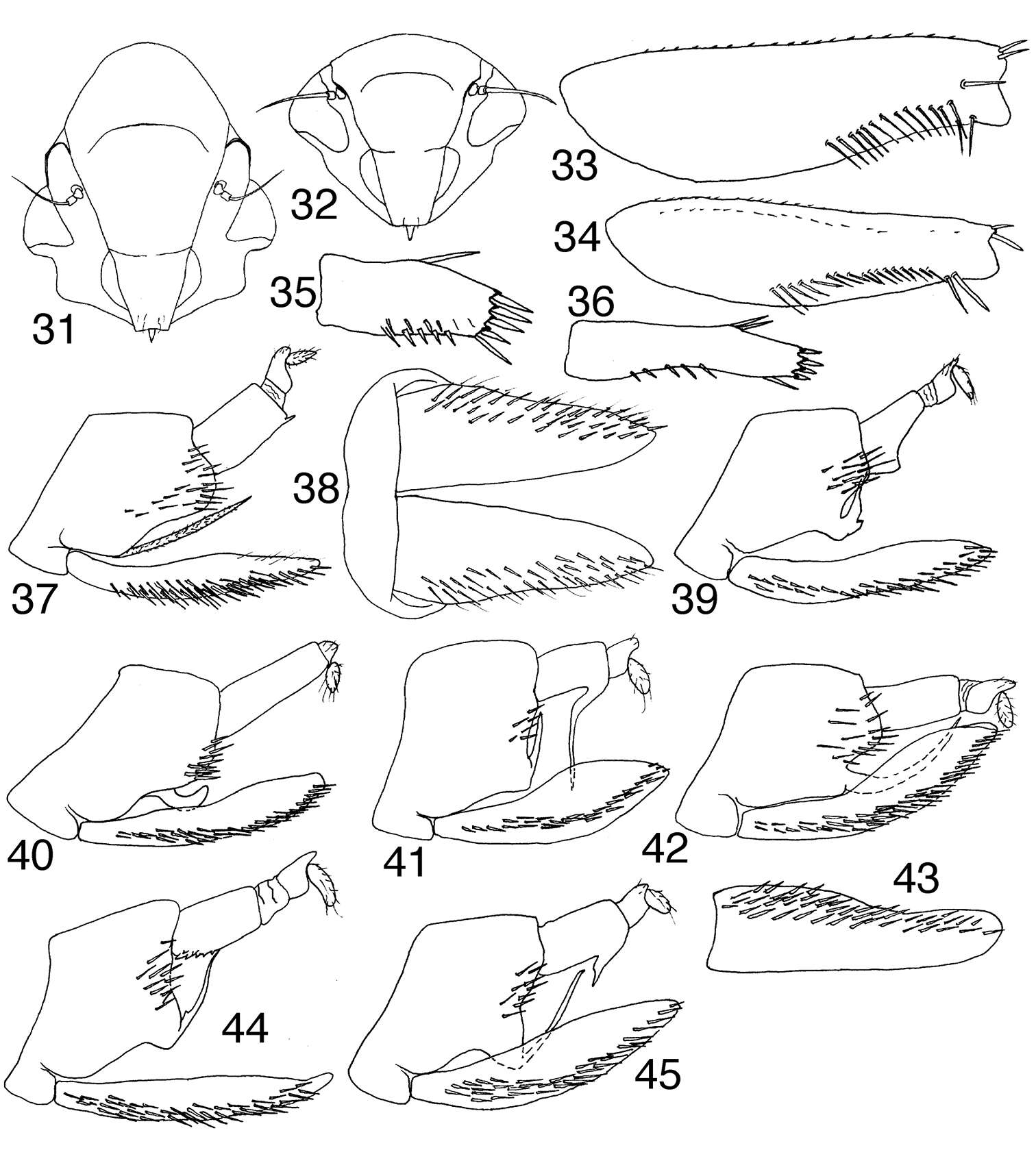
Figures 31–45.Tungurahualini 31–32 head, anteroventral view 31 Tungurahuala acuminata 32 Ilyapa viridis 33–34 prothoracic femur, anterior view 33 Tungurahuala acuminata 34 Ilyapa viridis 35–36 hind tarsomere I, ventral view 35 Tungurahuala acuminata 36 Ilyapa viridis 37 Tungurahuala acuminata, genital capsule, lateral view 38 same, valve and subgenital plates, ventral view 39 Ilyapa bifida, genital capsule, lateral view 40 same, Ilyapa loca 41 same, Ilyapa longispina 42 same, Ilyapa ochrescens 43 Ilyapa ochrescens, left subgenital plate, ventral view 44 Ilyapa recurvata, genital capsule, lateral view 45 same, Ilyapa viridis.
-
Carolina Cuezzo, David A. Nickle
Zookeys
Figures 8–15.Sinqasapatermes sachae. 8–11 worker gut in situ respectively from dorsal, right, ventral and left views 12–13 Malpighian tubules attachment 14 worker enteric valve armature 15 scheme of worker enteric valve configuration. C crop G gizzard M mesenteron, stippled P1 first proctodeal segment P2 enteric valve P3 paunch P4 colon P5 rectum TM Malpighian tubules.
-
Juli Pujade-Villar, Paul Hanson, Claudia A. Medina, Miguel Torres, George Melika
Zookeys
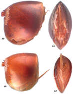
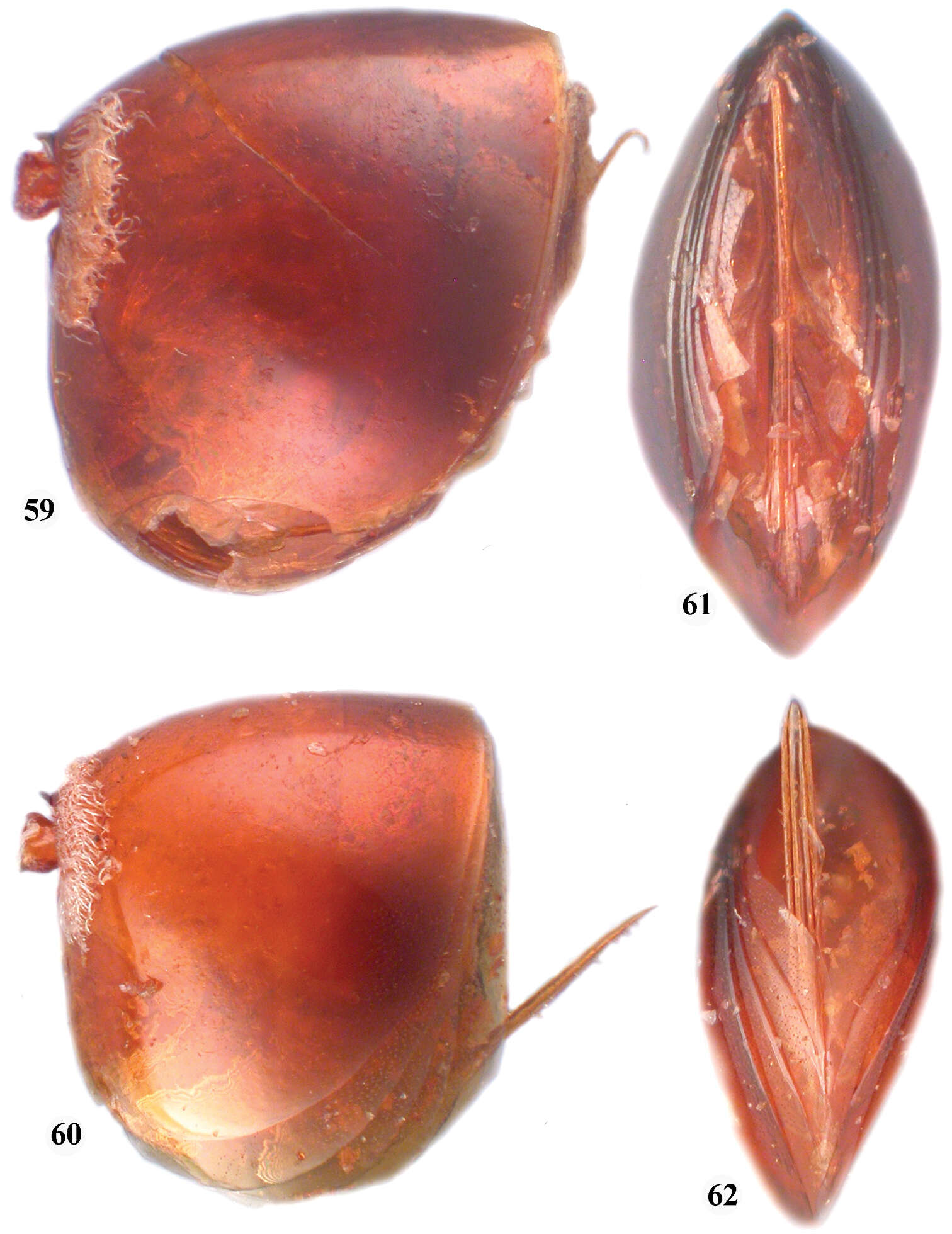
Figures 59–62.59 Zapatella cryptica, metasoma, female (lateral view) 60 Zapatella quercussimilis, metasoma, female (lateral view) 61 Zapatella cryptica, ventral spine of hypopygium 62 Zapatella quercussimilis, ventral spine of hypopygium.
-
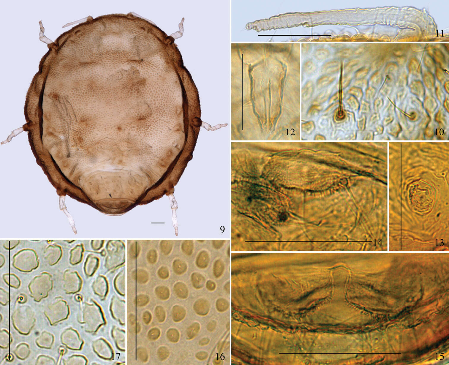
Figures 9–17.(9–15) Neonipponaphis pustulosis sp. n. Apterous viviparous female: 9 dorsal view of body 10 dorsal setae (long, thick, and stiff seta in left, fine and pointed seta in right) 11 antenna 12 ultimate rostral segment 13 siphunculus 14 cauda 15 anal plate. (16–17) Dorsal pustules on the same scale: 16 Neonipponaphis pustulosis sp. n. 17 Neonipponaphis shiiae Takahashi. Scale bars = 0.10 mm.
-
Boonsatien Boonsoong, Dietrich Braasch
Zookeys
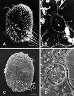
Figure 8.A–B General outline (A) and micropyle (B) of the egg of Rhithrogena tonkinensisSoldán & Braasch, 1986 C–D General outline (C) and micropyle (D) of the egg of Asionurus namnaoensis Braasch & Boonsoong, 2010. Scale bars 20 µm for A and C; 5 µm for B and D.
-
Jia Long, Ren Guo-Dong, Yu You-Zhi
Zookeys
Figure 2.1–9 Opatrum (Opatrum) subaratum Faldermann, 1835 1 Pupal habitus in dorsal view 2 Pupal habitus in lateral view 3 Pupal habitus in ventral view 4 Pronotum 5 Head 6 Urogomphi in dorsal view 7 Urogomphi in ventral view 8 Lateral process of abdominal tergite V 9 Lateral process of abdominal tergite VII 10–18 Eumylada potanini (Reitter, 1889) 10 Pupal habitus in dorsal view 11 Pupal habitus in lateral view 12 Pupal habitus in ventral view 13 Pronotum 14 Head 15 Urogomphi in dorsal view 16 Urogomphi in ventral view 17 Lateral process of abdominal tergite V 18 Lateral process of abdominal tergite VII.
-
Felipe N. Soto-Adames, Steven J. Taylor
Zookeys
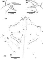
Figures 50–52.Trogolaphysa belizeana (50, 51) and Trogolaphysa jataca (52) 50 Prothoracic claw 51 Metathoracic claw 52 Dorsal chaetotaxy of head.