-
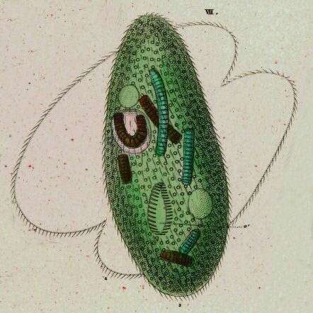
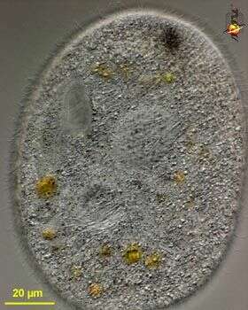
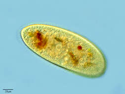
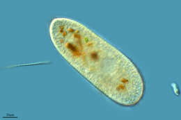
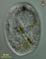
Ovoid, 60-100 micron long. Numerous large trichocysts.
-
Franceses, Canary Islands, Spain
-
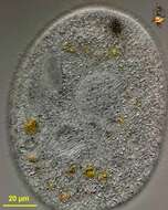
Matute, La Rioja, Spain
-
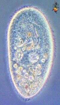
Right dorsolateral surface view of the hymenostome ciliate, Frontonia angusta (Kahl, 1931). Very similar in overall apppearance to F. acuminata (Ehrenberg,1833)Buetschli,1889. F. angusta lacks the anterior apical collection of pigmented granules seen in F. acuminata and its contractile vacuole has 3-4 excretory pores (4 in this case).The approximately 6 µm long extrusomes are clearly visible. Ingested diatoms and green algae are present. Collected from a freshwater pond near Boise, Idaho.DIC.
-
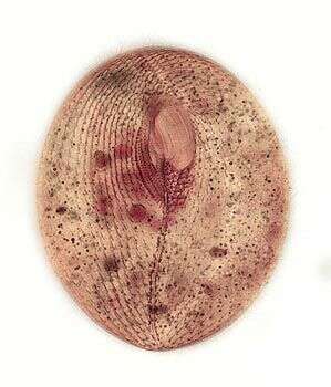
Ventral infraciliature of the hymenostome ciliate, Frontonia angusta (Kahl, 1931). Very similar in overall apppearance to F. acuminata (Ehrenberg,1833)Buetschli,1889. F. angusta lacks the anterior apical collection of pigmented granules seen in F. acuminata and its contractile vacuole has 3-4 excretory pores (not visible here).The prominent preoral and postoral sutures are visible. The 3 curved adoral membranelles are seen on the viewer's right of the oral apparatus. The vestibular ciliary rows are seen to the viewer's left of the the oral apparatus.The postoral ciliary field is seen abutting the posterior margin of the peristome to the viewer's right of the postoral suture.Stained by the silver carbonate technique (see Foissner, W. Europ. J. Protistol., 27:313-330;1991).Collected from a freshwater pond near Boise, Idaho.Brightfield.
-
-
-
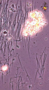
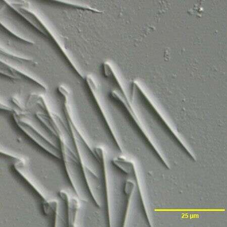
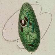
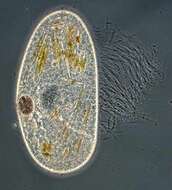
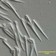
Frontonia (front-own-ee-a) is a peniculine ciliate and as such is closely related to the familiar Paramecium. It has many crystalline inclusions called trichocysts (a special form of extrusome). When stressed the crystalline structure of these changes, and they are expelled in large numbers and forceably from the cell. This action can force the cell away from the noxious stimulus. The expelled, the trichocysts look like little spears attached to the slide or to the substrate. Phase contrast.
-
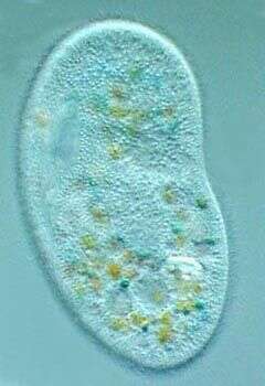
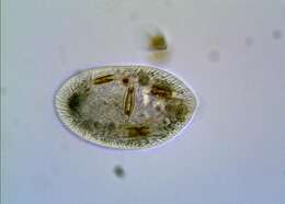
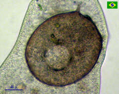
Frontonia (front-own-ee-a) is a peniculine ciliate and as such is closely related to the familiar Paramecium. The mouth is supported by strong rods which assists Frontonia in ingesting its preferred food - diatoms and other moderate sized algae. A diatom can be seen inside the cell. The mouth is located at about 10 o clock. Like many peniculines the cell has many extrusomes lying just under the cell surface, and these are expelled when the cells are challenged. Large grey area is the nucleus. Phase contrast.
-
Frontonia (front-own-ee-a) is a peniculine ciliate and as such is closely related to the familiar Paramecium. The mouth is supported by strong rods which assists Frontonia in ingesting its preferred food - diatoms and other moderate sized algae. Like many peniculines the cell has many extrusomes lying just under the cell surface, and these are expelled when the cells are challenged. Large grey area is the nucleus. Phase contrast.
-
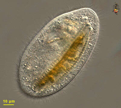
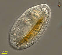
Frontonia (front-own-ee-a) is a peniculine ciliate and as such is closely related to the familiar Paramecium. The mouth is supported by strong rods which assists Frontonia in ingesting its preferred food - diatoms and other moderate sized algae. The mouth is located at about 10 o clock. Like many peniculines the cell has many extrusomes lying just under the cell surface, and these are expelled when the cells are challenged. Differential interference contrast.
-
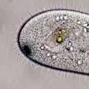
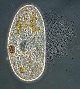
Frontonia (front-own-ee-a) is a peniculine ciliate and closely related to the familiar Paramecium. This image emphasises the cilia associated with the mouth. Like many peniculines the cell has many extrusomes lying just under the cell surface, and these are expelled when the cells are challenged. This species has black pigment granules. Differential interference contrast.
-
Frontonia (front-own-ee-a) is a peniculine ciliate and closely related to the familiar Paramecium. The mouth (upper left) is supported by strong rods which assists Frontonia in ingesting its preferred food - diatoms (as here) and other moderate sized algae. Like many peniculines the cell has many extrusomes lying just under the cell surface, and these are expelled when the cells are challenged. This species has black pigment granules. Differential interference contrast.
-
Frontonia (front-own-ee-a) is a peniculine ciliate and closely related to the familiar Paramecium. The mouth (upper left) is supported by strong rods which assists Frontonia in ingesting its preferred food - diatoms and other moderate sized algae. Like many peniculines the cell has many extrusomes lying just under the cell surface, and these are expelled when the cells are challenged. This species has black pigment granules. Differential interference contrast.
-
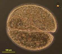
Frontonia (front-own-ee-a) is a peniculine ciliate and closely related to the familiar Paramecium. Two cells seen joined at the mouth like this are mating (undergoing conjugation, a process in which haploid nuclei are exchanged between the partners). Phase contrast.,
-
-
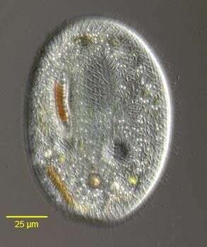
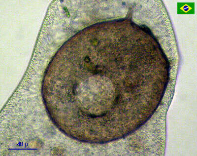
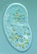
Frontonia, a ciliate that is common in freshwater habitats, eats diatoms. Just below the surface of the cell are hundreds of rod-shaped extrusomes that can transform explosively into stiff filaments used for defense from other predators or to push the cell away from undesirable sites.
-
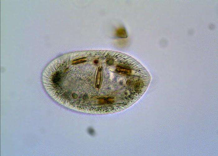
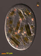
Frontonia likes to eat diatoms. The cytoplasmic contents of the diatom will be digested and the undigested siliceous frustule will be ejected. Differential interference contrast optics.
-
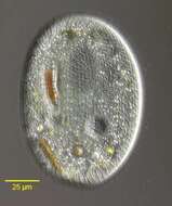
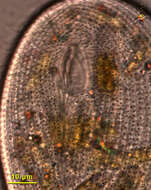
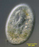
Detail of ventral surface, showing kineties (the dots are the tips of extrusomes)and the mouth. This cell has been eating diatoms. Differential interference contrast optics.
-
This cell has been eating diatoms. Differential interference contrast optics.
-
These are testate-eating ciliates. Be careful as they will destroy your testate amoebae cultures.
-
An unfortunate Centropyxis aculeata eaten by a Frontonia.
-
Frontonia (EHRENBERG,1838) is a peniculine ciliate and as such is closely related to the familiar Paramecium. It has many crystalline inclusions called trichocysts (a special form of extrusome). When stressed the crystalline structure of these changes, and they are expelled in large numbers and forceably from the cell. This action can force the cell away from the noxious stimulus. The expelled, the trichocysts look like little spears attached to the slide or to the substrate. DIC.
-
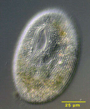
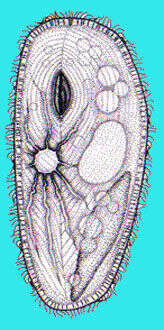
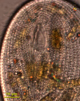
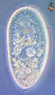
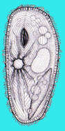
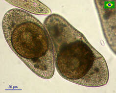
Portrait of the hymenostome ciliate, Frontonia acuminata (coronal optical section). F. acuminata is dorsoventrally flattened. The cell outline is broadly ellipsoid, tapering very slightly posteriorly. F. acuminata has a small cluster of dark brown cytoplasmic granules anteriorly (the similarly shaped F. angusta lacks such granules and is less flattened). The oral aperture is roughly triangular with the base posterior and the anterior apex terminating at a thin preoral suture. There is an undulating membrane on the right and three adoral membranelles on the left. There is a narrow postoral suture to the right of which lie prominent vestibular ciliary rows and to the left of which lie postoral kineties. The round macronucleus is seen here anteriorly with the prominent micronucleus indenting the left margin. Numerous extrusomes form a peripheral fringe(extrusomes seen here posterior and left of oral aperture). The single contractile vacuole is subequatorial. Probably omnivorous. Collected from a slow-moving freshwater stream near Boise, Idaho in July 2003. DIC optics.