-

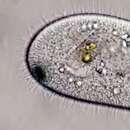
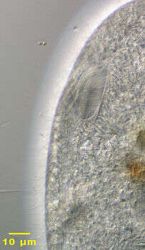
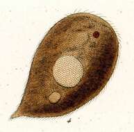
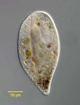
Portrait of the hymenostome ciliate, Frontonia acuminata (ventral view). F. acuminata is dorsoventrally flattened. The cell outline is broadly ellipsoid, tapering very slightly posteriorly. F. acuminata has a small cluster of dark brown cytoplasmic granules anteriorly (the similarly shaped F. angusta lacks such granules and is less flattened). The oral aperture is roughly triangular with the base posterior and the anterior apex terminating at a thin preoral suture. There is an undulating membrane on the right and three adoral membranelles on the left. There is a narrow postoral suture to the right of which lie prominent vestibular ciliary rows and to the left of which lie postoral kineties. The round macronucleus is seen here anteriorly with the prominent micronucleus indenting the left margin. Numerous extrusomes form a peripheral fringe. The single contractile vacuole is subequatorial. Probably omnivorous. Collected from a slow-moving freshwater stream near Boise, Idaho in July 2003. DIC optics
-
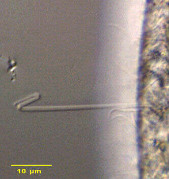
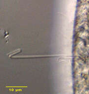
Detail of ejected extrusome of the hymenostome ciliate, Frontonia acuminata (EHRENBERG,1833) BUETSCHLI,1889. Numerous extrusomes form a peripheral fringe. The ejected extrusomes of this genus have a distinctive hook-shaped distal end. Collected from a slow-moving freshwater stream near Boise, Idaho in July 2003. DIC optics.
-
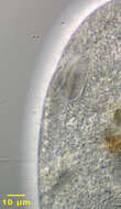
Detail of oral aperture of the hymenostome ciliate, Frontonia acuminata. The oral aperture is roughly triangular with the base posterior and the anterior apex terminating at a thin preoral suture. There is an undulating membrane on the right and three adoral membranelles on the left. There is a narrow postoral suture to the right of which lie prominent vestibular ciliary rows and to the left of which lie postoral kineties. Collected from a slow-moving freshwater stream near Boise, Idaho in July 2003. DIC optics.
-
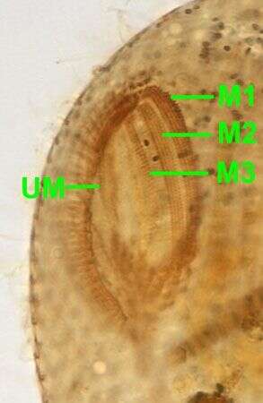
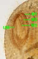
Oral infraciliature of Frontonia acuminata (EHRENBERG, 1833) BUETSCHLI, 1889. There are three slightly curved parallel adoral membranelles on the left of the oral aperture (M1,M2 and M3). There is an inconspicuous undulating membrane on the right side of the oral aperture (UM). Stained by the silver carbonate technique (Foissner,W. Europ. J. Protistol.27:313-330;1991).Brightfield.
-
Ventral infraciliature of Frontonia acuminata (EHRENBERG, 1833) BUETSCHLI, 1889. Stained by the silver carbonate technique (Foissner,W. Europ. J. Protistol.27:313-330;1991).Brightfield.
-
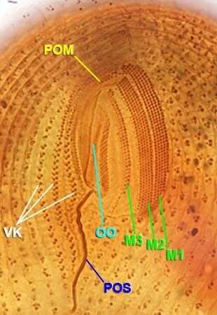
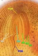
Oral infraciliature of Frontonia acuminata (EHRENBERG, 1833) BUETSCHLI, 1889. There are three slightly curved parallel adoral membranelles on the left of the oral aperture (M1,M2 and M3). There is a paraoral membrane on the right side of the oral aperture (POM). The oral opening (OO) is situated between the POM and M3. There are 3 vestibular kineties (VK) to the right of the POM. A sinuous densely argyrophilic line follows the course of the postoral suture (POS).Stained by the silver carbonate technique (Foissner,W. Europ. J. Protistol.27:313-330;1991).Brightfield.
-
Originally described by Ehrenberg under the name Ophryoglena acuminata,
-
Originally described by Ehrenberg under the name Ophryoglena acuminata,
-
Originally described by Ehrenberg under the name Ophryoglena acuminata,
-

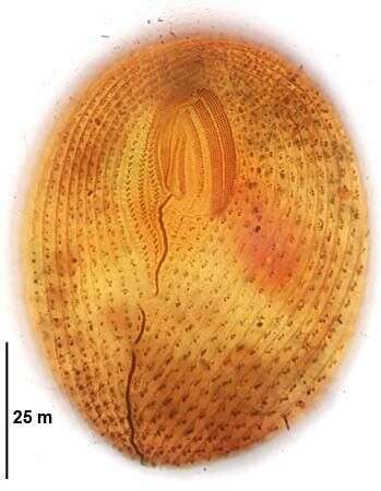
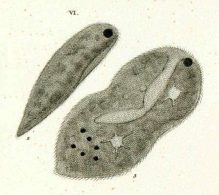
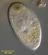
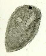
Portrait of the widely distributed hymenostome ciliate, Frontonia atra. This species has a distinctive dorsoventrally flattened teardrop shape. The anterior is broadly rounded, the posterior tapering to a blunt point. F. atra may be confused with Disematostoma buetschlii (the latter has a distinctive cross-striated pre and postoral ciliary suture and contains zoochlorellae and/or kleptoplasts). F. atra has a dense aggregate of dark brown cytoplasmic granules anteriorly (possibly endosymbiotic bacteria). The oral aperture is roughly triangular with the base posterior and the anterior apex terminating at a thin preoral suture. There is an undulating membrane on the right and three adoral membranelles on the left. There is a narrow postoral suture to the right of which lie prominent vestibular ciliary rows and to the left of which lie postoral kineties. The round macronucleus is seen here anterior and to the left of the oral aperture. Numerous extrusomes form a peripheral fringe. The single contractile vacuole (not seen here) is subequatorial on the right. Probably omnivorous. Often found feeding on diatoms and green algae. Collected from freshwater pond near Boise, Idaho in June 2003. DIC optics.
-
Portrait of the widely distributed hymenostome ciliate, Frontonia atra (Ehrenberg, 1833) Buetschli, 1889. This species has a distinctive dorsoventrally flattened teardrop shape. The anterior is broadly rounded, the posterior tapering to a blunt point. F. atra may be confused with Disematostoma buetschlii (the latter has a distinctive cross-striated pre and postoral ciliary suture and contains zoochlorellae and/or kleptoplasts). F. atra often has a dense aggregate of dark brown cytoplasmic granules anteriorly (possibly endosymbiotic bacteria). The oral aperture is roughly triangular with the base posterior and the anterior apex terminating at a thin preoral suture. There is an undulating membrane on the right and three adoral membranelles on the left. There is a narrow postoral suture to the right of which lie prominent vestibular ciliary rows and to the left of which lie postoral kineties. The ellipsoid macronucleus is seen here. Numerous extrusomes form a peripheral fringe. The single contractile vacuole has 2-6 excretory pores (5 in this case;seen to the viewrs right of the posterior end of the macronucleus). Often found feeding on diatoms and green algae. Collected from freshwater pond near Boise, Idaho in June 2003. DIC optics.
-
Originally described by Ehrenberg under the name Ophryoglena atra.
-
Originally described by Ehrenberg under the name Ophryoglena atra.
-
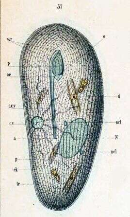
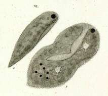
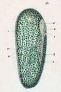
Key to Schewiakoff's abbreviations: a-- Anus cv--Contractile vacuole ccv--Canal of the contratile vacuole d--Ingested diatoms ek--Ectoplasm N--Macronucleus ncl--Micronucleus oe--Throat p--Pellicle P--Peristome tr -- Trichocysts wr -- Cilia series
-
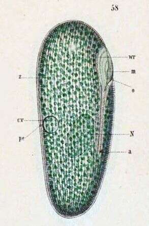
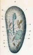
An individual that contains Zoochlorellae. a -- Anus cv -- Contractile vacuole m -- Undulating membrane N -- Macronucleus o -- Mouth pe -- Excretory pore of the contractile vacuole wr -- Cilia series Z -- Zoochlorellae
-
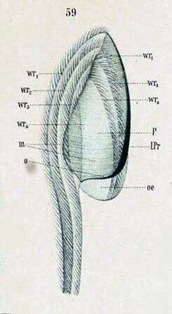
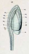
Detail of the oral apparatus. l. pr -- Left edge of peristome m -- Undulating membrane o -- Mouth oe -- Throat P -- Peristome wr1-wr4 -- Cilia series (peniculi and paroral)
-
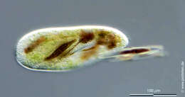
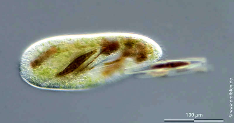
Sampling date 04/2016. Scale bars indicate 100 µm.Frontonia leucas feeding on diatoms.Place name: Pond Domänental near Kronshagen (Kiel, Germany) Latitude: 54.33211 Longitude: 10.060821Microscope Zeiss Axioplan, camera Olympus OM-D M5 MKII. DOF images.© Wolfgang Bettighofer,images under Creative Commons License V 3.0 (CC BY-NC-SA).For permission to use of (high resolution) images please contact
postmaster@protisten.de.For further information about the image, please click here:
Link to protisten.de page
-
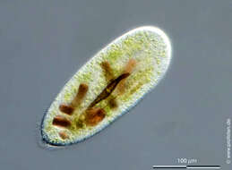
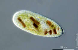
Sampling date 04/2016. Scale bars indicate 100 µm.Frontonia leucas feeding on diatoms.Place name: Pond Domänental near Kronshagen (Kiel, Germany) Latitude: 54.33211 Longitude: 10.060821Microscope Zeiss Axioplan, camera Olympus OM-D M5 MKII. DOF images.© Wolfgang Bettighofer,images under Creative Commons License V 3.0 (CC BY-NC-SA).For permission to use of (high resolution) images please contact
postmaster@protisten.de.For further information about the image, please click here:
Link to protisten.de page