Ecology
provided by NMNH Marine Dinoflagellates
P. hoffmannianum is a benthic species. Cells are motile or attached to detritus by mucilage (Faust 1990b).
- bibliographic citation
- Faust, Maria A. and Rose A. Gulledge. Identifying Harmful Marine Dinoflagellates. Smithsonian Contributions from the United States National Herbarium, volume 42: 1-144 (including 48 plates, 1 figure and 1 table).
Etymology
provided by NMNH Marine Dinoflagellates
This species is named in honor of Dr. Robert S. Hoffmann, Assistant Secretary for Research, Smithsonian Institution, for his encouragement, support and scientific leadership (Faust 1990b).
- bibliographic citation
- Faust, Maria A. and Rose A. Gulledge. Identifying Harmful Marine Dinoflagellates. Smithsonian Contributions from the United States National Herbarium, volume 42: 1-144 (including 48 plates, 1 figure and 1 table).
Habitat and Locality
provided by NMNH Marine Dinoflagellates
Populations of P. hoffmannianum are often associated with floating detritus in tropical coastal regions of the Caribbean Sea (Faust 1990b).
- bibliographic citation
- Faust, Maria A. and Rose A. Gulledge. Identifying Harmful Marine Dinoflagellates. Smithsonian Contributions from the United States National Herbarium, volume 42: 1-144 (including 48 plates, 1 figure and 1 table).
Morphology and Structure
provided by NMNH Marine Dinoflagellates
P. hoffmannianum is a photosynthetic species containing golden-brown chloroplasts, a centrally located pyrenoid, and a large posterior nucleus (Fig. 5) (Faust 1990b).
- bibliographic citation
- Faust, Maria A. and Rose A. Gulledge. Identifying Harmful Marine Dinoflagellates. Smithsonian Contributions from the United States National Herbarium, volume 42: 1-144 (including 48 plates, 1 figure and 1 table).
Nomenclatural Types
provided by NMNH Marine Dinoflagellates
Holotype: Prorocentrum hoffmannianum Faust, 1990: figs. 13, 14
Type Locality: Caribbean Sea: Twin Cays, Belize, Central America
- bibliographic citation
- Faust, Maria A. and Rose A. Gulledge. Identifying Harmful Marine Dinoflagellates. Smithsonian Contributions from the United States National Herbarium, volume 42: 1-144 (including 48 plates, 1 figure and 1 table).
Remarks
provided by NMNH Marine Dinoflagellates
In Carlson (1984), P. concavum identified on Plate 5, figs. n-s, is P. hoffmannianum based on thecal surface morphology, periflagellar area and intercalary band characteristics. In addition, the illustration of P. concavum (fig. 17) by Steidinger (1983) is neither P. concavum nor P. hoffmannianum, but is an unidentified species (Faust 1990b).
- bibliographic citation
- Faust, Maria A. and Rose A. Gulledge. Identifying Harmful Marine Dinoflagellates. Smithsonian Contributions from the United States National Herbarium, volume 42: 1-144 (including 48 plates, 1 figure and 1 table).
Reproduction
provided by NMNH Marine Dinoflagellates
P. hoffmannianum reproduces asexually by binary fission.
- bibliographic citation
- Faust, Maria A. and Rose A. Gulledge. Identifying Harmful Marine Dinoflagellates. Smithsonian Contributions from the United States National Herbarium, volume 42: 1-144 (including 48 plates, 1 figure and 1 table).
Species Comparison
provided by NMNH Marine Dinoflagellates
P. hoffmannianum is similar in shape to P. lima, but larger and broader with dense areolae. P. hoffmannianum is often misidentified as P. concavum, but can be distinguished by its ovoid shape and presence of areolae in the center of the valve (Fukuyo 1981; Faust 1990b; 1991). The architecture of the periflagellar area of P. hoffmannianum is similar to P. lima, P. concavum (Fukuyo 1981) and P. playfairi (Croome & Tyler 1987); however, P. hoffmannianum has a more complex platelet configuration (Faust 1990b). P. reticulatum (Faust 1997), P. sabulosum (Faust 1994), P. belizeanum (Faust 1993a) and P. hoffmannianum (Faust 1990b) share a distinct feature in the periflagellar area: three small accessory pores adjacent to a periflagellar pore (Faust 1997). Both P. hoffmannianum and P. belizeanum have a prominent flared curved apical collar on the left valve bordering the periflagellar area, although the curved apical collar of the latter species is rounder, whereas that of the former is flatter (Faust 1993a).
- bibliographic citation
- Faust, Maria A. and Rose A. Gulledge. Identifying Harmful Marine Dinoflagellates. Smithsonian Contributions from the United States National Herbarium, volume 42: 1-144 (including 48 plates, 1 figure and 1 table).
Species Overview
provided by NMNH Marine Dinoflagellates
P.hoffmannianum is an armoured, marine, benthic dinoflagellate species. This species is associated with floating detritus and sediment in tropical embayments of the Caribbean Sea.
- bibliographic citation
- Faust, Maria A. and Rose A. Gulledge. Identifying Harmful Marine Dinoflagellates. Smithsonian Contributions from the United States National Herbarium, volume 42: 1-144 (including 48 plates, 1 figure and 1 table).
Synonyms
provided by NMNH Marine Dinoflagellates
Exuviaella hoffmannianum (Faust) McLachlan, Boalch and Jahn, 1997
- bibliographic citation
- Faust, Maria A. and Rose A. Gulledge. Identifying Harmful Marine Dinoflagellates. Smithsonian Contributions from the United States National Herbarium, volume 42: 1-144 (including 48 plates, 1 figure and 1 table).
Taxonomic Description
provided by NMNH Marine Dinoflagellates
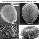
P. hoffmannianum is a bivalvate species often observed in valve view. Cells are ovoid, broadest in mid-region, tapering slightly apically (Figs. 1, 2, 5, 6). Cells are 45-55 µm long and 40-45 µm wide. Both valves are slightly concave in the center. The intercalary band is smooth and appears as a flared ridge around the cell (Figs. 1, 2, 5). Observed under LM, the marginal areolae can give the appearance of a striated intercalary band (Fig. 5) (Faust 1990b). The valve surface is deeply areolate; areolae are dense, large, and round to oblong (Figs. 1-4). Small round to ovoid pores are found within deep areolae; these pores have smooth margins, are 1.0-1.5 µm in diameter, and many bear trichocyst pores (Fig. 3). There are approximately 650-700 areolae on each valve (Faust 1990b). The periflagellar area is a wide triangle situated apically on the right valve (Figs. 1, 4). It houses eight periflagellar platelets and two periflagellar pores: a flagellar pore and auxiliary pore (equal in size); accessory pores are also present. The flagellar pore is surrounded by a small flared periflagellar collar (Fig. 4). Both left and right valves are apically excavated (Figs. 1, 4). The left valve exhibits a flared and flattened curved apical collar that borders the periflagellar area (Figs. 1, 2) (Faust 1990b).
- bibliographic citation
- Faust, Maria A. and Rose A. Gulledge. Identifying Harmful Marine Dinoflagellates. Smithsonian Contributions from the United States National Herbarium, volume 42: 1-144 (including 48 plates, 1 figure and 1 table).
Toxicity
provided by NMNH Marine Dinoflagellates
This species is considered toxic producing fast-acting toxin (FAT) and diarrhetic shellfish poison (DSP) toxin: okadaic acid (OA) (Aikman et al. 1993).
- bibliographic citation
- Faust, Maria A. and Rose A. Gulledge. Identifying Harmful Marine Dinoflagellates. Smithsonian Contributions from the United States National Herbarium, volume 42: 1-144 (including 48 plates, 1 figure and 1 table).