Description of Paraphysomonas vestita
provided by BioPedia
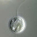
Ovoid to elongate cells, from 6 to 15 microns long. Two flagella insert in an apical depression. One flagellum is very short and held laterally while the other, two to three times the length of the cell, is held anteriorly and beats with short wavelength and high amplitude wave pattern. Cells often contain granules. Scales are visible in light microscopy as delicate spines. In electron microscopy, the scales have a plain circular base (1-2 microns) with a well-marked rim and a tapering spine 3 to 5 microns long.