-
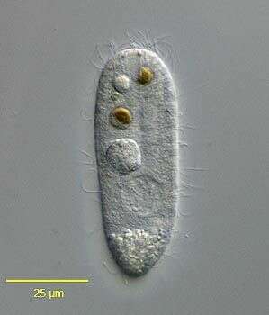
Longitude (deg): 1.2. Latitude (deg): 52.6. Longitude (deg/min): 1° 10' E. Latitude (deg/min): 52° 40' N. Vice county name: East Norfolk. Vice county no.: 27. Country: England. Identified by: Malcolm Storey. Comment: From shallow pools on chalky clay. Category: standard photograph or close-up. Photographic equipment used: "35mm transparencies (on a variety of films, but Agfa CT18 in the 1960's to early 1980's followed by Fujichrome in the late 1980's.) Transparencies scanned with Minolta Dimage Scan Dual II AF-2820U transparency scanner.".
-
Lardero, La Rioja, Spain
-
Herrera, Castille and Leon, Spain
-
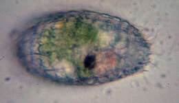
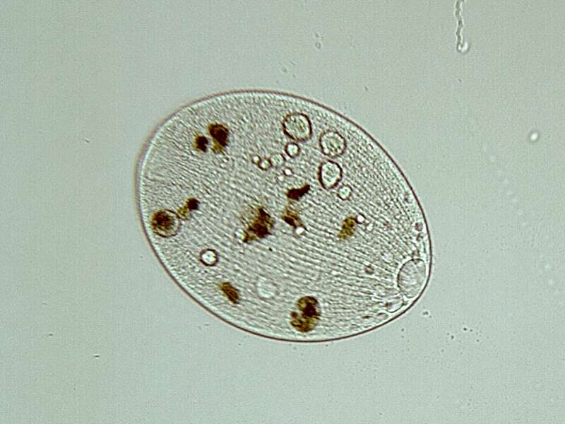
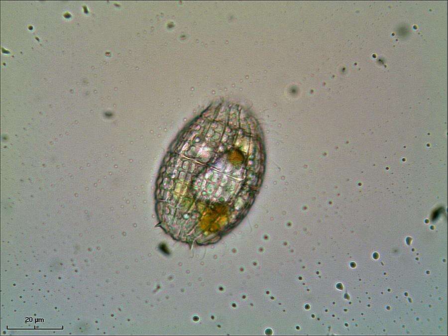
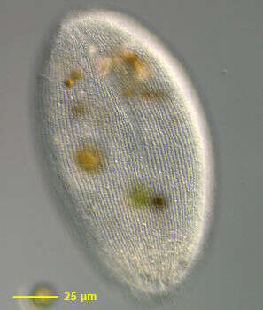
Ovoid to cylindrical; ciliation uniform; oral basket made up of double trichites which end up deep in ectoplasm. Macronucleus ovoid, reniform or ellongate. Cell body 80-200 micron long.
-
Cuelgamuros, Madrid, Spain
-
Villar del Pedroso, Extremadura, Spain
-
Los Cotos, Madrid, Spain
-
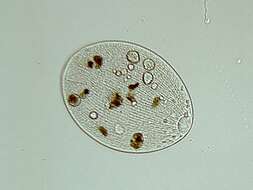
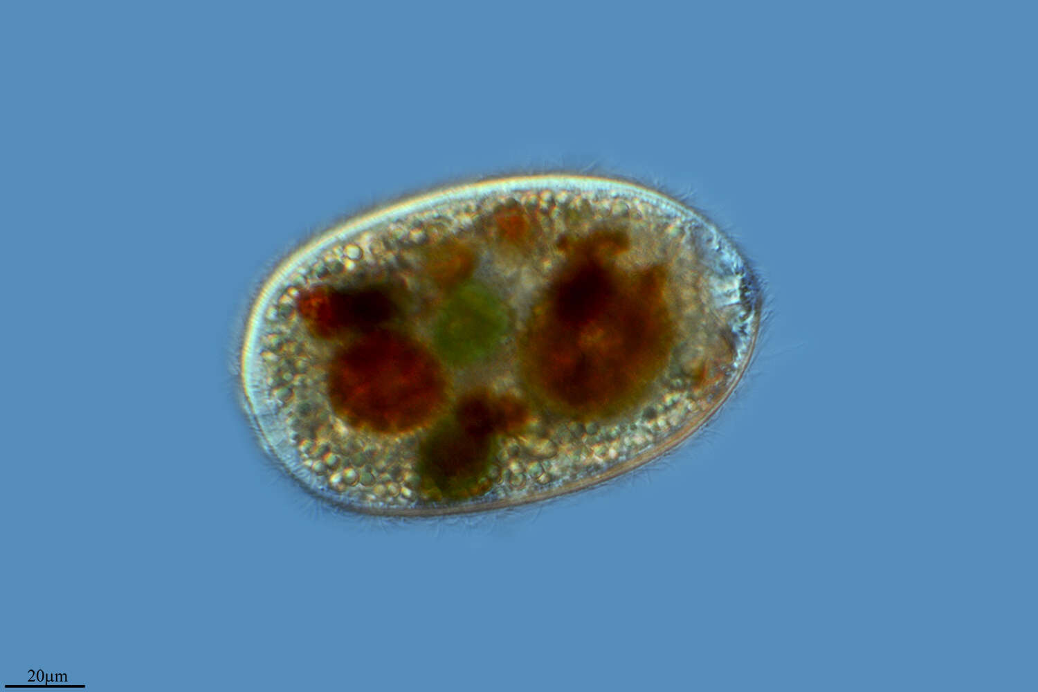
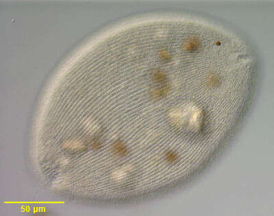
Body with regurarly arranged ectoplasmic plates. Cytostome at anterior end, surrounded by slightly longer cilia . Often spinous projection at or near posterior end.
-
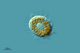
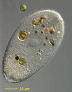
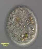
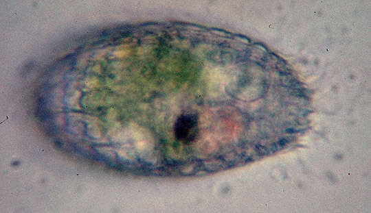
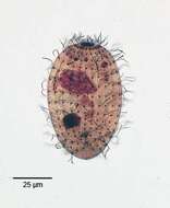
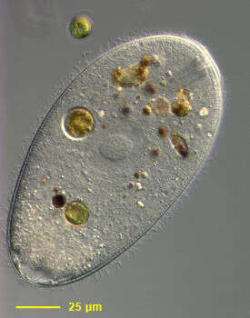
Portrait (ventral surface) of Holophrya discolor (Ehrenberg,1833) a common prostome ciliate. Body shape variable from nearly spheroid to oblong. Some species with zoochlorellae. The oral aperture is an anterior apical or somewhat offset invagination (seen here). The cytopharynx is supported by fine trichites (not visible here). Ciliature is dense and holotrichous with three double rows of kinetids forming a "dorsal brush" extending posterior from the oral aperture about 1/3 the body length. There are some longer caudal cilia. The contractile vacuole is posterior. Holophrya sp. are distinguished from the very similar genus, Prorodon, by their spherical or ellipsoid macronuclei. Prorodon sp. typically have sausage shaped or ribbon-shaped macronuclei. Omnivorous. From organically enriched freshwater pond near Boise, Idaho. DIC optics.
-
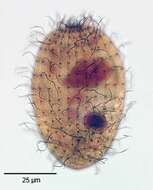
Portrait(dorsal surface) of Holophrya discolor (Ehrenberg,1833),a common prostome ciliate. Body shape variable from nearly spheroid to oblong. Some species with zoochlorellae. The oral aperture is an anterior apical or somewhat offset invagination. The cytopharynx is supported by fine trichites (not visible here). Ciliature is dense and holotrichous with three double rows of kinetids forming a "dorsal brush" extending posterior from the oral aperture about 1/3 the body length (seen in this image). There are some longer caudal cilia. The contractile vacuole is posterior. Holophrya sp. are distinguished from the very similar genus, Prorodon, by their spherical or ellipsoid macronuclei. Prorodon sp. typically have sausage shaped or ribbon-shaped macronuclei. Omnivorous. From organically enriched freshwater pond near Boise, Idaho. DIC optics.
-
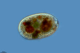
Portrait (optical coronal section) of Holophrya discolor (Ehrenberg,1833), a common prostome ciliate. Body shape variable from nearly spheroid to oblong. Some species with zoochlorellae. The oral aperture is an anterior apical or somewhat offset invagination. The cytopharynx is supported by fine trichites (seen here). Ciliature is dense and holotrichous with three double rows of kinetids forming a "dorsal brush" extending posterior from the oral aperture about 1/3 the body length. There are some longer caudal cilia. The spherical macronucleus located in the mid-body has a characteristic large nucleolus. The micronucleus is attached to the right posterior of the macronucleus in this image. Small peripheral mucocysts are present. The contractile vacuole is posterior. Numerous food vacuoles are seen. Holophrya sp. are distinguished from the very similar genus, Prorodon, by their spherical or ellipsoid macronuclei. Prorodon sp. typically have sausage shaped or ribbon-shaped macronuclei. Omnivorous. From organically enriched freshwater pond near Boise, Idaho. DIC optics.
-
Infraciliature of the prostome ciliate Holophrya discolor (Ehrenberg,1833). Densely stained nematodesmata ring the anterior apical cytostome.The dorsal brush of the "enklitoloph dexiotrop" type is seen anteriorly to the viewer's right (See: Hiller, S. and Bardele, C. Arch. Protistenk. 136:213-236, 1988). Collected from a freshwater aquaculture tub near Boise, Idaho.August 2004. Stained byt the silver carbonate technique (see Foissner, W. Europ. J. Protistol., 27:313-330;1991).Brightfield.
-
Anterolateral view of Holophrya discolor (Ehrenberg,1833)showing the elliptical anterior apical cytostome.DIC.
-
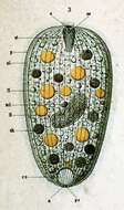
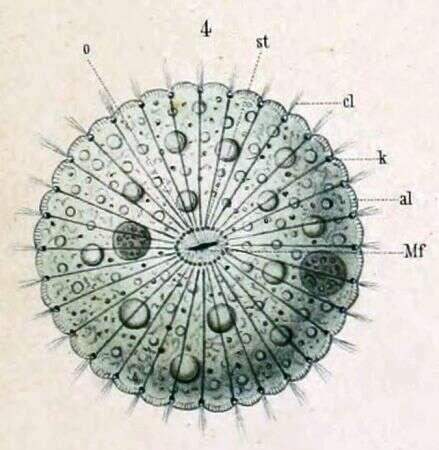
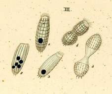
a -- Anus al -- Pellicular alveoli cv -- Contractile vacuole ft -- Fat droplets N -- Macronucleus ncl -- Micronucleus nk -- Food particle o -- Mouth oe-- Throat p -- Pellicle pe -- Excretory pore of the contractile vacuole st -- Cytopharyngeal basket
-
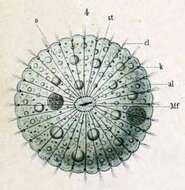
Key to Schewiakoff's abbreviations: al -- Pellicular alveoli cl -- Cilia k -- Plasmatic collar of the rod apparatus Mf -- Buccal are o -- Mouth st -- Cytopharyngeal basket
-
-
-
Oral Infraciliature of Nolandia nolandi (KAHL, 1930) SMALL & LYNN, 1985.Collected from a freshwater pond near Boise, Idaho. June 2008.Stained by the Protargol technique (Wilbert modification) (see Foissner, W. Europ. J. Protistol., 27:313-330;1991).Brightfield.
-
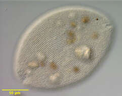
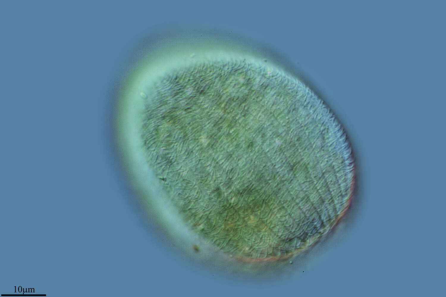
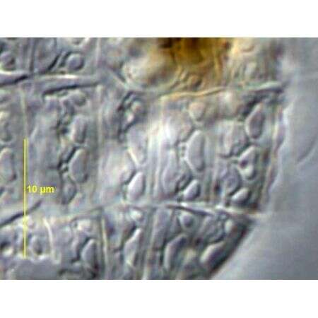
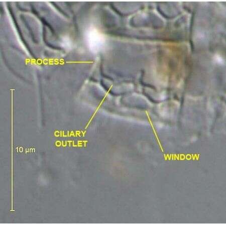
Calcified armor plates of Nolandia nolandi (KAHL, 1930) SMALL & LYNN, 1985.Collected from a freshwater pond near Boise, Idaho. June 2008.DIC.
-
Calcified armor plates of Nolandia nolandi (KAHL, 1930) SMALL & LYNN, 1985 .Collected from a freshwater pond near Boise, Idaho. June 2008.DIC.
-
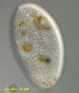
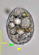
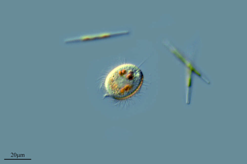
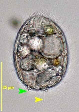
Nolandia nolandi (KAHL, 1930) SMALL & LYNN, 1985.Yellow arrowhead=caudal cilium.Green arrowhead=posterior spine.Collected from a freshwater pond near Boise, Idaho. June 2008.Brightfield,closed condenser.
-
Infraciliature of the planktonic protstomatid ciliate, Apsiktrata gracilis (Penard,1922)Foissner, Berger & Kohmann 1994. Morphologically quite similar to members of the genus Holophrya but lacking a "dorsal brush". The anterior apical cytostome and its circumoral dikinetids is seen here.There is a long caudal cilium in vivo (only its basal body is seen here at the posterior pole). Collected from a freshwater pond near Boise, Idaho. Silver carbonate stain (see Foissner, W. Europ. J. Protistol., 27:313-330;1991). Brightfield
-
Infraciliature of the planktonic protstomatid ciliate, Apsiktrata gracilis (Penard,1922)Foissner, Berger & Kohmann 1994. Morphologically quite similar to members of the genus Holophrya but lacking a "dorsal brush". The anterior apical cytostome and its circumoral dikinetids is seen here.The microfibrillar system associted with the basal bodies of the somatic kineties is visible here. Collected from a freshwater pond near Boise, Idaho. Silver carbonate stain (see Foissner, W. Europ. J. Protistol., 27:313-330;1991). Brightfield
-
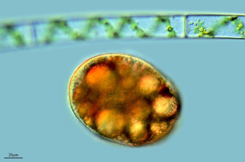
Portrait of the planktonic protstomatid ciliate, Apsiktrata gracilis (Penard,1922)Foissner, Berger & Kohmann 1994. Morphologically quite similar to members of the genus Holophrya but lacking a "dorsal brush". The anterior apical cytostome and its circumoral dikinetids is seen here.The microfibrillar system associted with the basal bodies of the somatic kineties is visible here. Collected from a freshwater pond near Boise, Idaho. DIC.