-
-
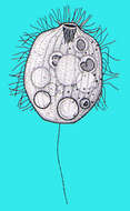
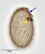
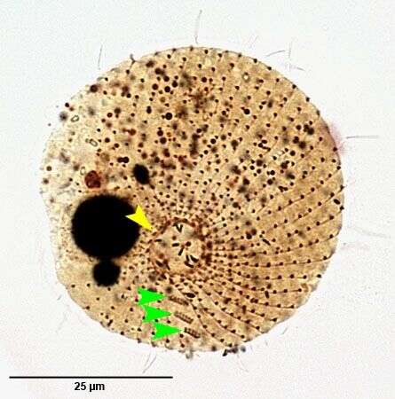
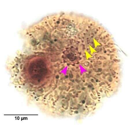
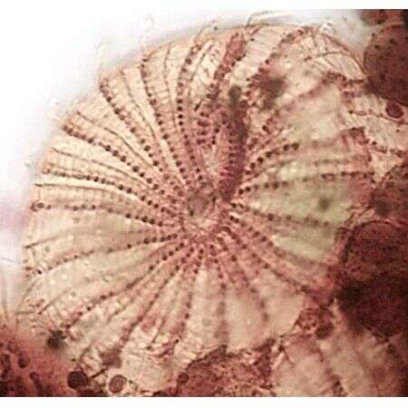
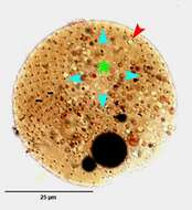
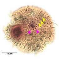
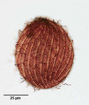
Infraciliature of Urotricha platystoma (STOKES,1886).The yellow arrowhead indicates one of the obliquely oriented dikinetids surronding the oral aperture. The green arrowheads indicate the three obliquely oriented adoral organelles, each composed of two rows of kinetids. Collected from the margin of a slow-moving outflow stream of a freshwater pond near Boise, Idaho.March 2007. Stained by the silver carbonate technique (Foissner,W. Europ. J. Protistol.27:313-330;1991).Brightfield.
-
Posterior view of the infraciliature of Urotricha platystoma(STOKES,1886).The somatic kineties terminate in the posterior 1/4 of the cell (light blue arrowheads). The posterior end of the cell is unciliated (asterisk) except for the single long caudal cilium. the red arrowhead indicates the excentric pore of the contractile vacuole.Collected from the margin of a slow-moving outflow stream of a freshwater pond near Boise, Idaho.March 2007. Stained by the silver carbonate technique (Foissner,W. Europ. J. Protistol.27:313-330;1991).Brightfield.
-
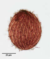
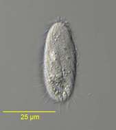
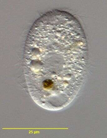
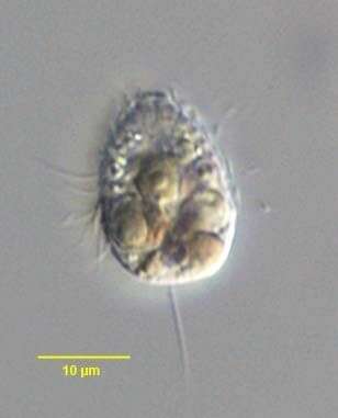
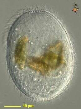
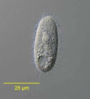
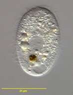
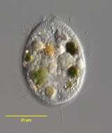
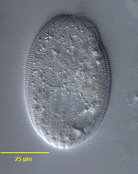
Urotricha platystoma (STOKES,1886).Collected from the margin of a slow-moving outflow stream of a freshwater pond near Boise, Idaho.March 2007. Phase contrast.
-
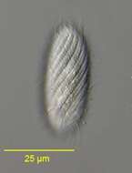
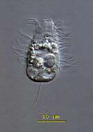
Urotricha platystoma (STOKES,1886). The long caudal cilium is out of the focal plane in this image.Collected from the margin of a slow-moving outflow stream of a freshwater pond near Boise, Idaho.March 2007.DIC
-
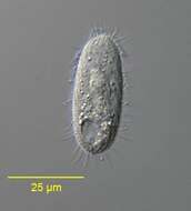
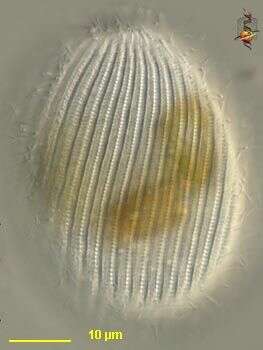
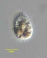
Urotricha platystoma (STOKES,1886). Collected from the margin of a slow-moving outflow stream of a freshwater pond near Boise, Idaho.March 2007.DIC
-
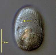
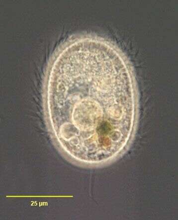
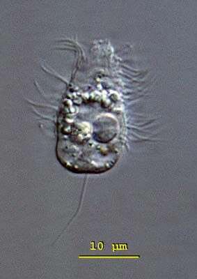
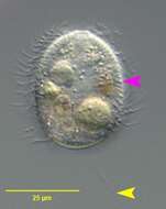
Urotricha platystoma (STOKES,1886). The yellow arrowhead indicates the single long caudal cilium.The pink arrowhead indicates the subcortical layer of fusiform extrusomes.Collected from the margin of a slow-moving outflow stream of a freshwater pond near Boise, Idaho.March 2007.DIC
-
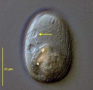
Urotricha (your-owe-trike-a) farcta: the anterior half of the body is conical and the posterior half trapeziform. The oral opening is at the anterior end of the cell. The contractile vacuole is located in the posterior end and the round macronucleus is in the middle of the body. The caudal cilium is slightly eccentric. This is a fast swimming cilate, the motion of which is interrupted by jumps. The species is mostly 15 - 30 microns long, this cell 22 microns. Differential interference contrast.
-
Oral infraciliature of Urotricha farcta (CLAPARÃDE&LACHMAN,1859). The pink arrowheads mark dikinetids of the undulating membrane (oral flaps).The yellow arrowheads mark the three minute adoral organelles. Collected from submerged dead leaves at the margin of a slow-flowing freshwater stream near Boise, Idaho.March 2007.Stained by the silver carbonate technique (Foissner,W. Europ. J. Protistol.27:313-330;1991).Brightfield.
-
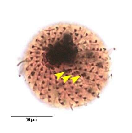
Infraciliature of Urotricha farcta (CLAPARÃDE&LACHMAN,1859). The yellow arrowheads mark the three minute adoral organelles. Collected from submerged dead leaves at the margin of a slow-flowing freshwater stream near Boise, Idaho.March 2007.Stained by the silver carbonate technique (Foissner,W. Europ. J. Protistol.27:313-330;1991).Brightfield.
-
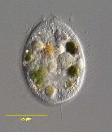
Urotricha farcta (CLAPARÃDE&LACHMAN,1859). Collected from submerged dead leaves at the margin of a slow-flowing freshwater stream near Boise, Idaho.March 2007.DIC.
-
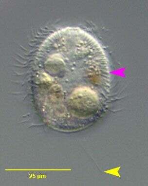
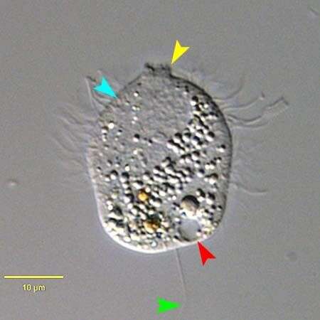
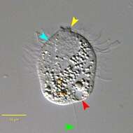
Urotricha farcta (CLAPARÃDE&LACHMAN,1859).This is a larger individual,slightly compressed.The yellow arrowhead marks the undulating membrane (oral flaps) that surrounds the anterior apical cytostome.The green arrowhead marks the single long eccentric posterior cilium.The red arrowhead marks the single subterminal posterior contractile vacuole.The light blue arrowhead marks the inconspicuous subpellicular extrusomes.Collected from submerged dead leaves at the margin of a slow-flowing freshwater stream near Boise, Idaho.March 2007.DIC.
-
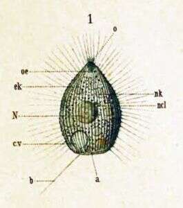
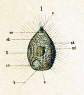
Key to Schewiakoff's abbreviations a -- Anus b -- Sensory bristle c.v -- Contractile vacuole ek -- Ectoplasm oe -- Throat N -- Macronucleus ncl -- Micronucleus o - Mouth nk -- Food particle
-
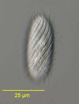
Placus (plake-us) is a barrel-shaped ciliate. Kineties of cilia run in a slightly spiral course from the anterior end - which is where the mouth is, to the posterior end of the body. Kineties have adjacent sharp-walled gutters. Eats detritus, such as algae. Differential interference contrast.
-
Placus (plake-us) is a barrel-shaped ciliate. Kineties of cilia run in a slightly spiral course from the anterior end - which is where the mouth is, to the posterior end of the body. Kineties have adjacent sharp-walled gutters. Eats detritus, such as algae. Differential interference contrast.
-
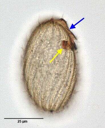
Portrait (right lateral view) of the prorodontid ciliate, Placus luciae (Kahl,1926) Kahl,1930. The cell is ovoid and slightly laterally compressed. The pellicle has broad slightly spiral ridges separated by narrow furrows. The small oval oral aperture is apical anterior, surrounded by a ring of unciliated kinetosomes. The cytopharynx is supported by fine trichites. The somatic ciliature uniform, with cilia.There is a "brush" composed of a double parallel row of kinetosomes bearing longer cilia. This is about 1/4 to 1/3 cell length arising adjacent to the oral aperture. It terminates at a ventral circular "pit" (seen here as a slight depression anteriorly on viewer's right) which has a supporting ring of transversely oriented trichites. The function of this structure is unknown. The ovoid macronucleus is centrally located. The contractile vacuole is poster lateral. Collected from a slow-flowing freshwater stream near Boise, Idaho. DIC.
-
Optical section (right lateral view) of the prorodontid ciliate, Placus luciae (Kahl,1926) Kahl,1930. The cell is ovoid and slightly laterally compressed. The pellicle has broad slightly spiral ridges separated by narrow furrows. The small oval oral aperture is apical anterior, surrounded by a ring of unciliated kinetosomes. The cytopharynx is supported by fine trichites. The somatic ciliature uniform, with cilia. There is a "brush" composed of a double parallel row of kinetosomes bearing longer cilia. This is about 1/4 to 1/3 cell length arising adjacent to the oral aperture. It terminates at a ventral circular "pit" (seen here as a slight depression anteriorly on viewer's right) which has a supporting ring of transversely oriented trichites (faintly visible here). the function of this structure is unknown . The ovoid macronucleus is centrally located. The contractile vacuole is posterolateral (seen in this image). Collected from a slow-flowing freshwater stream near Boise, Idaho. DIC.
-
Infraciliature (right lateral view) of the prorodontid ciliate, Placus luciae (Kahl,1926) Kahl,1930. The cell is ovoid and slightly laterally compressed. The pellicle has broad slightly spiral ridges separated by narrow furrows. The small oval oral aperture is apical anterior, surrounded by a ring of unciliated kinetosomes. The cytopharynx is supported by fine trichites. The somatic ciliature uniform, with cilia. There is a "dorsal brush" (blue arrow), about 1/4 to 1/3 cell length, composed of a double parallel row of kinetosomes bearing longer cilia. It arises adjacent to the oral aperture and terminates at a ventral circular "pit" or "fossette" which has a supporting ring of transversely oriented trichites (yellow arrow). The function of this structure is unknown. The ovoid macronucleus is centrally located. The contractile vacuole is posterolateral. Collected from a slow-flowing freshwater stream near Boise, Idaho.Stained by the silver carbonate technic (see Foissner, W.Europ. J. Protistol.27,313-330;1991). Brightfield.
-
Infraciliature (dorsal view) of the prorodontid ciliate, Placus luciae (Kahl,1926) Kahl,1930. The cell is ovoid and slightly laterally compressed. The pellicle has broad slightly spiral ridges separated by narrow furrows.In this preparation the reticulate pattern of the ridges is seen. Fine transverse striations cross the narrow furrows between the somatic kineties to a darkly stained longitudinal fibril on the right margin of the ridges. The small oval oral aperture is apical anterior, surrounded by a ring of unciliated kinetosomes. The cytopharynx is supported by fine trichites. The somatic ciliature uniform, with cilia. There is a "brush" , about 1/4 to 1/3 cell length, composed of a double parallel row of kinetosomes bearing longer cilia. This arises adjacent to the oral aperture and terminates at a ventral circular "pit" which has a supporting ring of transversely oriented trichites. The function of this structure is unknown . The ovoid macronucleus is centrally located. The contractile vacuole is posterolateral. Collected from a slow-flowing freshwater stream near Boise, Idaho.Stained by the silver carbonate technique (see Foissner, W.Europ. J. Protistol.27,313-330;1991) . Brightfield.
-
Anterior view of the infraciliature of the prorodontid ciliate, Placus luciae (Kahl,1926) Kahl,1930. The cell is ovoid and slightly laterally compressed. The pellicle has broad slightly spiral ridges separated by narrow furrows. The small oval oral aperture is apical anterior, surrounded by a ring of unciliated kinetosomes (seen well here). The cytopharynx is supported by fine trichites. The somatic ciliature uniform, with cilia. There is a "brush" (seen here at 12 0'clock) composed of a double parallel row of kinetosomes bearing longer cilia. This is about 1/4 to 1/3 cell length arising adjacent to the oral aperture. It terminates at a ventral circular "pit" which has a supporting ring of transversely oriented trichites. The function of this structure is unknown . Collected from a slow-flowing freshwater stream near Boise, Idaho.Stained by the silver carbonate technic (see Foissner, W.Europ. J. Protistol.27,313-330;1991). Brightfield.
-
Portrait (left lateral view) of the prorodontid ciliate, Placus luciae (Kahl,1926) Kahl,1930. The cell is ovoid and slightly laterally compressed. The pellicle has broad slightly spiral ridges separated by narrow furrows. The small oval oral aperture is apical anterior, surrounded by a ring of unciliated kinetosomes. The cytopharynx is supported by fine trichites. The somatic ciliature uniform, with cilia.There is a "brush" composed of a double parallel row of kinetosomes bearing longer cilia. This is about 1/4 to 1/3 cell length arising adjacent to the oral aperture. It terminates at a ventral circular "pit" (arrow) which has a supporting ring of transversely oriented trichites. The function of this structure is unknown. The ovoid macronucleus is centrally located. The contractile vacuole is poster lateral. Collected from a slow-flowing freshwater stream near Boise, Idaho. DIC.
-
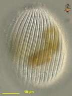
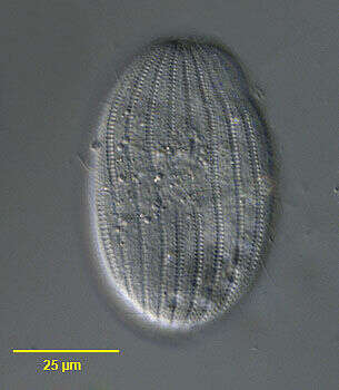
Surface detail of the prorodontid ciliate, Placus striatus (Cohn, 1866). The somatic kineties of P. striatus are distinctly more spiralled than those of P. luciae.DIC.
-
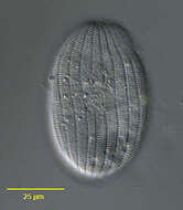
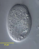
Portrait of the prorodontid ciliate, Placus striatus (Cohn,1866).DIC.
-
Portrait of the prorodontid ciliate, Placus striatus (Cohn,1866).DIC.