-
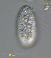
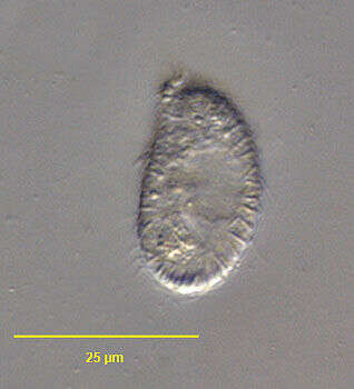
Portrait of the planktonic protstomatid ciliate, Apsiktrata gracilis (Penard,1922)Foissner, Berger & Kohmann 1994. Morphologically quite similar to members of the genus Holophrya but lacking a "dorsal brush". The anterior apical cytostome and its circumoral dikinetids is seen here.The microfibrillar system associted with the basal bodies of the somatic kineties is visible here. Collected from a freshwater pond near Boise, Idaho. Silver carbonate stain (see Foissner, W. Europ. J. Protistol., 27:313-330;1991). DIC.
-
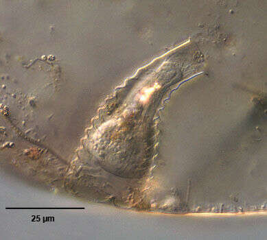
Portrait of free-swimming Vasicola ciliata (Tatem, 1869) that has vacated its lorica. Vasicola ciliata is a metacystid ciliate that produces a thin, transparent pseudochitinous vase-like lorica with shallow transverse corrugations (not seen in this image). In the lorica the cell body assumes a more globular shape. When swimming free the cell is more elongate. The circular cytostome is located in the center of the truncate anterior end. It opens into a cytopharynx supported by indistinct trichites. There are three concentric ciliary rings around the cytostome, the innermost with a single ciliary row, the middle with a double ciliary row and a third ring of four ciliary rows. The uniform longitudinal ciliary rows are formed of dikinetid kinetosomes the alignment which gives the appearance of transverse ciliary bands (paratenes). There is a sparse tuft of long caudal cilia (only one long eccentric posterior cilium is seen in the similar genus, Metacystis and another metacystid, Pelatractus, lacks caudal cilia). Multiple food vacuoles are visible in the cytoplasm. The central spherical macronucleus is seen in this image. The base of the lorica is partly filled with the expelled contents of defecation vacuoles. A single peripheral contractile vacuole is located in the posterior third of the cell (seen in this image on the viewer's left). A large, clear terminal vacuole may occur but is not seen in this image. The cell often vacates the lorica when disturbed. Vasicola ciliata is sapropelic and feeds on sulfur bacteria. Collected from stagnant freshwater sediment with strong smell of hydrogen sulfide near Boise, Idaho January 2004. DIC optics.
-
Anterior apical view of Vasicola ciliata (Tatem, 1869), a metacystid ciliate that produces a thin, transparent pseudochitinous vase-like lorica with shallow transverse corrugations (not seen in this image). The circular cytostome is located in the center of the truncate anterior end. It opens into a cytopharynx supported by indistinct trichites. There are three concentric ciliary rings around the cytostome, the innermost with a single ciliary row, the middle with a double ciliary row and a third ring of four ciliary rows. The uniform longitudinal ciliary rows are formed of dikinetids kinetosomes the alignment which gives the appearance of transverse ciliary bands (paratenes). . The cell often vacates the lorica when disturbed. When swimming free the cell is more elongate. The uniform longitudinal ciliary rows are formed of dikinetid kinetosomes the alignment which gives the appearance of transverse ciliary bands (paratenes). There is a sparse tuft of long caudal cilia (only one long eccentric posterior cilium is seen in the similar genus, Metacystis and another metacystid, Pelatractus, lacks caudal cilia). Vasicola ciliata is sapropelic and feeds on sulfur bacteria. Collected from stagnant freshwater sediment with strong smell of hydrogen sulfide near Boise, Idaho January 2004. DIC optics.
-
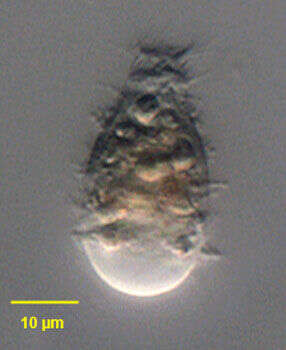
Portrait of Vasicola ciliata (Tatem, 1869), a metacystid ciliate that produces a thin, transparent pseudochitinous vase-like lorica with shallow transverse corrugations (seen in this image). In the lorica the cell body assumes a more globular shape. The circular cytostome is located in the center of the truncate anterior end. It opens into a cytopharynx supported by indistinct trichites. There are three concentric ciliary rings around the cytostome, the innermost with a single ciliary row, the middle with a double ciliary row and a third ring of four ciliary rows. The uniform longitudinal ciliary rows are formed of dikinetids kinetosomes the alignment which gives the appearance of transverse ciliary bands (paratenes). There is a sparse tuft of long caudal cilia (only one long eccentric posterior cilium is seen in the similar genus, Metacystis and another metacystid, Pelatractus, lacks caudal cilia). Multiple food vacuoles are visible in the cytoplasm. The central spherical macronucleus is not seen in this image. The base of the lorica is partly filled with the expelled contents of defecation vacuoles. A single peripheral contractile vacuole is located in the posterior third of the cell. A large, clear terminal vacuole may occur but is not seen in this image. The cell often vacates the lorica when disturbed. Vasicola ciliata is sapropelic and feeds on sulfur bacteria. Collected from stagnant freshwater pond sediment near Boise,Idaho;43°19'07.45"N 115°27'31.99"W, elev.4712 ft.;October2005. DIC optics.
-
-
Portrait of Vasicola ciliata (Tatem, 1869), a metacystid ciliate that produces a thin, transparent pseudochitinous vase-like lorica with shallow transverse corrugations (seen in this image). In the lorica the cell body assumes a more globular shape. The circular cytostome is located in the center of the truncate anterior end. It opens into a cytopharynx supported by indistinct trichites. There are three concentric ciliary rings around the cytostome, the innermost with a single ciliary row, the middle with a double ciliary row and a third ring of four ciliary rows. The uniform longitudinal ciliary rows are formed of dikinetids kinetosomes the alignment which gives the appearance of transverse ciliary bands (paratenes). There is a sparse tuft of long caudal cilia (only one long eccentric posterior cilium is seen in the similar genus, Metacystis and another metacystid, Pelatractus, lacks caudal cilia). Multiple food vacuoles are visible in the cytoplasm. The central spherical macronucleus is not seen in this image. The base of the lorica is partly filled with the expelled contents of defecation vacuoles. A single peripheral contractile vacuole is located in the posterior third of the cell. A large, clear terminal vacuole may occur but is not seen in this image. The cell often vacates the lorica when disturbed. Vasicola ciliata is sapropelic and feeds on sulfur bacteria. Collected from stagnant freshwater pond sediment near Boise,Idaho;43°19'07.45"N 115°27'31.99"W, elev.4712 ft.;October2005. Phase contrast.
-
-
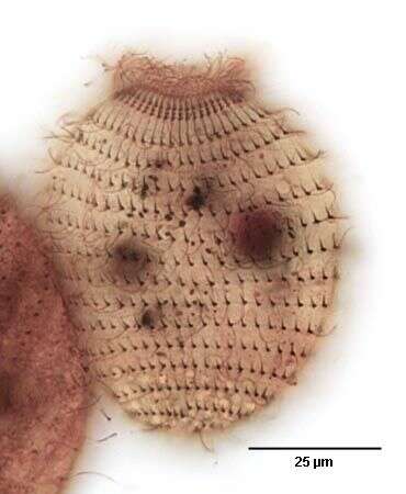
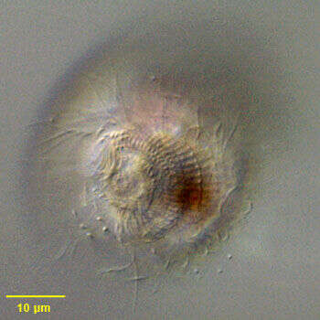
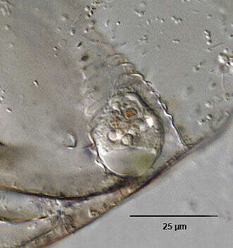
Anterior apical view of the infraciliature of Vasicola ciliata (Tatem, 1869). The cytostome is at the base of a shallow funnel-shaped depressin. There are three groups of cilia encircling the cytostome 1)the circumoral kinety (yellow arrowhead) is composed of a single row of transversely oriented dikinetids 2) the anterior row of "membranelles" composed of a single row of longitudinally oriented dikinetids (blue arrowhead 3) the posterior row of "membranelles", a band of four longitudinal orietd kinetids (green arrowhead). The second somatic kinety is indicated by the red arrowhead. Longitudinal fibrils extend from the cytostome to the right of the kinetids. Stained by the silver carbonate technique (see Foissner, W. Europ. J. Protistol., 27:313-330;1991). Brightfield.
-
Anterior apical view of the infraciliature of Vasicola ciliata (Tatem, 1869). The cytostome is at the base of a shallow funnel-shaped depressin. There are three groups of cilia encircling the cytostome 1)the circumoral kinety is composed of a single row of transversely oriented dikinetids 2) the anterior row of "membranelles" composed of a single row of longitudinally oriented dikinetids 3) the posterior row of "membranelles", a band of four longitudinal orietd kinetids. Longitudinal fibrils extend from the cytostome to the right of the kinetids. Stained by the silver carbonate technique (see Foissner, W. Europ. J. Protistol., 27:313-330;1991). Brightfield.
-
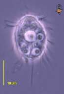
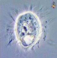
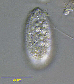
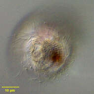
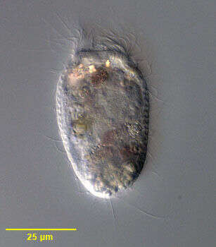
This ciliate was found in anoxic samples in relative abundance. No loricae were observed and there was a considerable range of size. The identity is tentative. Observations and image by Jeffrey Cole.
-
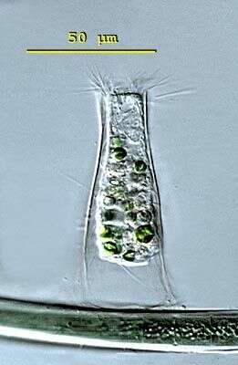
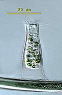
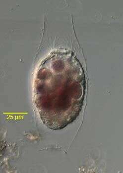
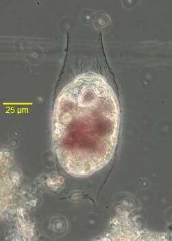
Metacystis (met-ah-sis-tiss) lagenula has a transparent lorica which is formed like an Erlenmeyer flask. The oral aperture is equiped with long pectinelles. There is a conspicuous caudal cilium and the contractile vacuole can be seen in the posterior third of the cell. This specimen was collected in freshwater ponds near Konstanz, Germany. Differencial interference contrast.
-
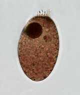
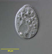
Portrait of the Prostome ciliate, Plagiocampa chaetophorae (Kahl, 1930). The cells are ellipsoid. Free-swimming cells are ovoid to cylindrical in shape. The oral aperture is an apical slit bordered on the right by multiple finger-like protoplasmic projections (seen well in this image). These projections bear extrusomes (not visible in vivo). The cytopharynx is supported by short fine trichites. The longitudinal somatic kineties are uniform. This species lacks a long caudal cilium. P. chaetophorae is distinguished from other species of Plagiocampa by a prominent layer of rod-shaped extrusomes lying perpendicular to the pellicle. The ovoid macronucleus and the adjacent micronucleus are centrally located. A single contractile vacuole is located at the posterior terminus. A food vacuole is visible posteriorly in this cell. Plagiocampa feeds on bacteria, flagellates and other ciliates. Kahl found this species associated with the thalli of the green alga Chaetophora. Collected from en ephemeral freshwater pool without Chaetophora near Boise, Idaho November 2004. DIC.
-

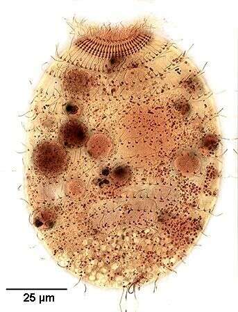
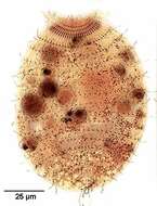
Anterior apical view of the Prostome ciliate, Plagiocampa rouxi (Kahl, 1926). Free-swimming cells are ovoid to cylindrical in shape. The oral aperture is an apical slit bordered dorsally by 8 finger-like protoplasmic projections (the dark basal bodies of these are seen well in this image but the projections themselves have been lost during fixation) and ventrally by 3 short obliquely oriented adoral membranelles (seen well here). The finger-like projections bear extrusomes, which are not visible in vivo. The cytopharynx is supported by short fine trichites (not visible in this image). The longitudinal somatic kineties (13-18 in number) are uniform (seen here). There is a single long caudal cilium. The ovoid macronucleus and the adjacent micronucleus are located just posterior to the center (densely stained here). A single contractile vacuole (not seen in this image) is located at the posterior terminus. Plagiocampa feeds on bacteria, flagellates and other ciliates. Collected from a polysaprobic farm pond with abundant Anabaena near Boise, Idaho August 2004. Silver carbonate stain (see Foissner, W.Europ. J. Protistol.27,313-330;1991). Brightfield.
-

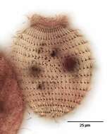
Dorsal aspect of the Prostome ciliate, Plagiocampa rouxi (Kahl, 1926). Free-swimming cells are ovoid to cylindrical in shape. The oral aperture is an apical slit bordered dorsally by 8 finger-like protoplasmic projections (seen here) and ventrally by 3 short obliquely oriented adoral membranelles (not seen in this view). The finger-like projections bear extrusomes, which are not visible in vivo. The cytopharynx is supported by short fine trichites (not visible in this image). The longitudinal somatic kineties (13-18 in number) are uniform (not seen in this image). There is a single long caudal cilium (not seen in this image). The ovoid macronucleus and the adjacent micronucleus are located just posterior to the center (densely stained here). A single contractile vacuole (not seen in this image) is located at the posterior terminus. Plagiocampa feeds on bacteria, flagellates and other ciliates. Collected from a polysaprobic farm pond with abundant Anabaena near Boise, Idaho August 2004. Silver carbonate stain (see Foissner, W.Europ. J. Protistol.27,313-330;1991). Brightfield.
-
Portrait of the Prostome ciliate, Plagiocampa rouxi(Schewiakoff, 1892). This slightly squashed individual is one of the several Plagiocampa species that lack a long caudal cilium. Free-swimming cells are ovoid to cylindrical in shape. The oral aperture is an apical slit bordered on the right by multiple finger-like protoplasmic projections (seen well in this image). These projections bear extrusomes (not visible in vivo). The cytopharynx is supported by short fine trichites (seen well in this image). The longitudinal somatic kineties are uniform. The ovoid macronucleus and the adjacent micronucleus are located just posterior to the center. A single contractile vacuole (seen in this image) is located at the posterior terminus. Multiple food vacuoles are visible in the cytoplasm of this cell. Plagiocampa feeds on bacteria, flagellates and other ciliates. Collected from a freshwater agricultural irrigation canal near Boise, Idaho November 2003. DIC optics.
-
Anterior detail of the Prostome ciliate, Plagiocampa rouxi(Schewiakoff, 1892). This slightly squashed individual is one of the several Plagiocampa species that lack a long caudal cilium. Free-swimming cells are ovoid to cylindrical in shape. The oral aperture is an apical slit bordered on the right by multiple finger-like protoplasmic projections (seen well in this image). These projections bear extrusomes (not visible in vivo). The cytopharynx is supported by short fine trichites (not seen here). The longitudinal somatic kineties are uniform. The ovoid macronucleus and the adjacent micronucleus are located just posterior to the center. A single contractile vacuole (not seen in this image) is located at the posterior terminus. Multiple food vacuoles are visible in the cytoplasm of this cell. Plagiocampa feeds on bacteria, flagellates and other ciliates. Collected from a freshwater agricultural irrigation canal near Boise, Idaho November 2003. DIC optics.
-
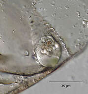
Portrait of Metacystis recurva (Penard,1922), a loricate prostomatid ciliate. The body is elongate but quite contractile. The anterior is bluntly truncate and the posterior broader and rounded. There is usually a distinctive large clear protuberant posterior vacuole but this may be lacking (as in this case) leading to confusion with the similar genus, Vasicola. The oral aperture is apical, surrounded by four rows of peribuccal cilia. The kinetids of the longitudinal kineties line up with one another to form horizontal rows called paratenes. The cell surface may be transversely furrowed along these paratenes. There is often a long laterally located posterior cilium (not seen here). The central macronucleus and posterior contractile vacuole are not well seen here. The highly refractile material in the neck of the cell is an aggregate of cytoplasmic crystals. The lorica is a narrow curved truncate cone shape open at the anterior end with 12-15 transverse corrugations (thanks to Martin Kreutz for his translation of Kahlâs species description). The lorica is nearly colorless in young individuals and becomes sepia color with age, presumably due to deposition of minerals. The overlying cladocercan shell distorts the color in this image. Loricae are often found inside the vacant shells of cladocercans. Metacystis is said to feed on sulfur bacteria. From sapropelic freshwater aquaculture tank near Boise, Idaho. DIC optics.
-
Portrait of Metacystis recurva (Penard,1922), a prostomatid ciliate. This individual has fled its lorica and is swimming free. In the lorica the body is usually elongate but quite contractile. The free-swimming individuals are typically contracted. The anterior is bluntly truncate and the posterior broader and rounded. The distinctive large, clear, protuberant posterior vacuole is seen in this image but this may sometimes be lacking leading to confusion with the similar genus, Vasicola. The oral aperture is apical, surrounded by four rows of peribuccal cilia. The kinetids of the longitudinal kineties line up with one another to form horizontal rows called paratenes. The cell surface may be transversely furrowed along these paratenes. There is often a long laterally located posterior cilium (not seen here). The central macronucleus and posterior contractile vacuole are not well seen here. The lorica is a narrow curved truncate cone shape with 12-15 transverse corrugations (thanks to Martin Kreutz for his translation of Kahlâs species description). Metacystis is said to feed on sulfur bacteria. From sapropelic freshwater aquaculture tank near Boise, Idaho. DIC optics.
-
Portrait of Metacystis recurva (Penard,1922), a loricate prostomatid ciliate. The body is elongate but as seen in this image quite contractile. The anterior is bluntly truncate and the posterior broader and rounded. There is usually a distinctive large clear protuberant posterior vacuole as seen here but this may be lacking in some individuals leading to confusion with the similar genus, Vasicola. The lorica is a narrow curved truncate cone shape with 12-15 transverse corrugations (thanks to Martin Kreutz for his translation of Kahlâs species description). The lorica is nearly colorless in young individuals and becomes sepia color with age, presumably due to deposition of minerals. The oral aperture is apical, surrounded by four rows of peribuccal cilia. The kinetids of the longitudinal kineties line up to form horizontal rows called paratenes. The cell surface may be transversely furrowed along the paratenes. There is often a long laterally located posterior cilium (not seen here). The central macronucleus and posterior contractile vacuole are not well seen here. The lorica is a narrow curved truncate cone shape open at the anterior end with 12-15 transverse corrugations (thanks to Martin Kreutz for his translation of Kahlâs species description). The lorica is nearly colorless in young individuals and becomes sepia color with age, presumably due to deposition of minerals. Loricae are often found inside the vacant shells of cladocercans. Said to feed on sulfur bacteria. From sapropelic freshwater aquaculture tank near Boise, Idaho. DIC optics.
-
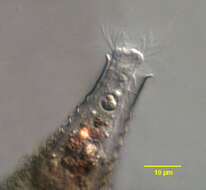
Anterior detail of Metacystis recurva (Penard,1922), a prostomatid ciliate. The oral aperture is apical, surrounded by four rows of long peribuccal cilia. The kinetids of the longitudinal kineties line up with one another to form horizontal rows called paratenes. The cell surface may be transversely furrowed along these paratenes. The highly refractile material in the neck of the cell is an aggregate of cytoplasmic crystals. The lorica is a narrow curved truncate cone shape with 12-15 transverse corrugations (thanks to Martin Kreutz for his translation of Kahlâs species description). The lorica is nearly colorless in young individuals and becomes sepia color with age, presumably due to deposition of minerals. The overlying cladocercan shell distorts the color in this image. Loricae are often found inside the vacant shells of cladocercans. ). Metacystis is said to feed on sulfur bacteria. From sapropelic freshwater aquaculture tank near Boise, Idaho. DIC optics.
-
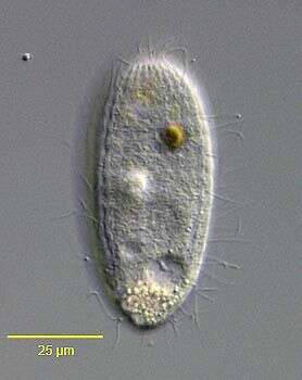
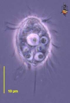
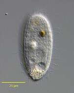
Urotricha (your-owe-trike-a) This ciliate has cilia all over the body surface, typically has a long caudal cilium, and has an ingestion area at the anterior end of the cell. It eats detritus. Common, with many species which are hard to distinguish from each other. Phase contrast.
-
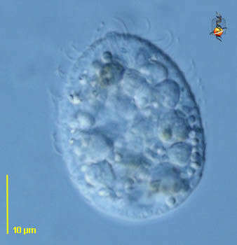
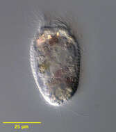
Urotricha (your-owe-trike-a) This ciliate has cilia all over the body surface, typically has a long caudal cilium, and has an ingestion area at the anterior end of the cell. It eats detritus. Common, with many species which are hard to distinguish from each other. Differential interference contrast.
-
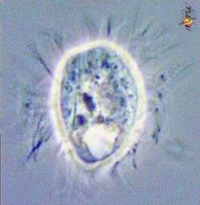
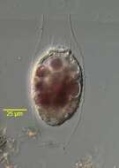
Urotricha (your-owe-trike-a) This ciliate has cilia all over the body surface, typically has a long caudal cilium, and has an ingestion area at the anterior end of the cell. It eats detritus. Common, with many species which are hard to distinguish from each other. Phase contrast.
-
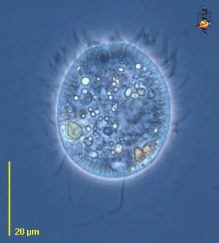
Urotricha (your-owe-trike-a) This ciliate has cilia all over the body surface, typically has a long caudal cilium, and has an ingestion area at the anterior end of the cell. It eats detritus. Common, with many species which are hard to distinguish from each other. Phase contrast.