-
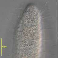
Detail view of Dexiotricha granulosa (Kent, 1881) Foissner, 1994 showing the prominent ring-shaped cytoplasmic glycogen granules. From freshwater pond near Boise, Idaho. DIC.
-
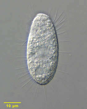
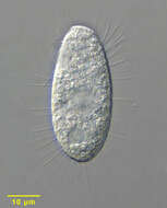
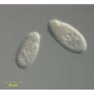
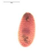
Portrait of the hymenostome ciliate, Dexiotricha granulosa (Kent, 1881) Foissner, 1994, synonymous with Loxocephulus granulosa. The cell is ovoid, broadly rounded posteriorly and truncate anteriorly. Regular longitudinal kineties terminate at a subapical band of circumferential kineties demarcating a cilia-free truncate apical area or frontal plate. There is a single long caudal cilium. The oral aperture is small and difficult to visualize in vivo. It is located in the anterior quarter with an undulating membrane on the right (seen faintly here) and 3 membranelles (not seen here). The macronucleus is spheroid and located in the mid-cell. The contractile vacuole is seen here to the left of the macronucleus. The spherical micronucleus is not seen here. The cytoplasm contains many small refractile ring-shaped glycogen granules, which are diagnostic for the species (see detail images). Dexitricha is bactiverous. From freshwater pond near Boise, Idaho. Differential interference contrast.
-
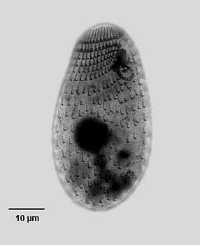
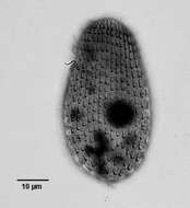
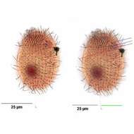
Ventrolateral view of the infraciliature of the hymenostome ciliate, Dexiotricha granulosa (Kent, 1881) Foissner, 1994. Synonym of Loxocephulus granulosa. The cell is ovoid, broadly rounded posteriorly and truncate anteriorly. Regular longitudinal kineties terminate at a subapical band of circumferential kineties demarcating a cilia-free truncate apical area or frontal plate. Fibrils radiate anteriorly from the kinetids of the anteriormost paratene (seen here). There is a single long caudal cilium. The oral aperture is small and difficult to visualize in vivo. It is located in the anterior quarter with an undulating membrane on the right (seen here) and 3 membranelles (the posterior most seen here). The macronucleus is spheroid and located in the mid-cell. Single contractile vacuole. From freshwater pond near Boise, Idaho. Silver carbonate stain (see Foissner, W. Europ. J. Protistol., 27:313-330;1991).Brightfield. Black and white.
-
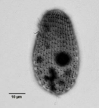
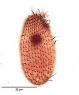
Dorsal view of the infraciliature of the hymenostome ciliate, Dexiotricha granulosa (Kent, 1881) Foissner, 1994. Synonym of Loxocephulus granulosa. The cell is ovoid, broadly rounded posteriorly and truncate anteriorly. Regular longitudinal kineties terminate at a subapical band of circumferential kineties demarcating a cilia-free truncate apical area or frontal plate. Fibrils radiate anteriorly from the kinetids of the anteriormost paratene (seen here). There is a single long caudal cilium. The oral aperture is small and difficult to visualize in vivo. It is located in the anterior quarter with an undulating membrane on the right (seen here) and 3 membranelles (the posterior most seen here). The macronucleus is spheroid and located in the mid-cell. Single contractile vacuole. From freshwater pond near Boise, Idaho. Silver carbonate stain (see Foissner, W. Europ. J. Protistol., 27:313-330;1991).Brightfield. Black and white.
-
Ventral view of the infraciliature of late division of the hymenostome ciliate, Dexiotricha granulosa (Kent, 1881) Foissner, 1994. Synonym of Loxocephulus granulosa. The cell is ovoid, broadly rounded posteriorly and truncate anteriorly. Regular longitudinal kineties terminate at a subapical band of circumferential kineties demarcating a cilia-free truncate apical area or frontal plate. Fibrils radiate anteriorly from the kinetids of the anteriormost paratene (seen here). There is a single long caudal cilium. The oral aperture is small and difficult to visualize in vivo. It is located in the anterior quarter with an undulating membrane on the right (seen here) and 3 membranelles ( seen most clearly in the proter). The macronucleus is spheroid and located in the mid-cell. Single contractile vacuole. From freshwater pond near Boise, Idaho. Silver carbonate stain (see Foissner, W. Europ. J. Protistol., 27:313-330;1991).Brightfield. Black and white.
-
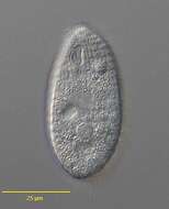
Portrait of the hymenostome ciliate, Dexiotricha granulosa (KENT,1881) FOISSNER, 1994, synonymous with Loxocephulus granulosus. The cell is ovoid, broadly rounded posteriorly and truncate anteriorly. Regular longitudinal kineties terminate at a subapical band of circumferential kineties demarcating a cilia-free truncate apical area or frontal plate. There is a single long caudal cilium. The oral aperture is small and difficult to visualize in vivo. It is located in the anterior quarter with an undulating membrane on the right (seen faintly here) and 3 membranelles (not seen here). The macronucleus is spheroid and located in the mid-cell. The contractile vacuole is seen here to the left of the macronucleus. The spherical micronucleus is not seen here. The cytoplasm contains many small refractile ring-shaped glycogen granules, which are diagnostic for the species (see detail images). Dexitricha is bactiverous. From freshwater pond near Boise, Idaho. Differential interference contrast.
-

Ventrolateral view of the infraciliature of the hymenostome ciliate, Dexiotricha granulosa (Kent, 1881) Foissner, 1994. Synonym of Loxocephulus granulosa. The cell is ovoid, broadly rounded posteriorly and truncate anteriorly. Regular longitudinal kineties terminate at a subapical band of circumferential kineties demarcating a cilia-free truncate apical area or frontal plate. Fibrils radiate anteriorly from the kinetids of the anteriormost paratene. There is a single long caudal cilium (green line). The oral aperture is small and difficult to visualize in vivo. It is located in the anterior quarter with a paraoral membrane on the right (red line) and 3 adoral membranelles (dark blue lines). Closely spaced basal bodies of the somatic kinaty to the right of the oral aperture form a "pseudomembrane" (light blue line). The macronucleus is spheroid and located in the mid-cell. Single contractile vacuole. From freshwater pond near Boise, Idaho. Silver carbonate stain (see Foissner, W. Europ. J. Protistol., 27:313-330;1991).Brightfield.
-
-
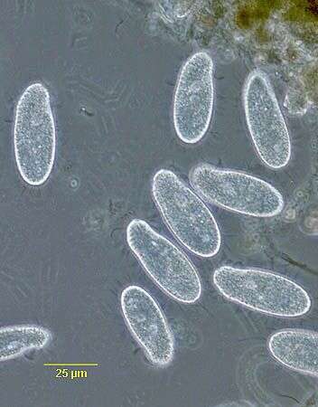
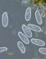
Dexiotricha tranquilla (Kahl,1926). DIC.
-
Dexiotricha tranquilla (Kahl,1926). Phase contrast.
-
Dexiotricha tranquilla (Kahl,1926).Brightfield.