-
-
-
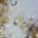
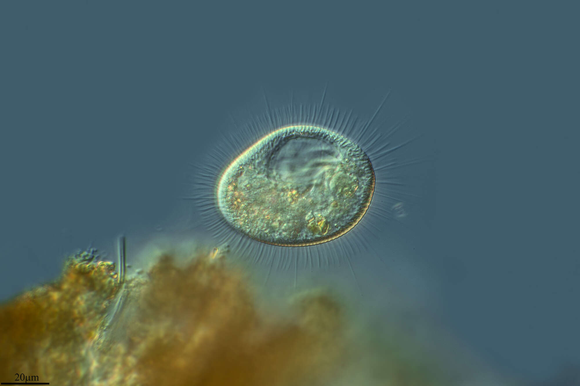
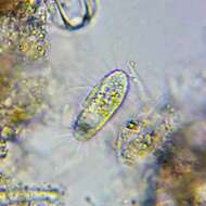
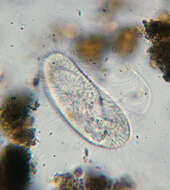
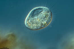
[taxonomy:genus=Cyclidium]
Date:
12 Aug 2011
Location:
Permanently wet longkang beside NUS Enterprise Incubator, under shade of large banyan tree. Drain full of leaf litter and shed banyan roots. Water clear, not green, grey-brown floc when bottom and litter stirred.
Microscope:
Bright-field with closed condenser aperture.
Camera:
Nikon D7000
Collector:
Brandon Seah
-
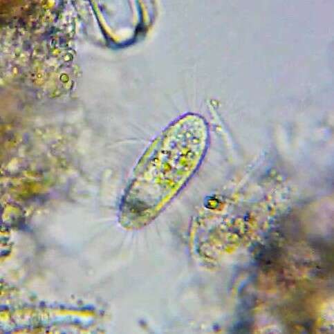
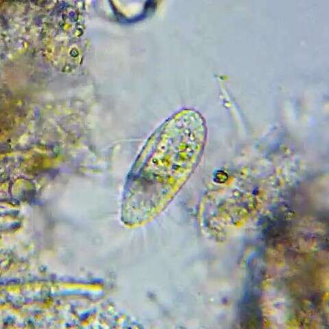
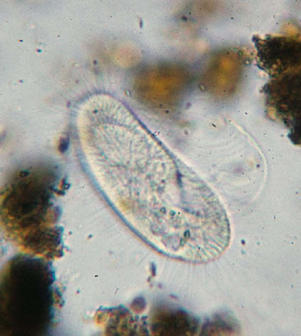
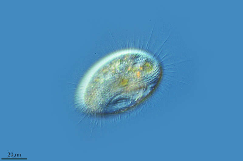
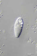
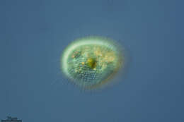
Longitude (deg): -0.7. Latitude (deg): 51.1. Longitude (deg/min): 0° 50' W. Latitude (deg/min): 51° 10' N. Vice county name: Surrey. Vice county no.: 17. Country: England. Associated species: Marchantia polymorpha. Identified by: Malcolm Storey. Comment: with Marchantia polymorpha. Category: standard photograph or close-up. Photographic equipment used: "35mm transparencies (on a variety of films, but Agfa CT18 in the 1960's to early 1980's followed by Fujichrome in the late 1980's.) Transparencies scanned with Minolta Dimage Scan Dual II AF-2820U transparency scanner.".
-
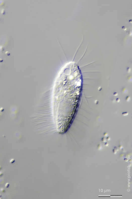
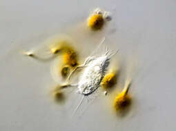
Ctedoctema acanthocryptum Scale bar indicates 10 µm. Collected from a pond near Lemkendorf, isle of Fehmarn (Baltic Sea). The image was built up using several photomicrographic frames with manual stacking technique. The images were taken using Zeiss Axioplan with Canon EOS 70D.Image under Creative Commons License V 3.0 (CC BY-NC-SA). Place name: Pond in Lemkendorf, isle Fehmarn (Baltic Sea, Germany) Latitude: 54.472296 Longitude: 11.095662 Multiebenen-Abbildung, manuell gestapelt. Der Messbalken markiert eine Länge von 10 µm. Probe aus dem Dorfteich Lemkendorf, Insel Fehmarn. Mikrotechnik: Zeiss Axioplan, Kamera: Canon EOS 70D. Creative Commons License V 3.0 (CC BY-NC-SA). For permission to use of (high-resolution) images please contact postmaster@protisten.de.
-
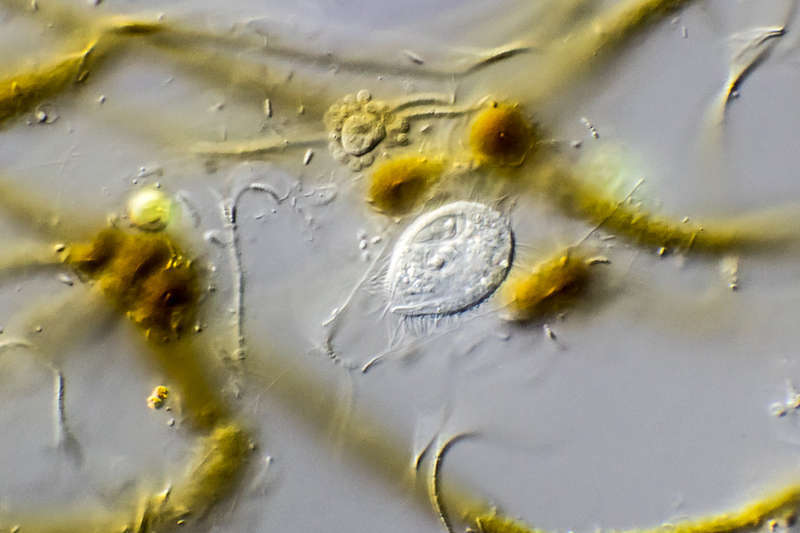
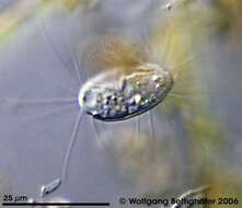
Cristigera setosa This small ciliat belonging to group of scuticociliates/hymenostomata is not easy to photograph. The picture shows the sail-like undulating membran. Sample collected from Simmelried near Konstanz (Baden-Wuerttemberg, Germany). This image was taken using Zeiss Universal with Olympus C7070 CCD camera.Image under Creative Commons License V 3.0 (CC BY-NC-SA). Place name: Bog Hegne Moor near Lake Constance (Germany) Latitude: 47.718106 Longitude: 9.093974 Dieser kleine Ciliat gehört zur Gruppe der Scuticociliaten / Hymenostomata, er ist nicht leicht zu fotografieren. Das Bild zeigt seine große adorale Membranellenzone, die wie ein Segel aussieht. Probe aus dem Simmelried nahe Konstanz. Mikrotechnik: Zeiss Universal, Kamera: Olympus C7070. Creative Commons License V 3.0 (CC BY-NC-SA). For permission to use of (high-resolution) images please contact postmaster@protisten.de.
-
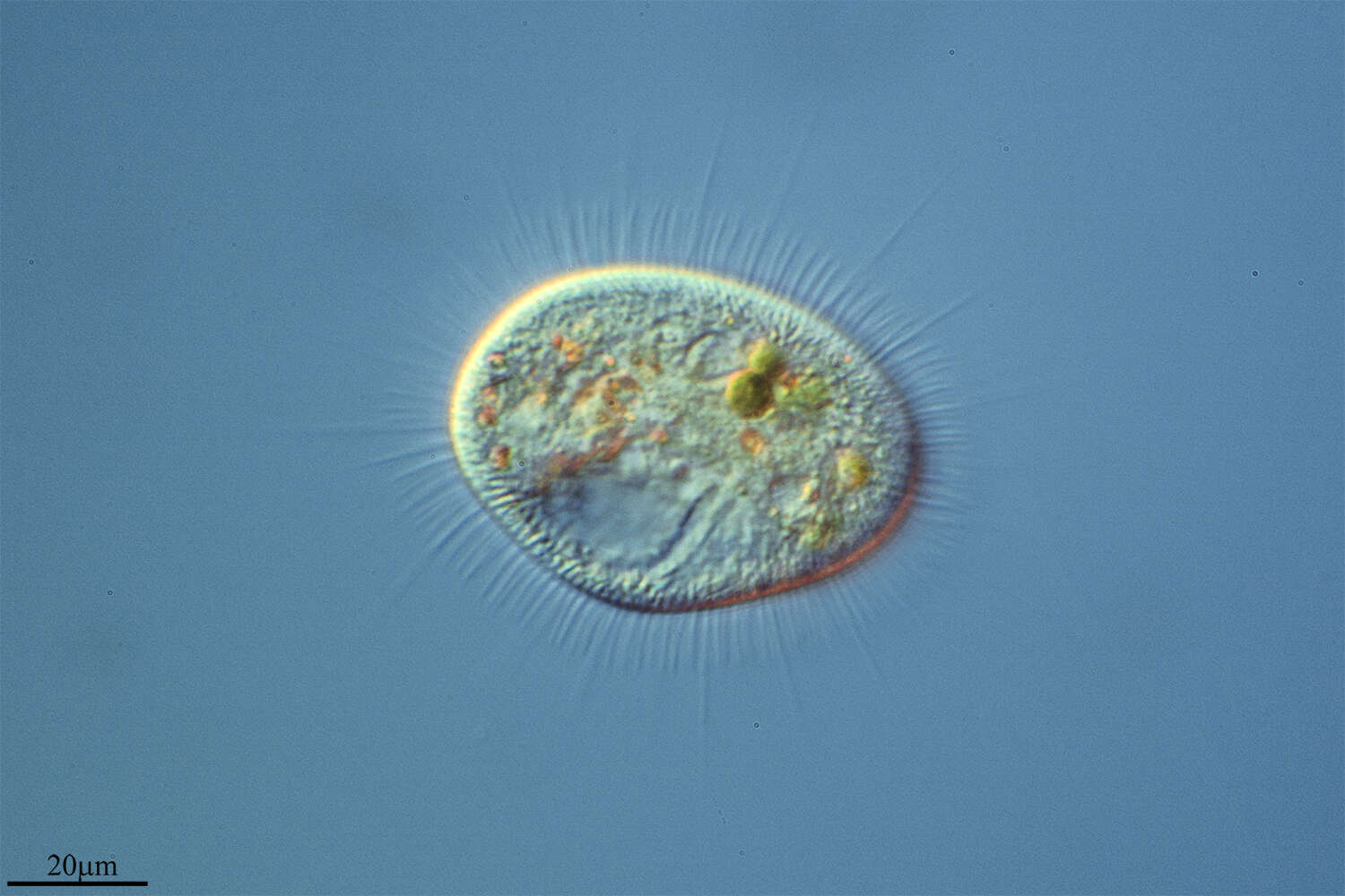
Calyptotricha lanuginosa The specimen was gathered in the pond Birkensee near Rödelsee (Lower Franconia, Germany). Sampling date 7/2018.Copyright Dr. Rainer Meisch, Würzburg, Germany.Images were taken using Zeiss Axioplan with Canon DSLR Image under Creative Commons License V 3.0 (CC BY-NC-SA). Place name: Pond Birkensee near Rödelsee (Lower Franconia, Germany) Latitude: 49.71819841 Longitude: 10.27807474 Probe aus dem Birkensee bei Rödelsee (Unterfranken). Datum der Aufsammlung: 7/2018. Copyright Dr. Rainer Meisch, Würzburg. Mikrotechnik: Zeiss Axioplan, Kamera: Canon DSLR. Creative Commons License V 3.0 (CC BY-NC-SA). For permission to use of (high-resolution) images please contact postmaster@protisten.de.
-
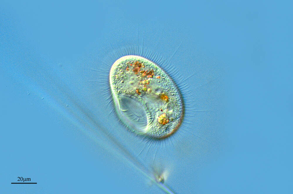
Calyptotricha lanuginosa The specimen was gathered in the pond Birkensee near Rödelsee (Lower Franconia, Germany). Sampling date 7/2018.Copyright Dr. Rainer Meisch, Würzburg, Germany.Images were taken using Zeiss Axioplan with Canon DSLR Image under Creative Commons License V 3.0 (CC BY-NC-SA). Place name: Pond Birkensee near Rödelsee (Lower Franconia, Germany) Latitude: 49.71819841 Longitude: 10.27807474 Probe aus dem Birkensee bei Rödelsee (Unterfranken). Datum der Aufsammlung: 7/2018. Copyright Dr. Rainer Meisch, Würzburg. Mikrotechnik: Zeiss Axioplan, Kamera: Canon DSLR. Creative Commons License V 3.0 (CC BY-NC-SA). For permission to use of (high-resolution) images please contact postmaster@protisten.de.
-
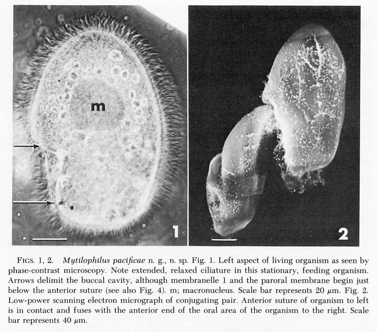
The species now known as Peniculistoma mytili was first described as Conchopthirus mytili by William De Morgan in 1925 in an article in the Journal of the Marine Biological Association (vol 13, 600-660).
-
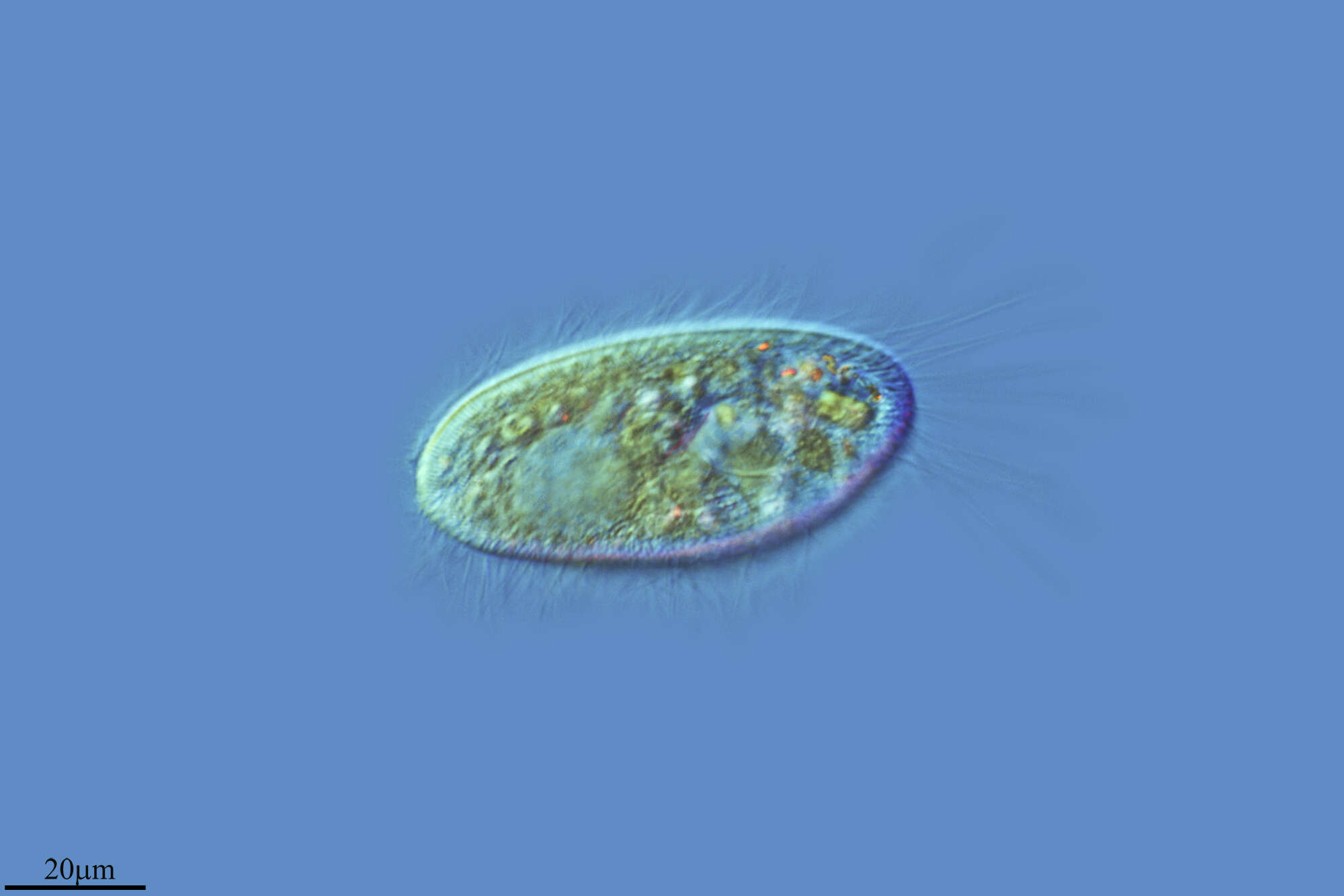
Image of Peniculistoma mytili (endocommensal of the blue mussel Mytilis edulis) using DIC optics. Image by G. A. Antipa.
-
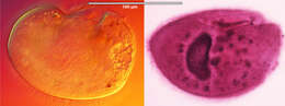
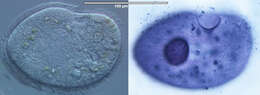
Left image: Live specimen imaged using DIC optics; Right image: protargol-stained specimen. Images by G. A. Antipa
-
Canencia, Madrid, Spain
-
Galende, Castille and Leon, Spain
-
Hoyo de Manzanares, Madrid, Spain
-
Ribadelago de Franco, Castille and Leon, Spain
-
Hoyo de Manzanares, Madrid, Spain
-
Canencia, Madrid, Spain
-
Hoyo de Manzanares, Madrid, Spain
-
Logroo, La Rioja, Espaa
-
Talveila, Castille and Leon, Spain
-
Images from the species description by Antipa & Dolan (1985)
-
Left panel: live specimen imaged with DIC optics (image by G.A. Antipa); right panel: protargol-stained specimen (image by D.H. Lynn).
-
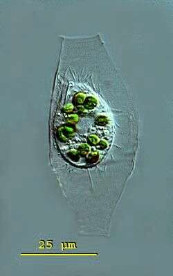
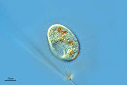
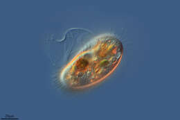
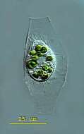
Calyptotricha (kah-lip-toe-trike-a) pleuronemoides is an ovoid to pyriform ciliate. The ciliate forms a transparent lorica. The lorica is tube-like and has apertures at both ends. The middle the tube can have parallel sides or a central bulbous region in which the ciliate is housed. The undulating membrane of the oral aperture stretches down the right side of the body to form a pouch in the posterior body half. Extrusomes are present. There is a conspicuous caudal cilium. Contractile vacuole in posterior body region. The macronucleus is spherical with attached micronuclei. Several endosymbiotic algae are visible and the conspicuous caudal cilium. Ciliate measuring 28 microns, lorica 64 microns. This specimen was collected in freshwater ponds near Konstanz, Germany. Differencial interference contrast.