-
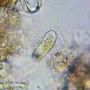
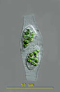
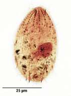
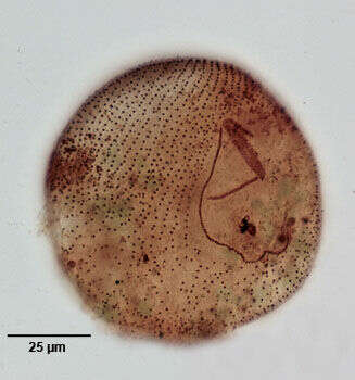
Calyptotricha (kah-lip-toe-trike-a) pleuronemoides is an ovoid to pyriform ciliate. The ciliate forms a transparent lorica. The lorica is tube-like and has apertures at both ends. The middle the tube can have parallel sides or a central bulbous region in which the ciliate is housed. The undulating membrane of the oral aperture stretches down the right side of the body to form a pouch in the posterior body half. Extrusomes are present. There is a conspicuous caudal cilium. Contractile vacuole in posterior body region. The macronucleus is spherical with attached micronuclei. This image taken shortly after cell division when there are two specimens in the lorica. Lorica measuring 68 microns. This specimen was collected in freshwater ponds near Konstanz, Germany. Differential interference contrast.
-
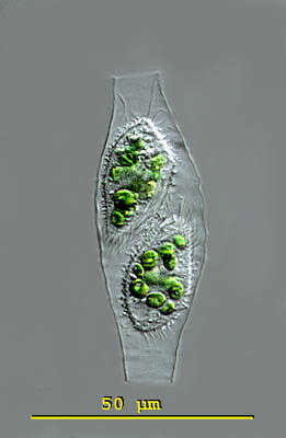
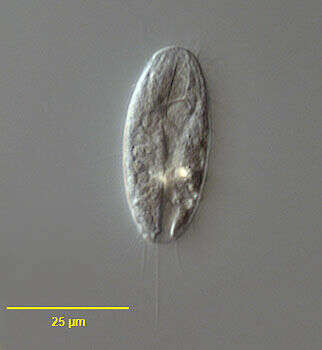
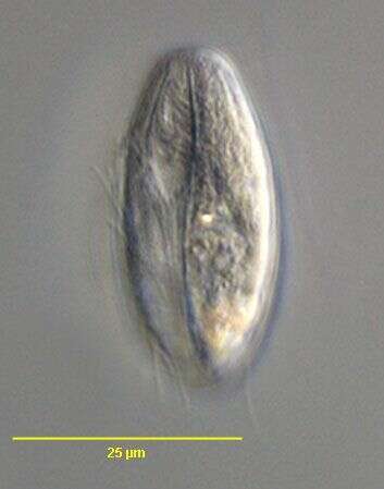
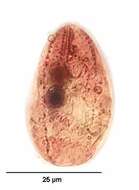
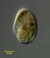
Portrait of the loricate pleuronematid ciliate, Calyptotricha pleuronemoides (Phillips, 1882). The transparent lorica of this species is open at both ends and dilated in the center where the cell resides. The cell is bluntly pointed anteriorly and broadly rounded posteriorly. The peristome is about 3/4 cell length. There is a prominent undulating membrane on the right margin of the peristome curving around its posterior end to form a shallow pouch (seen well here). There are three inconspicuous adoral membranelles. The longitudinal somatic kineties are uniformly distributed. There is a preoral and postoral suture. There is a single long caudal cilium. There is a single posterior contractile vacuole. The spherical macronucleus is centrally located. C. lanuginosa is similar in appearance of the cell except that it has two long anterior apical cilia and a cylindrical lorica with parallel sides. Collected from a freshwater dredge pond near Idaho City, Idaho June 2003. DIC.
-
Portrait of the loricate pleuronematid ciliate, Calyptotricha pleuronemoides (Phillips, 1882). The transparent lorica of this species is open at both ends and dilated in the center where the cell resides. The cell is bluntly pointed anteriorly and broadly rounded posteriorly. The peristome is about 3/4 cell length. There is a prominent undulating membrane on the right margin of the peristome curving around its posterior end to form a shallow pouch. There are three inconspicuous adoral membranelles. The longitudinal somatic kineties are uniformly distributed. There is a preoral and postoral suture. There is a single long caudal cilium. There is a single posterior contractile vacuole. The spherical macronucleus is centrally located. C. lanuginosa is similar in appearance of the cell except that it has two long anterior apical cilia and a cylindrical lorica with parallel sides. Collected from a freshwater dredge pond near Idaho City, Idaho June 2003. DIC.
-
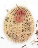
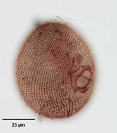
Dorsal infraciliature of Calyptotricha pleuronemoides (PHILLIPS,1882). Collected from organically enriched stagnant water at the edge of a freshwater stream near Boise, Idaho.Stained by the silver carbonate technique (Foissner,W. Europ. J. Protistol.27:313-330;1991).Brightfield.
-
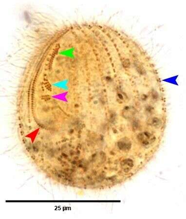
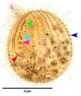
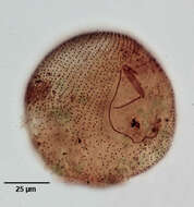
Ventral infraciliature of Calyptotricha pleuronemoides (PHILLIPS,1882). The red arrowhead marks the kinetids of the first adoral membranelle (M1). The light blue and pink arrowheads mark the kinetids of M2 and M3 respectively. the red arrowhead marks the right paraoral (undulating) membrane. The dark blue arrow head marks a dikinetid of a somatic kinety.Collected from organically enriched stagnant water at the edge of a freshwater stream near Boise, Idaho.Stained by the silver carbonate technique (Foissner,W. Europ. J. Protistol.27:313-330;1991).Brightfield.
-
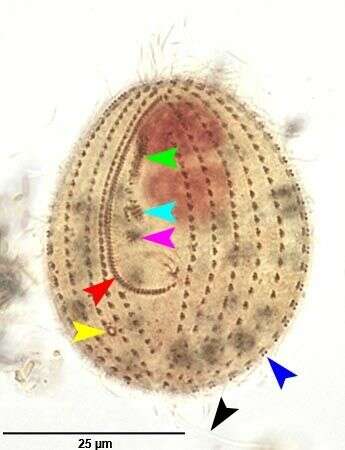
Ventral infraciliature of Calyptotricha pleuronemoides (PHILLIPS,1882). The red arrowhead marks the kinetids of the first adoral membranelle (M1). The light blue and pink arrowheads mark the kinetids of M2 and M3 respectively. the red arrowhead marks the right paraoral (undulating) membrane. The dark blue arrow head marks a dikinetid of a somatic kinety. the black arrowhead marks the single long caudal cilium. The yellow arrowhead marks the excretory pore of the contractile vacuole.Collected from organically enriched stagnant water at the edge of a freshwater stream near Boise, Idaho.Stained by the silver carbonate technique (Foissner,W. Europ. J. Protistol.27:313-330;1991).Brightfield.
-
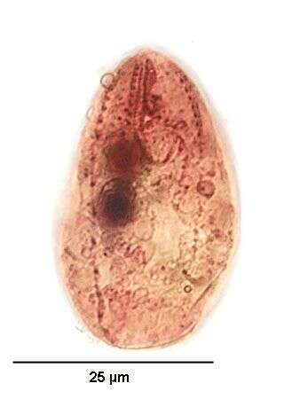
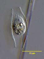
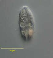
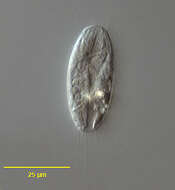
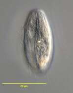
Portrait (ventral surface) of the pleuronematine scuticociliate, Cristigera phoenix (Penard, 1922). Collected from a freshwater aquaculture pond near Boise, Idaho November 2004. DIC.
-
Portrait (ventral surface) of the pleuronematine scuticociliate, Cristigera phoenix (Penard, 1922). Collected from a freshwater aquaculture pond near Boise, Idaho November 2004. DIC.
-
Stained by the silver carbonate technique (see Foissner, W. Europ. J. Protistol., 27:313-330;1991).Brightfield.
-
Stained by the silver carbonate technique (see Foissner, W. Europ. J. Protistol., 27:313-330;1991).Brightfield.
-
Stained by the silver carbonate technique (see Foissner, W. Europ. J. Protistol., 27:313-330;1991).Brightfield.
-
Portrait (ventral surface) of the pleuronematine scuticociliate, Cristigera phoenix (Penard, 1922). Collected from a freshwater aquaculture pond near Boise, Idaho November 2004. DIC.
-
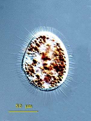
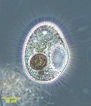
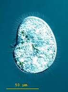
Histiobalantium is easy to distinguish from the similar genus Pleuronema by the stiff long cilia which intersperse the dense somatic ciliation because Histiobalantium have cilia distributed over the whole cell whereas at Pleuronema this cilia are confined to the posterior region. The body of Histiobalantium natans is elliptical, with the right side slightly concave and anterior end a little narrower than the posterior. The peristome is deep, equipped with a well-developed undulating membrane. Several micronuclei are attached to the two round macronuclei. The contractile vacuole is located in the posterior half of the body. 55 - 105 microns. From fresh water ponds and lakes. This free-swimming specimen of Histiobalantium natans was collected in freshwater ponds near Konstanz, Germany The contractile vacuole is located in the posterior. 95 microns. Differential interference contrast.
-
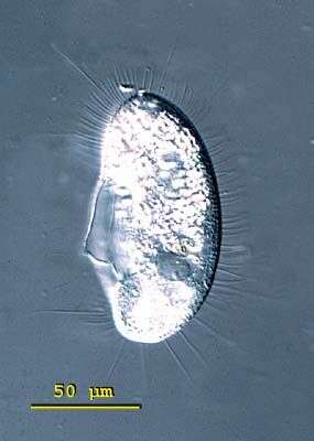
Histiobalantium is easy to distinguish from the similar genus Pleuronema by the stiff long cilia which intersperse the dense somatic ciliation because Histiobalantium have cilia distributed over the whole cell whereas at Pleuronema this cilia are confined to the posterior region. The body of Histiobalantium natans is elliptical, with the right side slightly concave and anterior end a little narrower than the posterior. The peristome is deep, equipped with a well-developed undulating membrane. Several micronuclei are attached to the two round macronuclei. The contractile vacuole is located in the posterior half of the body. 55 - 105 microns. From fresh water ponds and lakes. This specimen of Histiobalantium natans shows the stiff long cilia around the cell. 95 (m. From freshwater ponds near Konstanz, Germany. Differential interference contrast.
-
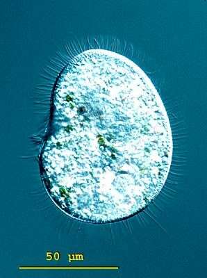
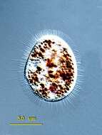
Histiobalantium is easy to distinguish from the similar genus Pleuronema by the stiff long cilia which intersperse the dense somatic ciliation because Histiobalantium have cilia distributed over the whole cell whereas at Pleuronema this cilia are confined to the posterior region. The body of Histiobalantium natans is elliptical, with the right side slightly concave and anterior end a little narrower than the posterior. The peristome is deep, equipped with a well-developed undulating membrane. Several micronuclei are attached to the two round macronuclei. The contractile vacuole is located in the posterior half of the body. 55 - 105 microns. From fresh water ponds and lakes. Well-fed specimen of Histiobalantium natans from a freshwater pond near Konstanz, Germany. 88 m. icrons Differential interference contrast.
-
Histiobalantium is easy to distinguish from the similar genus Pleuronema by the stiff long cilia which intersperse the dense somatic ciliation because Histiobalantium have cilia distributed over the whole cell whereas at Pleuronema this cilia are confined to the posterior region. The body of Histiobalantium natans is elliptical, with the right side slightly concave and anterior end a little narrower than the posterior. The peristome is deep, equipped with a well-developed undulating membrane. Several micronuclei are attached to the two round macronuclei. The contractile vacuole is located in the posterior half of the body. 55 - 105 microns. From fresh water ponds and lakes. Well-fed specimen of Histiobalantium natans from a freshwater pond near Konstanz, Germany. 88 microns. Differential interference contrast.
-
Histiobalantium is easy to distinguish from the similar genus Pleuronema by the stiff long cilia which intersperse the dense somatic ciliation. Histiobalantium have long cilia distributed over the whole cell whereas at Pleuronema this cilia are confined to the posterior region. The body of Histiobalantium natans is elliptical, with the right side slightly concave and anterior end a little narrower than the posterior. The peristome is deep, equipped with a well-developed undulating membrane. Several micronuclei are attached to the two round macronuclei. The contractile vacuole is located in the posterior half of the body. 55 - 105 microns long. From fresh water ponds and lakes. This free-swimming specimen of Histiobalantium natans was collected in freshwater ponds near Konstanz, Germany. The contractile vacuole is located in the posterior. 95 microns. Differential interference contrast.
-
Histiobalantium is easy to distinguish from the similar genus Pleuronema by the stiff long cilia which intersperse the dense somatic ciliation. Histiobalantium have long cilia distributed over the whole cell whereas at Pleuronema this cilia are confined to the posterior region. The body of Histiobalantium natans is elliptical, with the right side slightly concave and anterior end a little narrower than the posterior. The peristome is deep, equipped with a well-developed undulating membrane. Several micronuclei are attached to the two round macronuclei. The contractile vacuole is located in the posterior half of the body. 55 - 105 microns long. From fresh water ponds and lakes. This specimen of Histiobalantium natans shows the stiff long cilia around the cell. 95 microns. From freshwater ponds near Konstanz, Germany. Differential interference contrast.
-
Histiobalantium is easy to distinguish from the similar genus Pleuronema by the stiff long cilia which intersperse the dense somatic ciliation. Histiobalantium have long cilia distributed over the whole cell whereas at Pleuronema this cilia are confined to the posterior region. The body of Histiobalantium natans is elliptical, with the right side slightly concave and anterior end a little narrower than the posterior. The peristome is deep, equipped with a well-developed undulating membrane. Several micronuclei are attached to the two round macronuclei. The contractile vacuole is located in the posterior half of the body. 55 - 105 microns long. From fresh water ponds and lakes. This well-fed specimen of Histiobalantium natans is from a freshwater pond near Konstanz, Germany. 88 microns. Differential interference contrast.
-
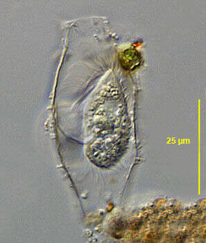
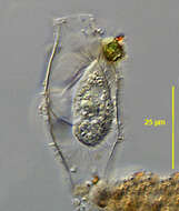
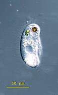
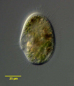
Histiobalantium natans(Claparede & Lachmann, 1858), a large scuticociliate - with long stiff cilia extending outwards from the body surface. This individual has ingested a Trachelomonas. Collected from a freshwater pond near Boise, Idaho.Phase contrast.
-
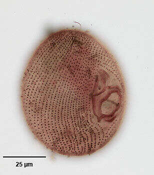
Ventral infraciliature of the hymenostome ciliate, Histiobalantium natans (Claparede & Lachmann, 1858). The cell is ovoid to reniform in outline with a broadly rounded posterior. The oval oral aperture is located in the mid-portion of the cell. There is an undulating membrane on the right margin of the peristome curving around the posteriorly located cytostome to form a cup or pouch. There are three adoral membranelles on the left side of the peristome. M1 and M2 are parallel and oriented obliquely to the undulating membrane while membranelle M3 is inclined slightly posteriorly between the posterior ends of M1 and M2 and the undulating membrane, forming a triangle. The somatic ciliature is composed of longitudinal kineties. Longer bristle-like cilia are interspersed with more numerous short cilia. Both pre- and post-oral sutures are present. A long caudal cilium absent. There are three contractile vacuoles. Macronucleus with an irregular shape, usually divided into two parts with several micronuclei. Similar in appearance to Pleuronema which may have long caudal cilia but whose other somatic cilia are of equal length. Stained by the silver carbonate technique (see Foissner, W. Europ. J. Protistol., 27:313-330;1991). Collected from a freshwater pond near Boise, Idaho, february 2005. Brightfield.
-
Portraitof the hymenostome ciliate, Histiobalantium natans (Claparede & Lachmann, 1858). The cell is ovoid to reniform in outline with a broadly rounded posterior. The oval oral aperture is located in the mid-portion of the cell. There is an undulating membrane on the right margin of the peristome curving around the posteriorly located cytostome to form a cup or pouch. There are three adoral membranelles on the left side of the peristome. M1 and M2 are parallel and oriented obliquely to the undulating membrane while membranelle M3 is inclined slightly posteriorly between the posterior ends of M1 and M2 and the undulating membrane, forming a triangle. The somatic ciliature is composed of longitudinal kineties. Longer bristle-like cilia are interspersed with more numerous short cilia. Both pre- and post-oral sutures are present. A long caudal cilium absent. There are three contractile vacuoles. Macronucleus with an irregular shape, usually divided into two parts with several micronuclei. Similar in appearance to Pleuronema which may have long caudal cilia but whose other somatic cilia are of equal length. Stained by the silver carbonate technique (see Foissner, W. Europ. J. Protistol., 27:313-330;1991). Collected from a freshwater pond near Boise, Idaho, february 2005.DIC.
-
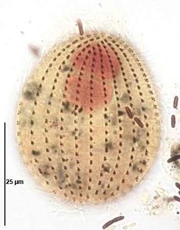
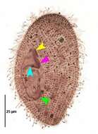
Oral infraciliature of the hymenostome ciliate, Histiobalantium natans (Claparede & Lachmann, 1858). The cell is ovoid to reniform in outline with a broadly rounded posterior. The oval oral aperture is located in the mid-portion of the cell. There is an undulating membrane on the right margin of the peristome curving around the posteriorly located cytostome to form a cup or pouch. There are three adoral membranelles on the left side of the peristome. M1 and M2 are parallel and oriented obliquely to the undulating membrane while membranelle M3 is inclined slightly posteriorly between the posterior ends of M1 and M2 and the undulating membrane, forming a triangle. The somatic ciliature is composed of longitudinal kineties. Longer bristle-like cilia are interspersed with more numerous short cilia. Both pre- and post-oral sutures are present. A long caudal cilium absent. There are three contractile vacuoles. Macronucleus with an irregular shape, usually divided into two parts with several micronuclei. Similar in appearance to Pleuronema which may have long caudal cilia but whose other somatic cilia are of equal length. Stained by the silver carbonate technique (see Foissner, W. Europ. J. Protistol., 27:313-330;1991). Collected from a freshwater pond near Boise, Idaho, february 2005. Brightfield.
-

Oral infraciliature of the hymenostome ciliate, Histiobalantium natans (Claparede & Lachmann, 1858). The cell is ovoid to reniform in outline with a broadly rounded posterior. The oval oral aperture is located in the mid-portion of the cell. There is an undulating membrane on the right margin of the peristome curving around the posteriorly located cytostome to form a cup or pouch (green arrowhead). There are three adoral membranelles on the left side of the peristome. M1 and M2 are parallel and oriented obliquely to the undulating membrane (yellow and pink arrowheads respectively) while membranelle M3 is inclined slightly posteriorly between the posterior ends of M1 and M2 and the undulating membrane, forming a triangle (light blue arrowhead). The somatic ciliature is composed of longitudinal kineties. Longer bristle-like cilia are interspersed with more numerous short cilia. Both pre- and post-oral sutures are present. A long caudal cilium absent. There are three contractile vacuoles. Macronucleus with an irregular shape, usually divided into two parts with several micronuclei. Similar in appearance to Pleuronema which may have long caudal cilia but whose other somatic cilia are of equal length. Stained by the silver carbonate technique (see Foissner, W. Europ. J. Protistol., 27:313-330;1991). Collected from a freshwater pond near Boise, Idaho, february 2005. Brightfield.