-
Anatoly B. Babenko, Ayuna B. Chimitova, Sophya K. Stebaeva
Zookeys
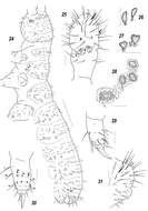
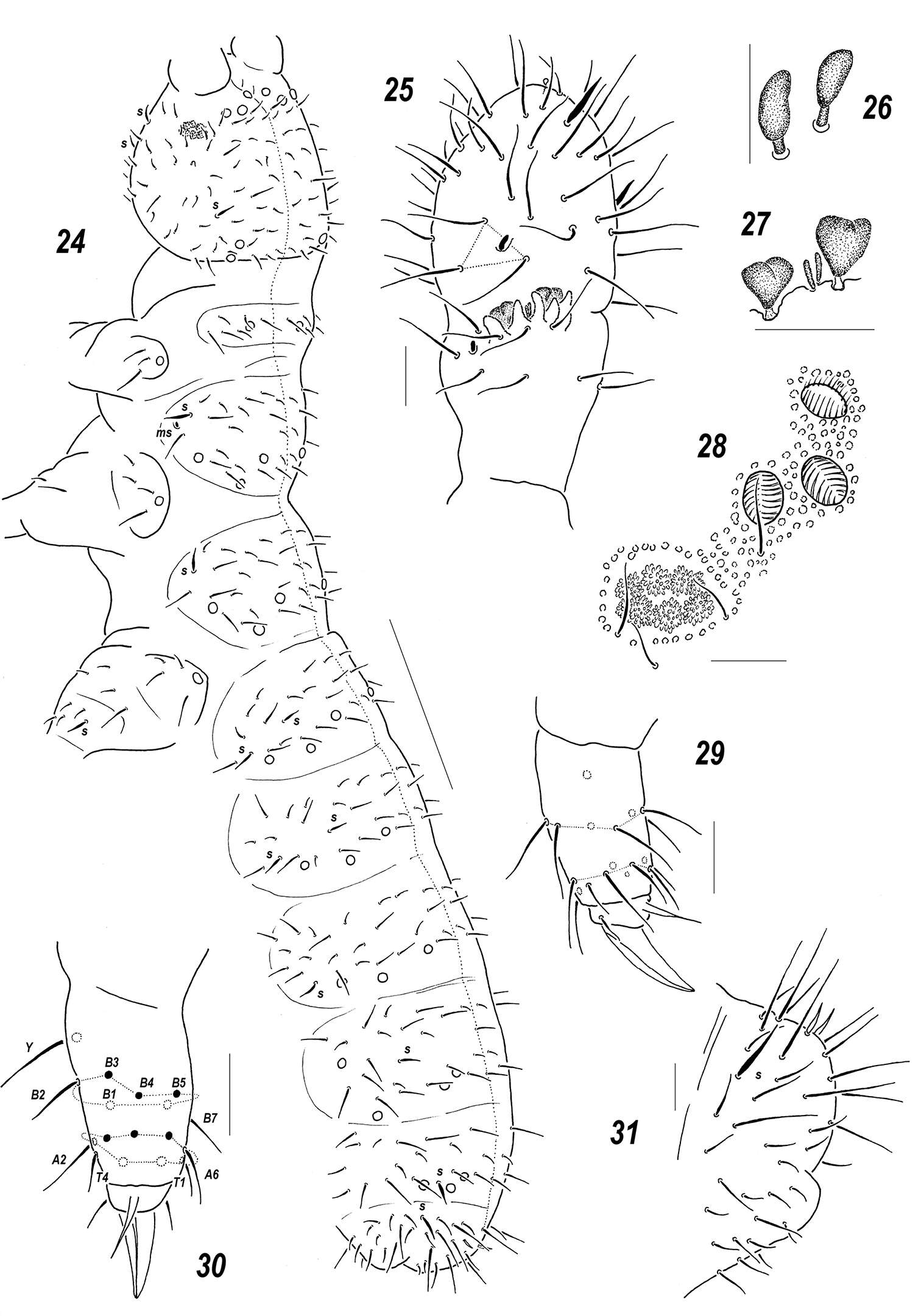
Figures 24–31.Sensillonychiurus amuricus sp. n. 24 dorsal chaetotaxy 25 Ant.3–4 26–27 sensorial elements of Ant.3 organ, different view 28 PAO and adjacent pso 29–30 tibiotarsus of Lg.3, different views 31 Abd.6. Scales: 24 – 0.1 mm, 25–31 – 0.01 mm.
-
José G. Palacios-Vargas, Hugo H. Mejía-Madrid
Zookeys
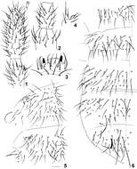
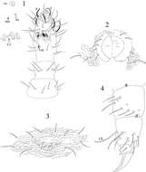
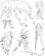
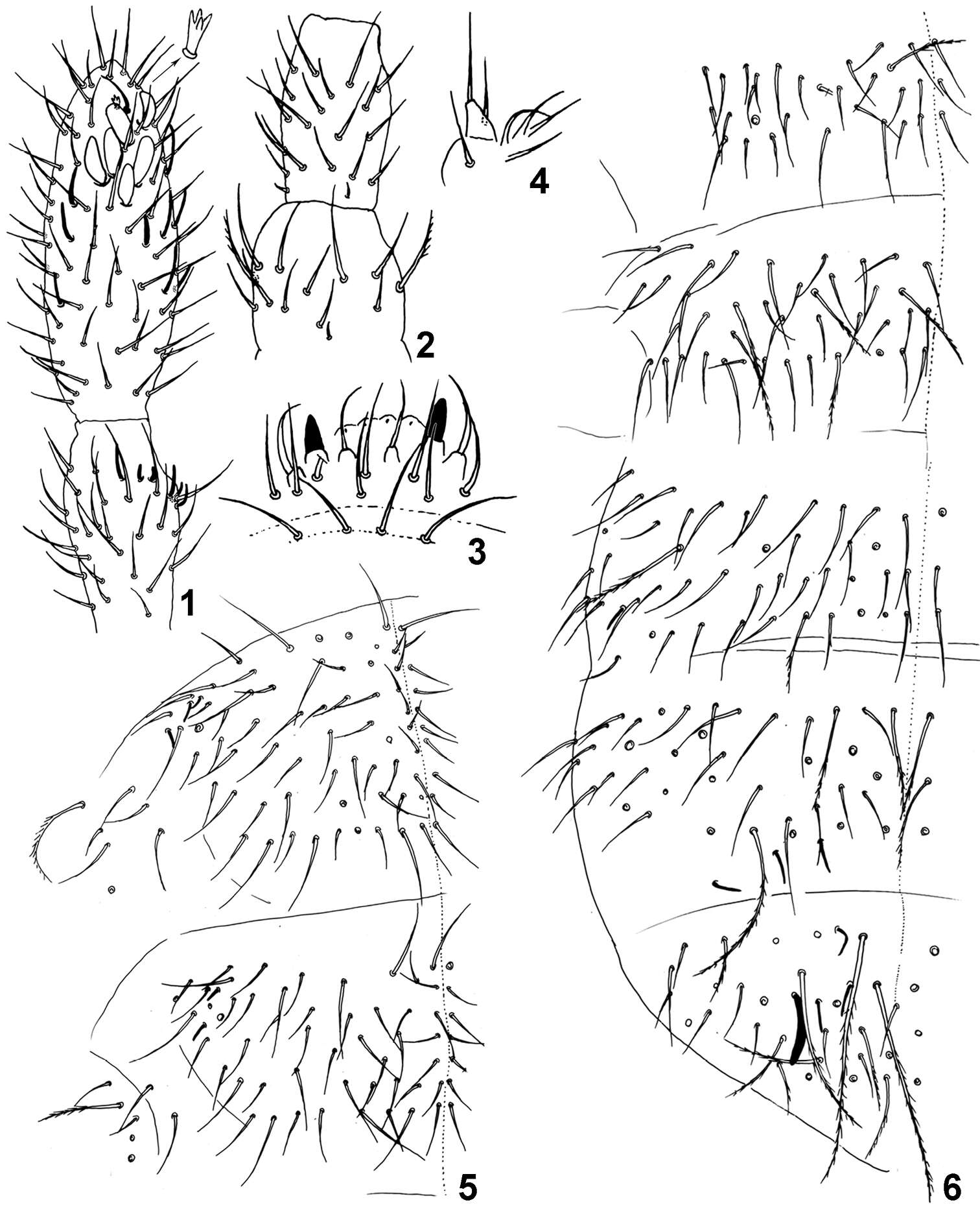
Figures 1–6.Pseudachorutes nica sp. n. 1 right antenna from II to IV dorsal view 2 Ant. III and IV in ventral view with magnification of some setae from ventral file 3 mandible 4 maxilla 5 labium 6 dorsal chaetotaxy of the head and thorax I (thorax has a drawing style represents granulation close to setae).
-
Maria Cleide de Mendonça, Eduardo A. Abrantes, Ana Carolina R. Neves
Zookeys
Figure 1–6.Isotomiella macedoi sp.n. 1 Ant III-IV Dorsal view, detail of the apical microsensillum 2 Ant I-II Dorsal view 3 Labral and prelabral chaetae 4 Outer lobe of maxilla 5 Dorsal chaetotaxy of Th II-III 6 Dorsal chaetotaxy of Abd I-VI.
-
Gabriel C. Queiroz, Maria Cleide de Mendonça
Zookeys
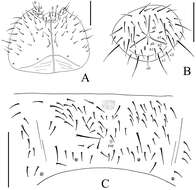
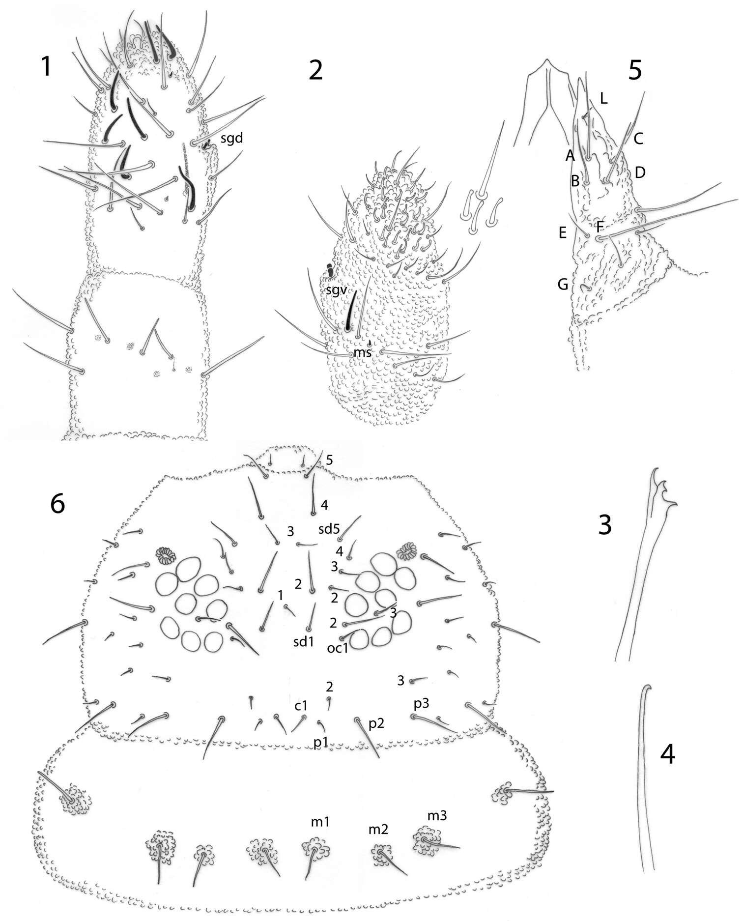
Figures 1–8.Micronella itacaman sp. n. 1 Dorsal view of Ant I–IV 2. Ventral view of Ant I–IV 3 PAO and its surrounding chaetae 4 Maxilla 5 Labium 6 Head chaetotaxy of specimen from Itatiaia 7 Head chaetotaxy of specimen from Teresópolis 8 Head chaetotaxy of specimen from Alto Caparaó. Scale bars: 10μm (1–5); 20 μm (6–8).
-
Xiang-Qun Yuan, Zhi-Xiang Pan
Zookeys
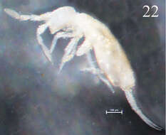
Figure 22.Habitus of Sinella triseta sp. n.
-
Xin Sun, Louis Deharveng, Donghui Wu
Zookeys
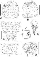
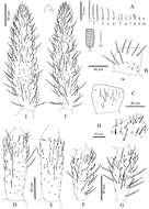
Figure 1.Thalassaphorura problematica sp. n. A dorsal side of body B ventral side of Abd. I–VI C PAO D clubs and papillae of AIIIO E Labium F Antenna. Scales: 0.1 mm (A–B, F), 0.01 mm (C–E).
-
Daoyuan Yu, Feng Zhang, Louis Deharveng
Zookeys
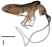
Figure 1.Tomocerus postantennalis sp. n. Appearance in alcohol. Scale bar: 1000 μm.
-
José G. Palacios-Vargas, Ana E. Salazar Martínez
Zookeys
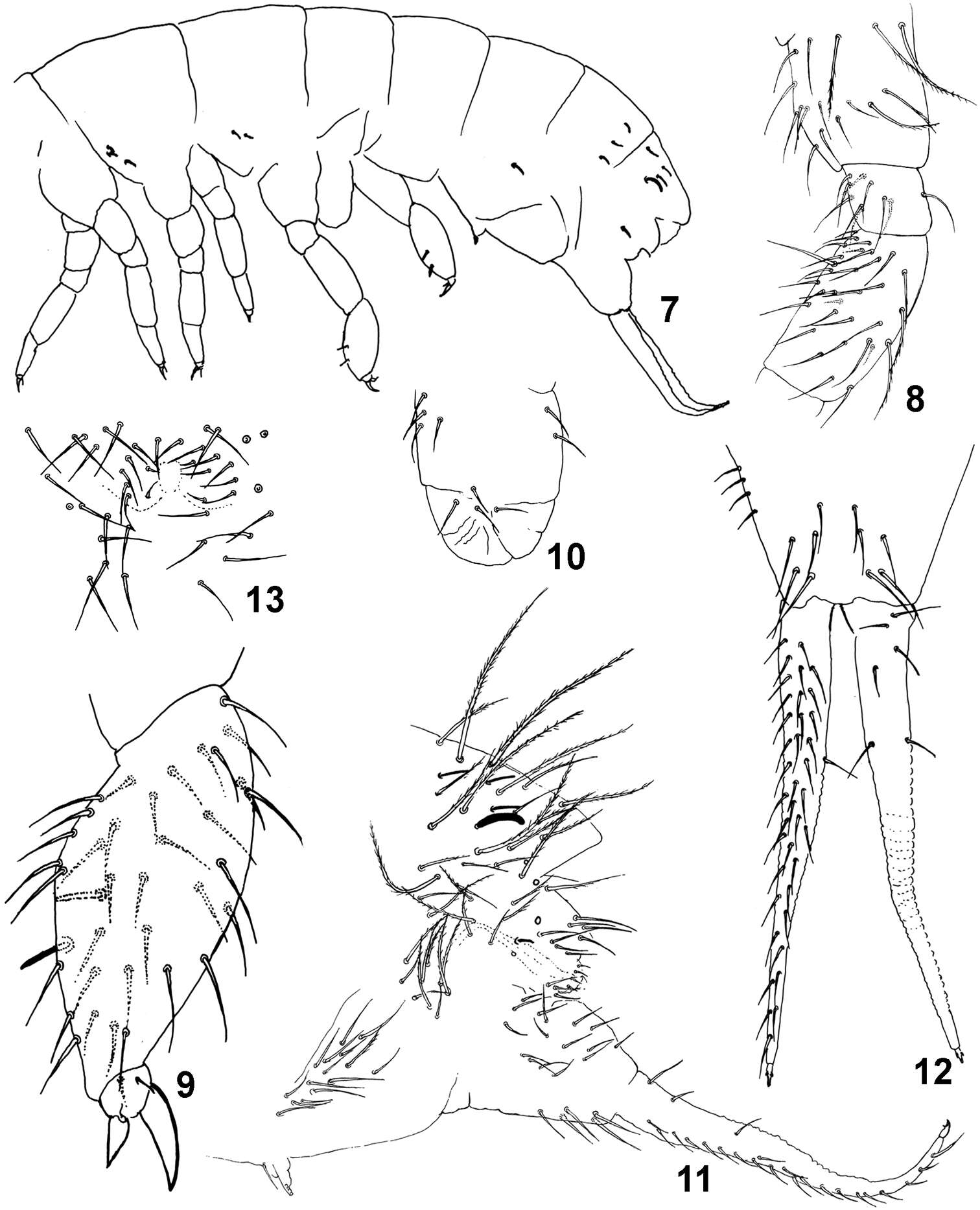
Figures 1–4.Tullbergia alcirae sp. n. 1 antennal segments I to IV with details of sensorial structures 2 ventral tube 3 female genital plate 4 femur and tibiotarsus III.
-
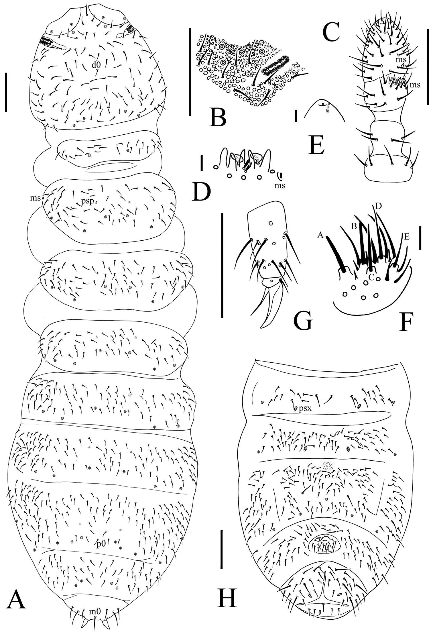
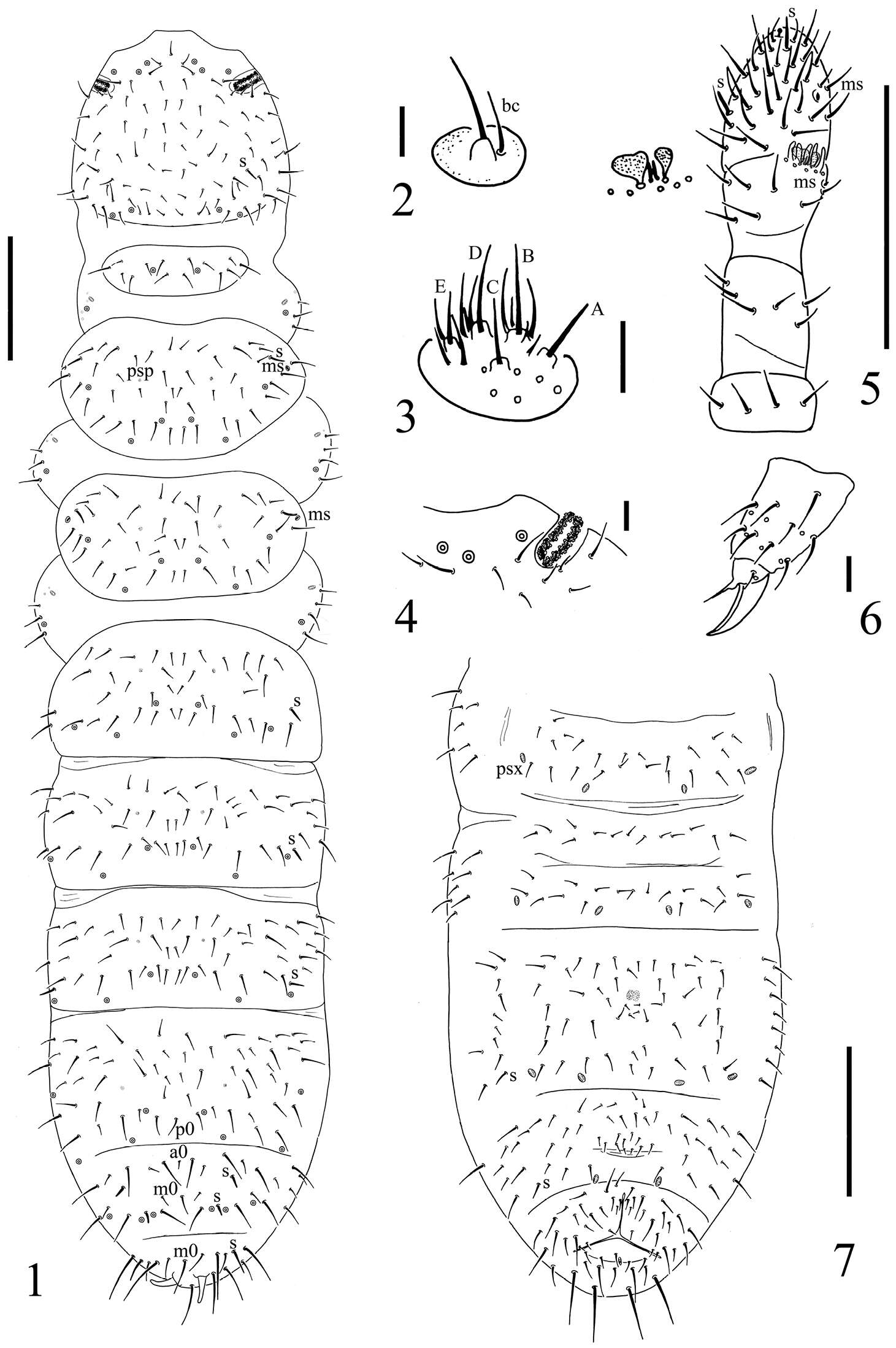
Figure 3.Onychiurus heilongjiangensis sp. n. A dorsal side of body B PAO C Ant. I–IV D Ant. III sensory organ E antennal tip F labium G distal part of leg III H Abd. II–VI sterna. Scale bars: 0.1 mm (A, C, G–H), 0.01 mm (B, D–F).
-
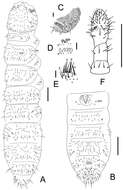
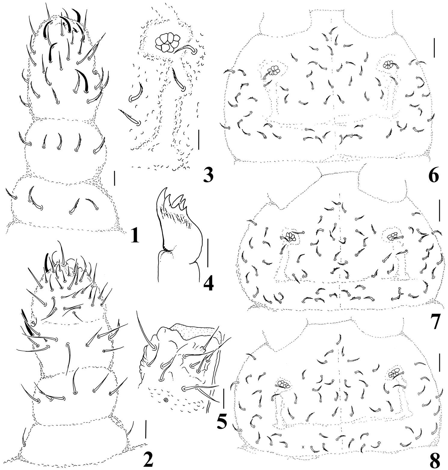
Figure 1.Spinonychiurus sinensis sp. n. A dorsal chaetotaxy of body B maxillary palp C labium D postantennal organ E antenna F distal part of leg III G chaetotaxy of Abd. II–VI sterna. Scale bars: 0.1 mm (A, E, G), 0.01 mm (B–D, F).
-
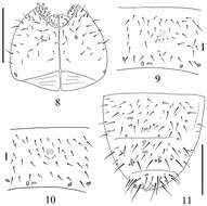
José G. Palacios-Vargas, Hugo H. Mejía-Madrid
Zookeys
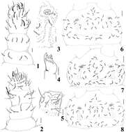
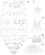
Figures 7–10.Pseudachorutes nica sp. n. 7 tibiotarsus III and unguis in ventral view 8 dorsal chaetotaxy of Th. II and III and Abd. I–VI (drawing style represents granulation close to setae) 9 furcula 10 female genital plate 11 male genital plate.
-
Maria Cleide de Mendonça, Eduardo A. Abrantes, Ana Carolina R. Neves
Zookeys
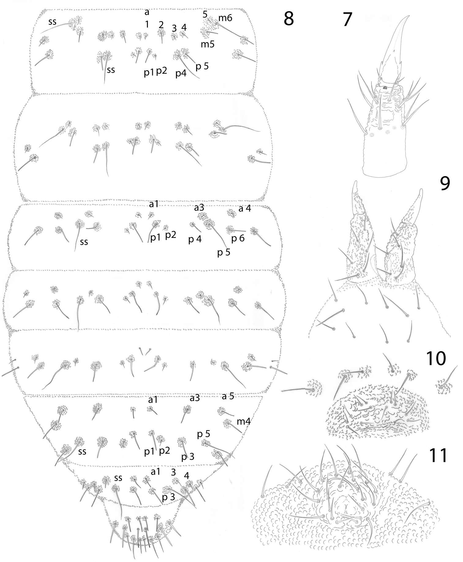
Figure 7–13.Isotomiella macedoi sp.n. 7 Sensillary pattern of the body 8 Subcoxa and femur of leg III 9 Tibiotarsus and unguis of leg III 10 Lateral view of ventral tube 11 Lateral view of abd. V-VI, subcoxa furcal, furca and tenaculum 12 Furca 13 Male genital plate.
-
Gabriel C. Queiroz, Maria Cleide de Mendonça
Zookeys
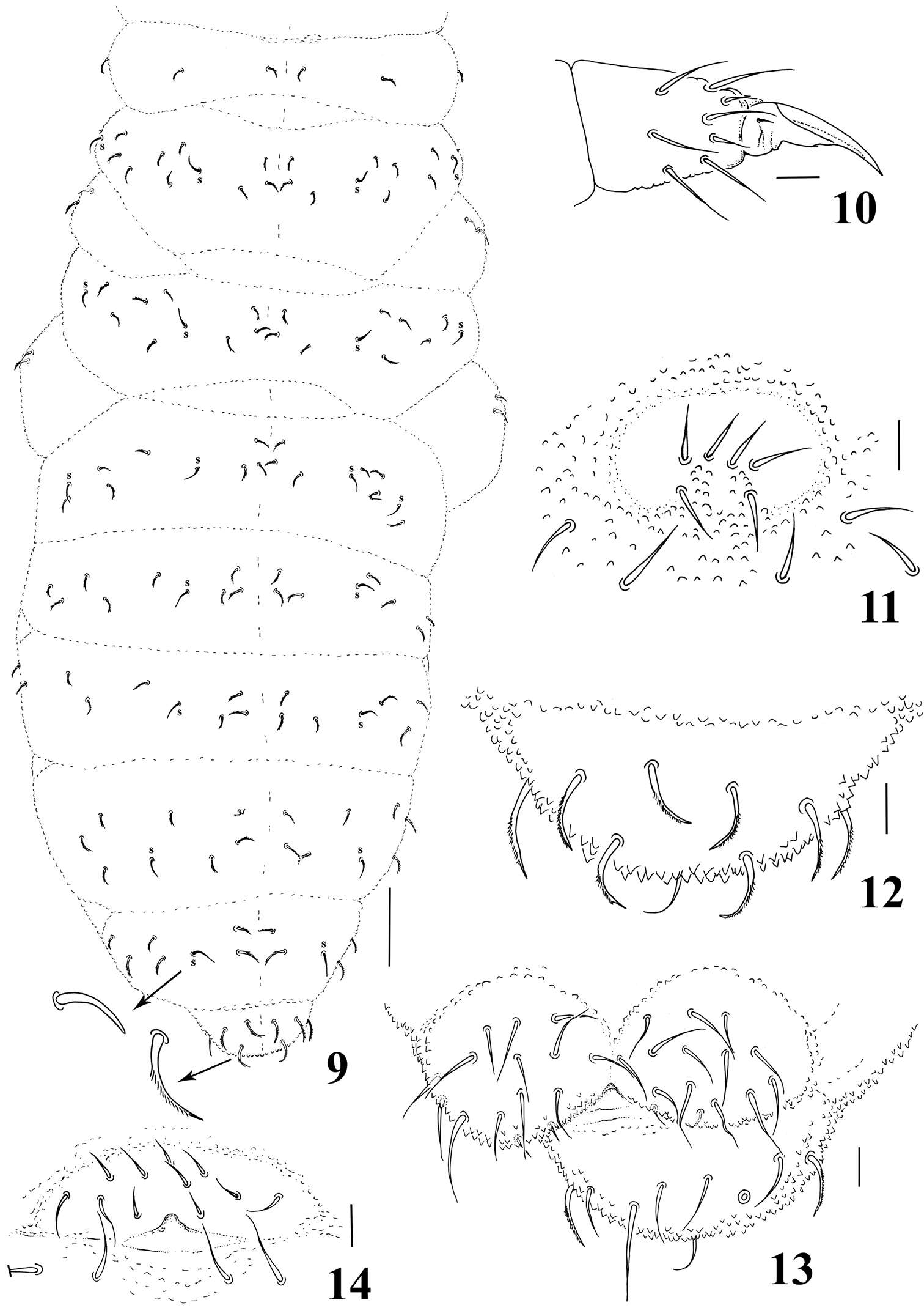
Figures 9–14.Micronella itacaman sp. n. 9. Dorsal body chaetotaxy with details of sensilla and chaetae 10 Tita of leg I 11 Furcal area 12 Dorsal view of Abd VI 13 Anal valves and ventral view of Abd VI 14 Female genital plate. Scale bars: 10μm (10–14); 50μm (9).
-
Xiang-Qun Yuan, Zhi-Xiang Pan
Zookeys
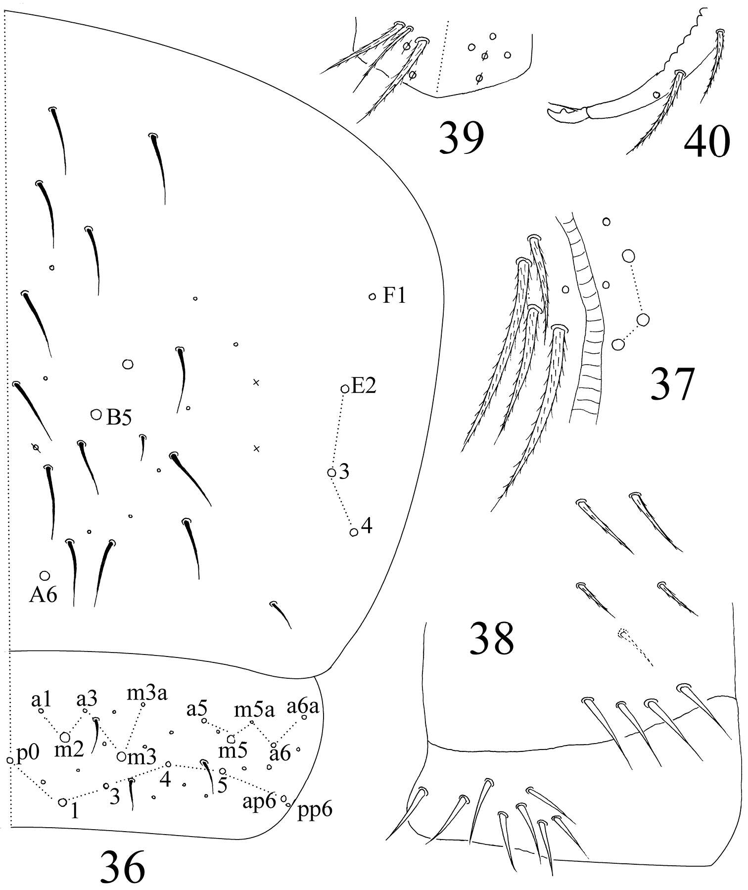
Figures 36–40.Sinella triseta sp. n. 36 dorsal chaetotaxy of Abd. IV–V 37 anterior face of VT 38 posterior face and lateral flap of VT 39 manubrial plaque 40 apical dentes and mucro.
-
Xin Sun, Louis Deharveng, Donghui Wu
Zookeys
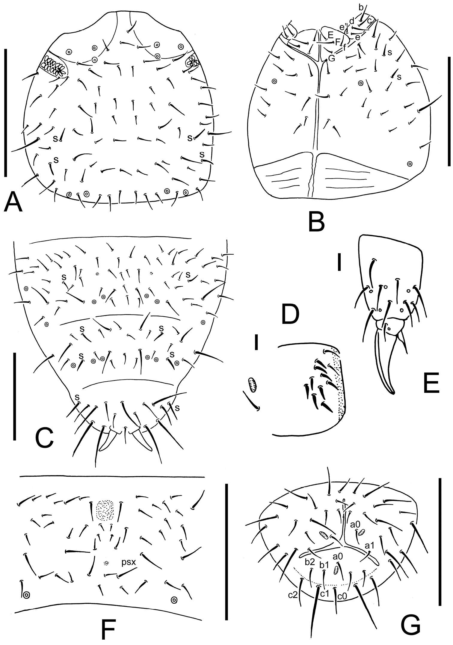
Figure 2.Thalassaphorura problematica sp. n. A dorsal side of head B ventral side of head C Abd. IV–VI terga D ventral tube (showing male ventral organ) E distal part of leg III F furca G anal valves. Scales: 0.1 mm (A–C and F–G), 0.01 mm (D–E)
-
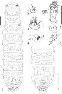
Daoyuan Yu, Feng Zhang, Louis Deharveng
Zookeys
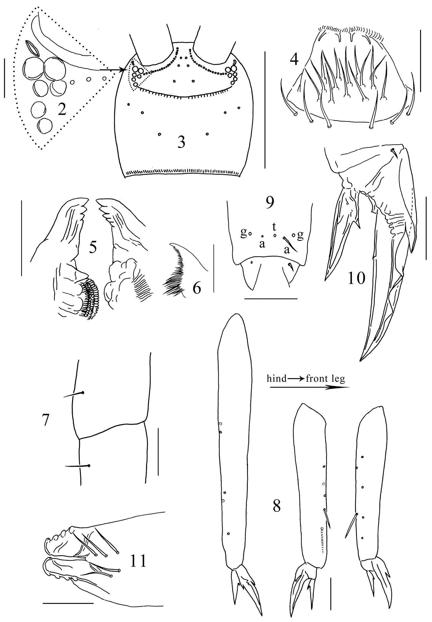
Figures 2–11.Tomocerus postantennalis sp. n. 2 PAO and ocelli 3 cephalic dorsal chaetotaxy 4 labrum 5 mandible 6 maxillary lamella five 7 trochanteral-femoral organ 8 tibiotarsus 9 anterior view of distal tibiotarsal chaetae (t: tenent hair, a: accessory chaetae, g: guard chaetae) 10 claw 11 tenaculum. Scale bars: 2, 7, 9, 10, 11 = 50 μm; 3 = 500 μm; 4, 5, 8 = 100 μm; 6= 20 μm.
-
José G. Palacios-Vargas, Ana E. Salazar Martínez
Zookeys
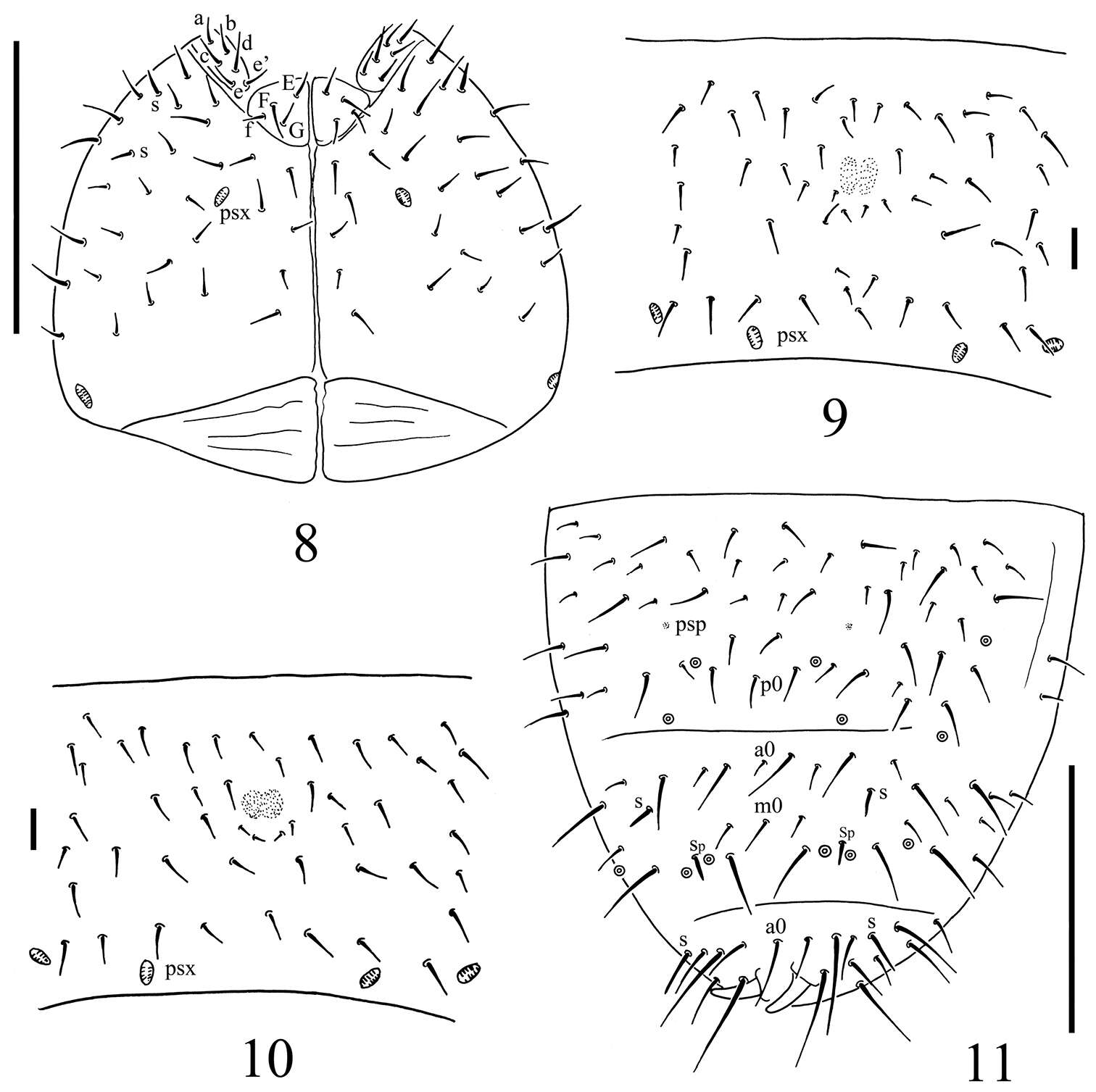
Figures 5–8.Tullbergia alcirae sp. n. 5 dorsal head and thoracic chaetotaxy 6 male genital plate 7 right half of labial and postlabial quetotaxy 8 dorsal abdominal cheatotaxy.
-
Figure 4.Onychiurus heilongjiangensis sp. n. A ventral side of head B anal valves C Abd. IV sternum. Scale bars: 0.1 mm (A–C).
-
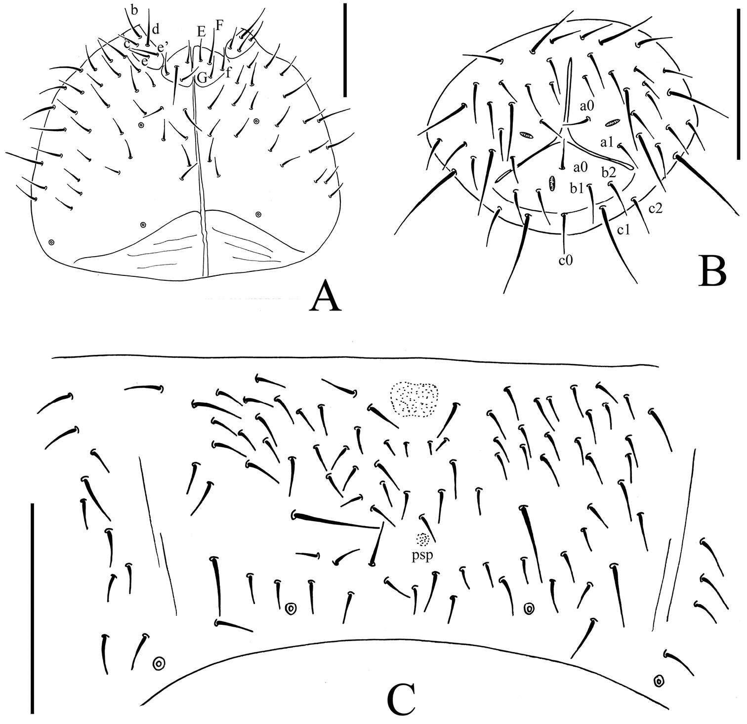
Figure 2.Spinonychiurus sinensis sp. n. A ventral chaetotaxy of head B–C central part of abdominal sternum IV D dorsal chaetotaxy of Abd. IV–VI. Scale bars: 0.1 mm (A, D), 0.01 mm (B–C).
-
Maria Cleide de Mendonça, Eduardo A. Abrantes, Ana Carolina R. Neves
Zookeys
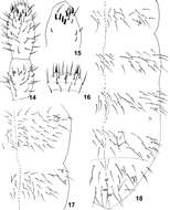
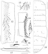
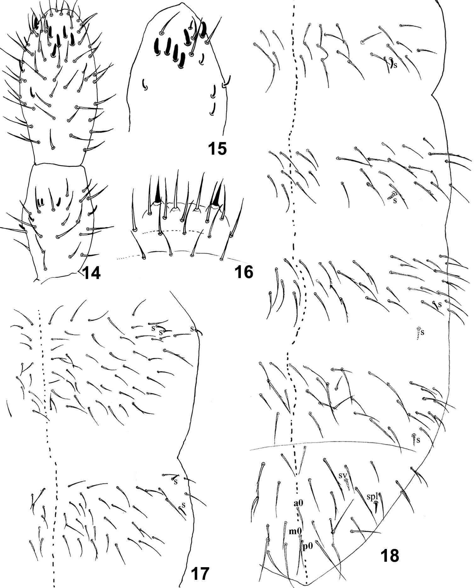
Figure 14–18.Isotomiella uaisp.n. 14 Ant III-IV Dorsal view 15 Sensillary pattern of Ant IV 16 Labral chaetae 17 Dorsal chaetotaxy of Th II-III 18 Dorsal chaetotaxy of Abd I-VI.
-
Gabriel C. Queiroz, Maria Cleide de Mendonça
Zookeys
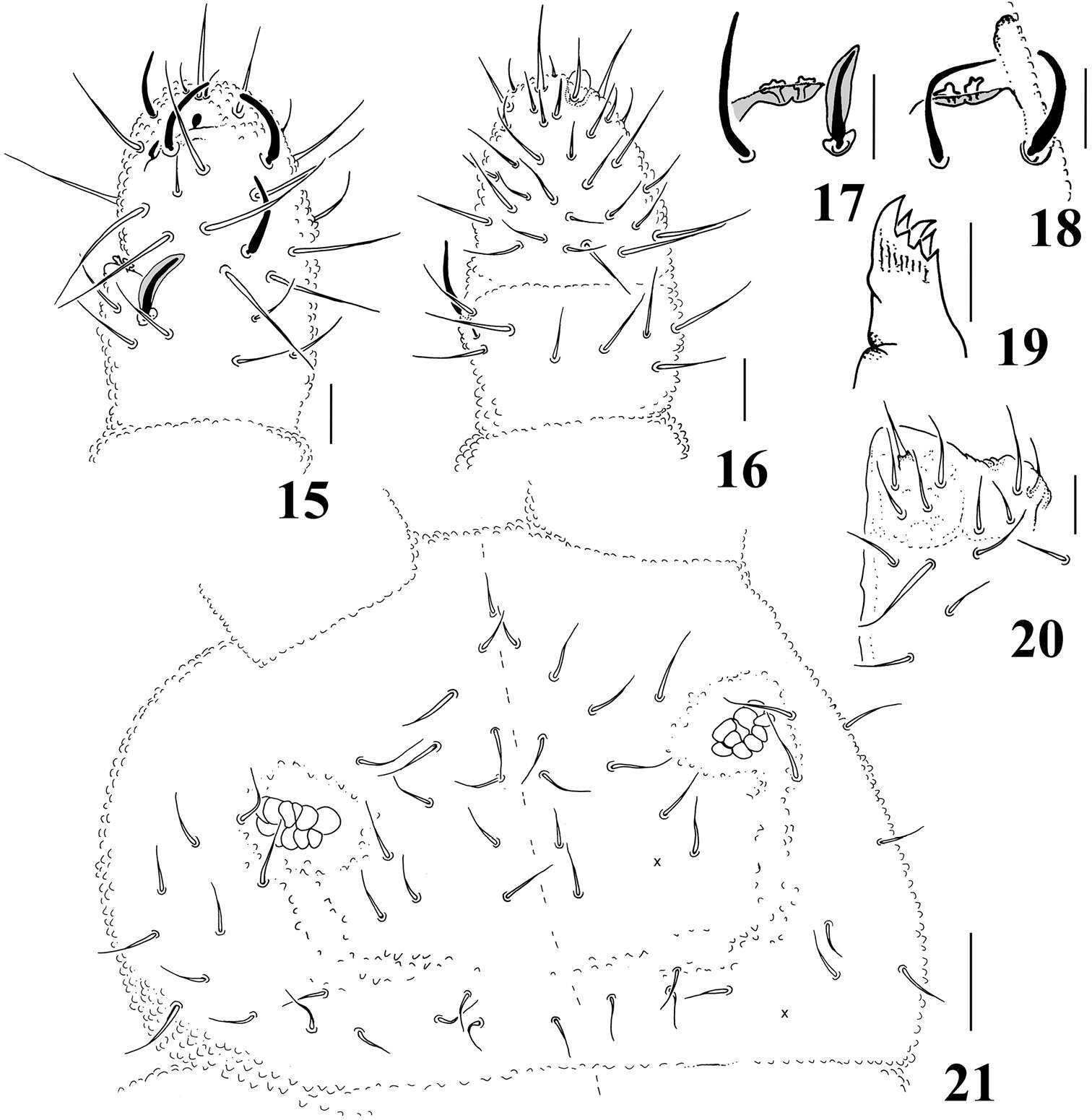
Figures 15–21.Micronella longisensilla sp. n. 15 Dorsal view of Ant III–IV 16 Ventral view of Ant III–IV 17 Detail of Ant III organ 18 Detail of Ant III organ (same specimen of Fig. 17, right antennae) 19 Maxilla 20 Labium 21 Head. Scale bars: 10μm (15–20); 20μm (21). x represents missing chaeta.
-
Xiang-Qun Yuan, Zhi-Xiang Pan
Zookeys
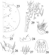
Figures 23–29.Sinella triseta sp. n. 23 dorsal cephalic chaetotaxy 24 basal chaetae of Ant. I 25 basal chaetae of Ant. II 26 Ant. III organ 27 clypeus 28 labrum 29 labial base 30 labial palp.
-
Sopark Jantarit, Chutamas Satasook, Louis Deharveng
Zookeys
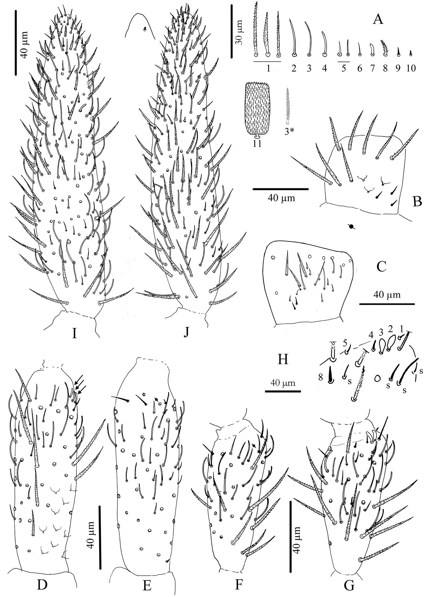
Figure 3.Cyphoderus songkhlaensis sp. n. continued A chaetae of antenna drawn from optical microscope, except 3* derived from SEM image B dorsal side of right Ant.I C ventral side of right Ant.I D dorsal side of right Ant.II; the apical swollen sens of type-7 are indicated by arrows E ventral side of right Ant.II with apical pseudopore F ventral side of right Ant.III with apical pseudopore G dorsal side of right Ant.III H distal organite of Ant.III I ventral side of Ant.IV J dorsal side of Ant.IV with separate view of the subapical organite (left).
-
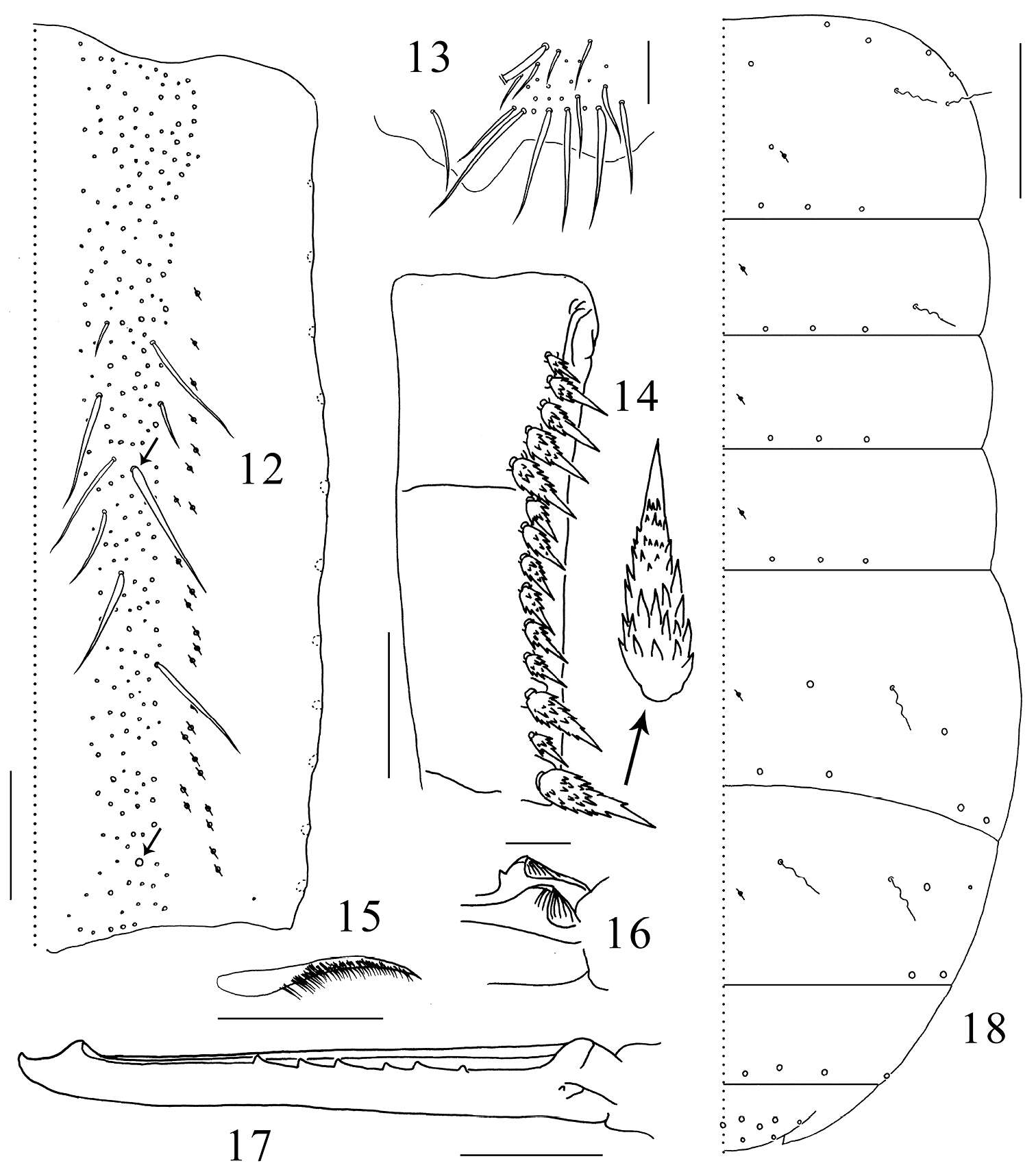
Daoyuan Yu, Feng Zhang, Louis Deharveng
Zookeys
Figures 12–18.Tomocerus postantennalis sp. n. 12 dorsal face of manubrium (right side; prominent chaetae arrowed) 13 disto-dorsal chaetae on manubrium (left side) 14 dental spines (left side) 15 feathered chaeta on dens 16 basal teeth of left mucro 17 right mucro 18 body chaetotaxy. Scale bars: 12, 14 = 100μm; 13, 17 = 50 μm; 15, 16 = 21 μm; 18 = 400 μm. Large circles: macrochaetae; small circles: mesochaetae; wavy lines: bothriotricha; circles with a slash: pseudopores.