-
Anatoly B. Babenko, Ayuna B. Chimitova, Sophya K. Stebaeva
Zookeys
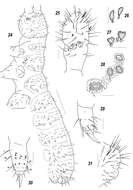
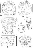
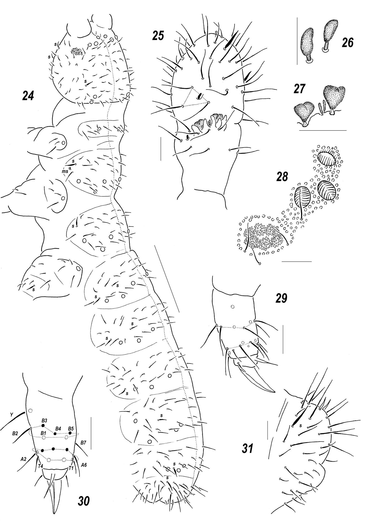
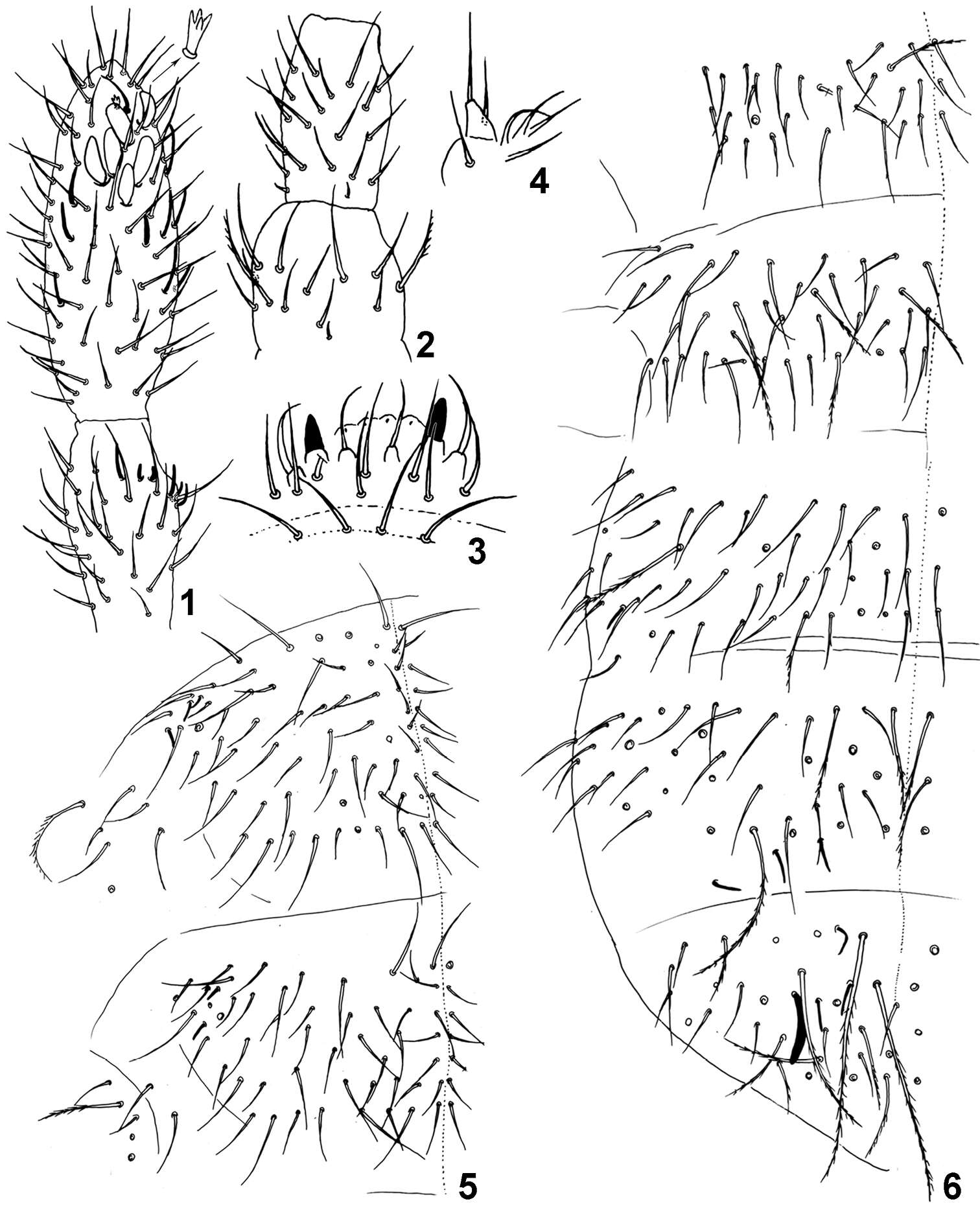
Figures 24–31.Sensillonychiurus amuricus sp. n. 24 dorsal chaetotaxy 25 Ant.3–4 26–27 sensorial elements of Ant.3 organ, different view 28 PAO and adjacent pso 29–30 tibiotarsus of Lg.3, different views 31 Abd.6. Scales: 24 – 0.1 mm, 25–31 – 0.01 mm.
-
José G. Palacios-Vargas, Hugo H. Mejía-Madrid
Zookeys
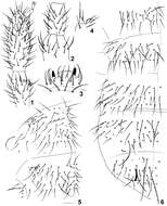
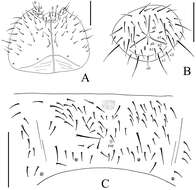
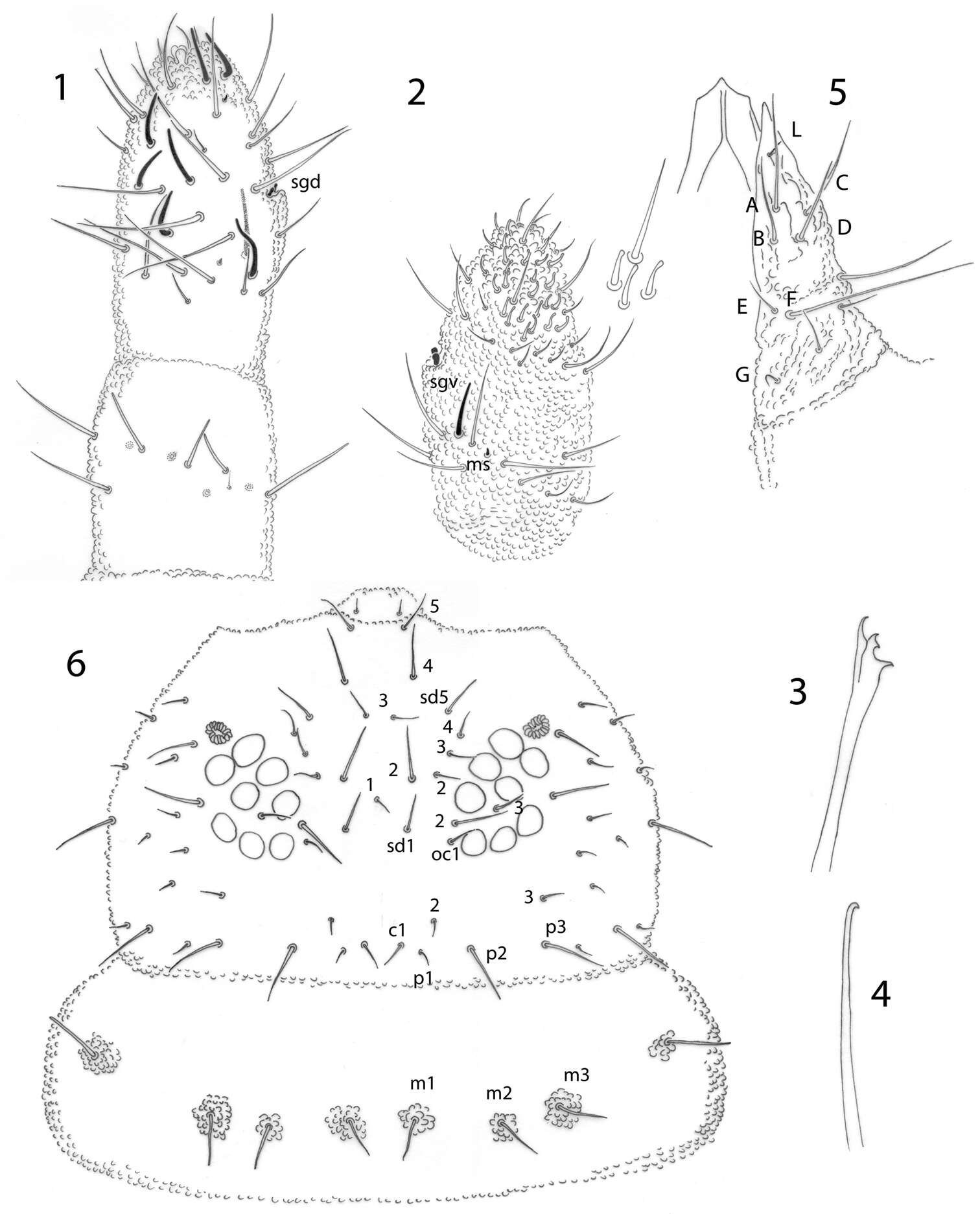
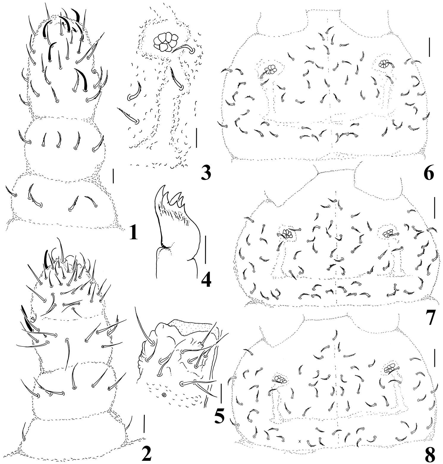
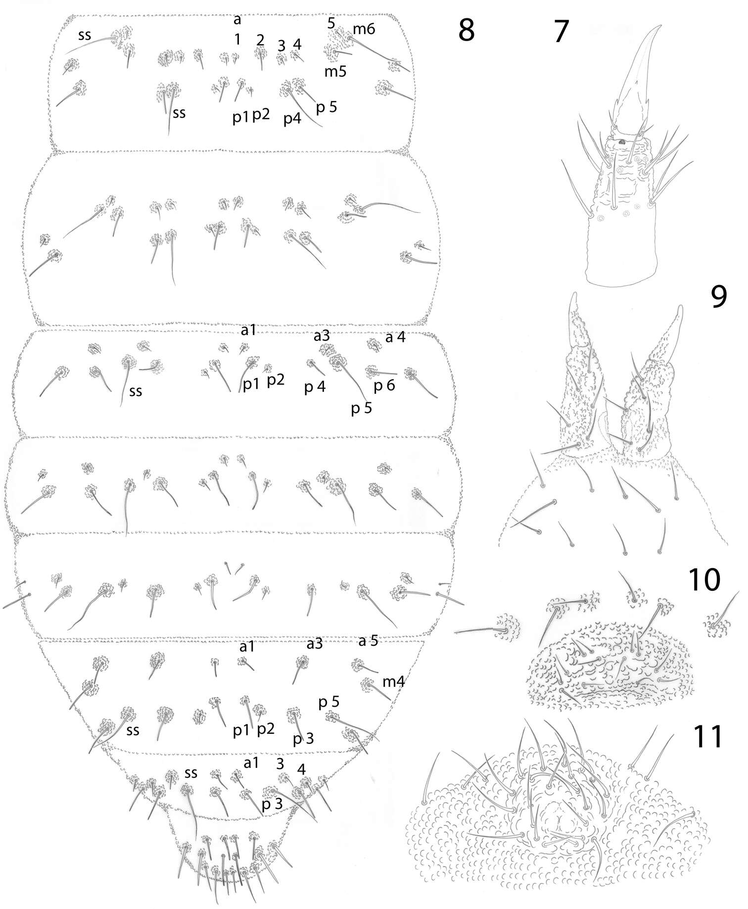
Figures 1–6.Pseudachorutes nica sp. n. 1 right antenna from II to IV dorsal view 2 Ant. III and IV in ventral view with magnification of some setae from ventral file 3 mandible 4 maxilla 5 labium 6 dorsal chaetotaxy of the head and thorax I (thorax has a drawing style represents granulation close to setae).
-
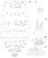
Maria Cleide de Mendonça, Eduardo A. Abrantes, Ana Carolina R. Neves
Zookeys
Figure 1–6.Isotomiella macedoi sp.n. 1 Ant III-IV Dorsal view, detail of the apical microsensillum 2 Ant I-II Dorsal view 3 Labral and prelabral chaetae 4 Outer lobe of maxilla 5 Dorsal chaetotaxy of Th II-III 6 Dorsal chaetotaxy of Abd I-VI.
-
Gabriel C. Queiroz, Maria Cleide de Mendonça
Zookeys
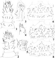
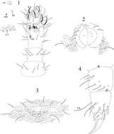
Figures 1–8.Micronella itacaman sp. n. 1 Dorsal view of Ant I–IV 2. Ventral view of Ant I–IV 3 PAO and its surrounding chaetae 4 Maxilla 5 Labium 6 Head chaetotaxy of specimen from Itatiaia 7 Head chaetotaxy of specimen from Teresópolis 8 Head chaetotaxy of specimen from Alto Caparaó. Scale bars: 10μm (1–5); 20 μm (6–8).
-
Xiang-Qun Yuan, Zhi-Xiang Pan
Zookeys
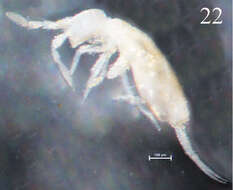
Figure 22.Habitus of Sinella triseta sp. n.
-
Xin Sun, Louis Deharveng, Donghui Wu
Zookeys
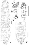
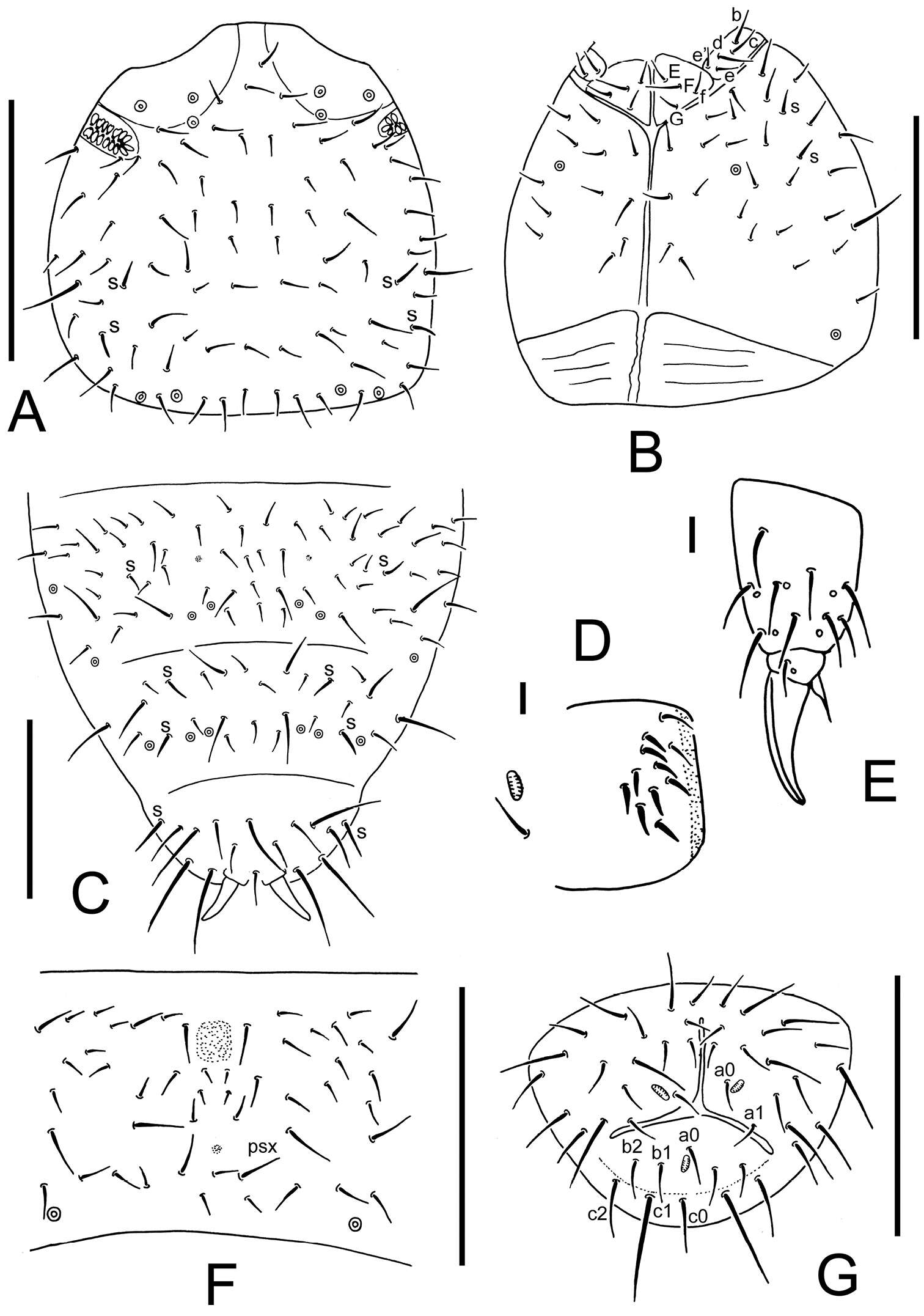
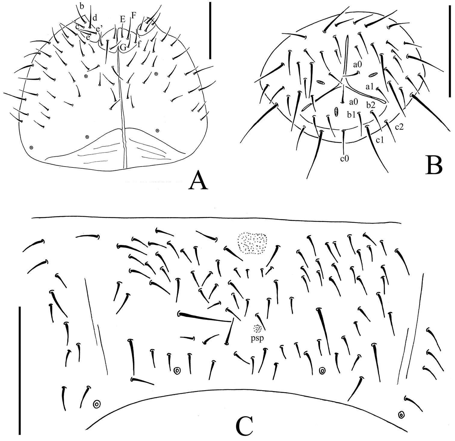
Figure 1.Thalassaphorura problematica sp. n. A dorsal side of body B ventral side of Abd. I–VI C PAO D clubs and papillae of AIIIO E Labium F Antenna. Scales: 0.1 mm (A–B, F), 0.01 mm (C–E).
-
Daoyuan Yu, Feng Zhang, Louis Deharveng
Zookeys
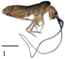
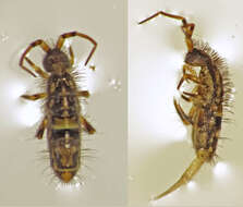
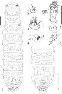
Figure 1.Tomocerus postantennalis sp. n. Appearance in alcohol. Scale bar: 1000 μm.
-
José G. Palacios-Vargas, Ana E. Salazar Martínez
Zookeys
Figures 1–4.Tullbergia alcirae sp. n. 1 antennal segments I to IV with details of sensorial structures 2 ventral tube 3 female genital plate 4 femur and tibiotarsus III.
-
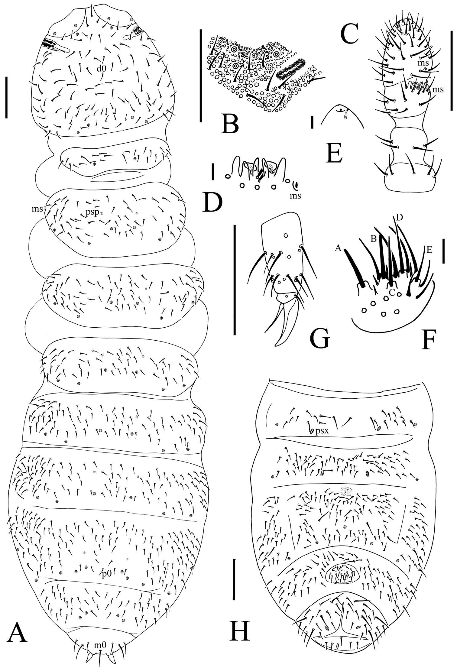
Figure 3.Onychiurus heilongjiangensis sp. n. A dorsal side of body B PAO C Ant. I–IV D Ant. III sensory organ E antennal tip F labium G distal part of leg III H Abd. II–VI sterna. Scale bars: 0.1 mm (A, C, G–H), 0.01 mm (B, D–F).
-
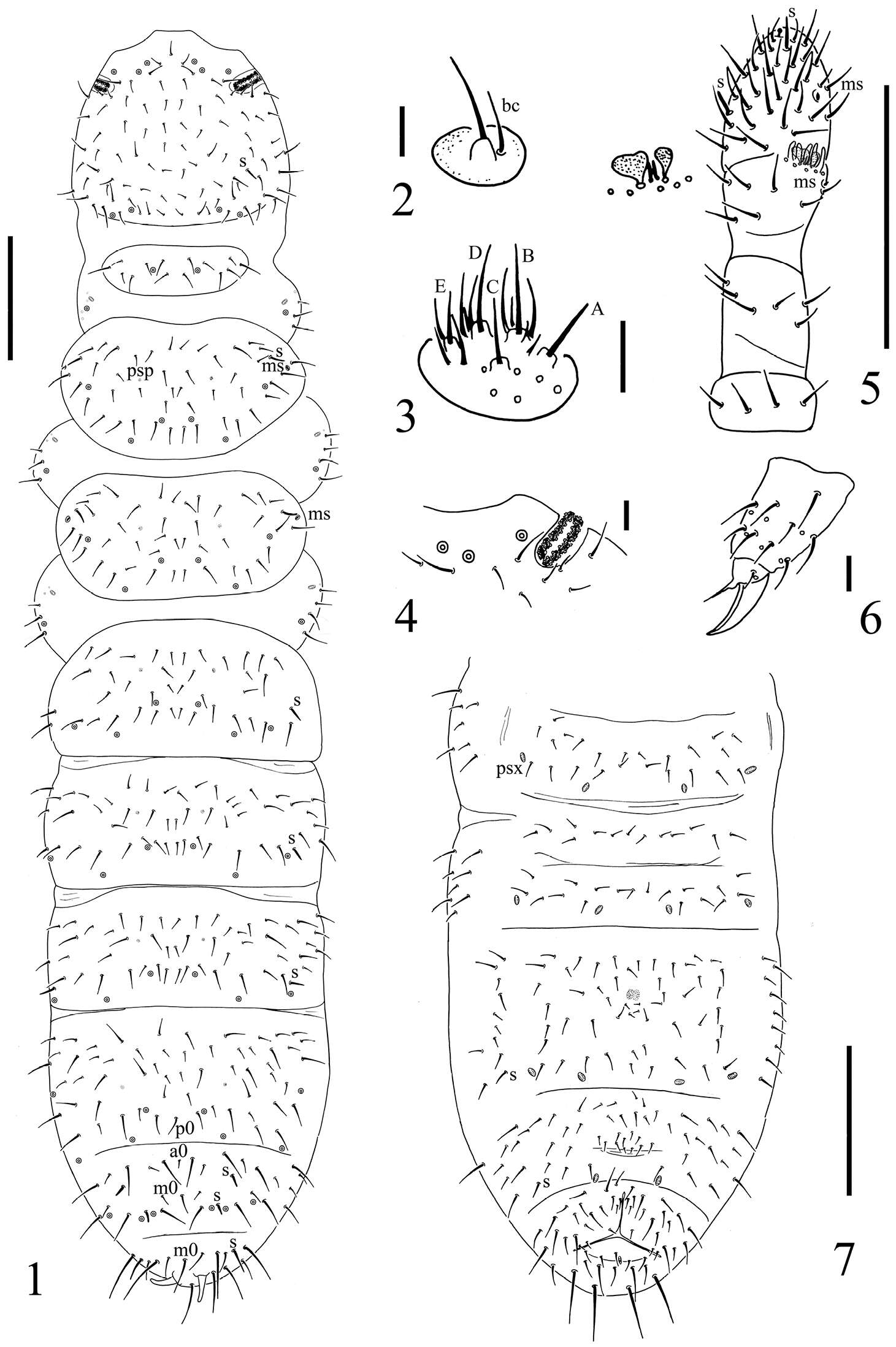
Figure 1.Spinonychiurus sinensis sp. n. A dorsal chaetotaxy of body B maxillary palp C labium D postantennal organ E antenna F distal part of leg III G chaetotaxy of Abd. II–VI sterna. Scale bars: 0.1 mm (A, E, G), 0.01 mm (B–D, F).
-
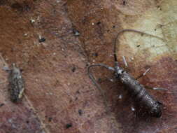
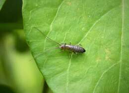
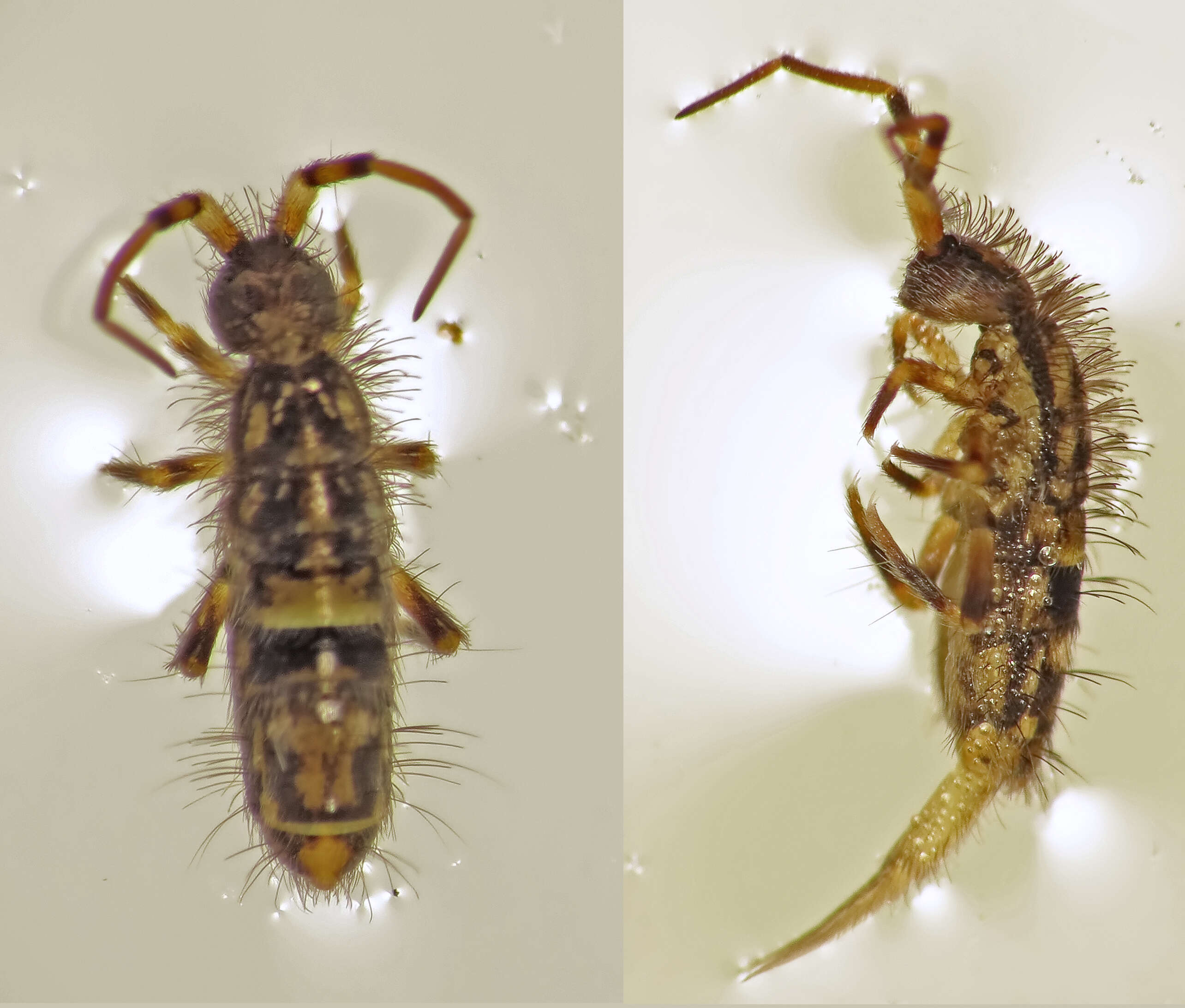
possibly: Genus: Pogonognathellus ???Family: Tomoceridae SCHFFER, 1896[det. Manuel Valdueza, 2012, based on this photo]Subclass: Collembola LUBBOCK, 1870 (Springschwnze, Springtails)Class: Entognatha Subphylum: HexapodaPhylum: Arthropoda2011-07-24_vic. Regensburg, Bavaria, GermanyIMG_3928
-
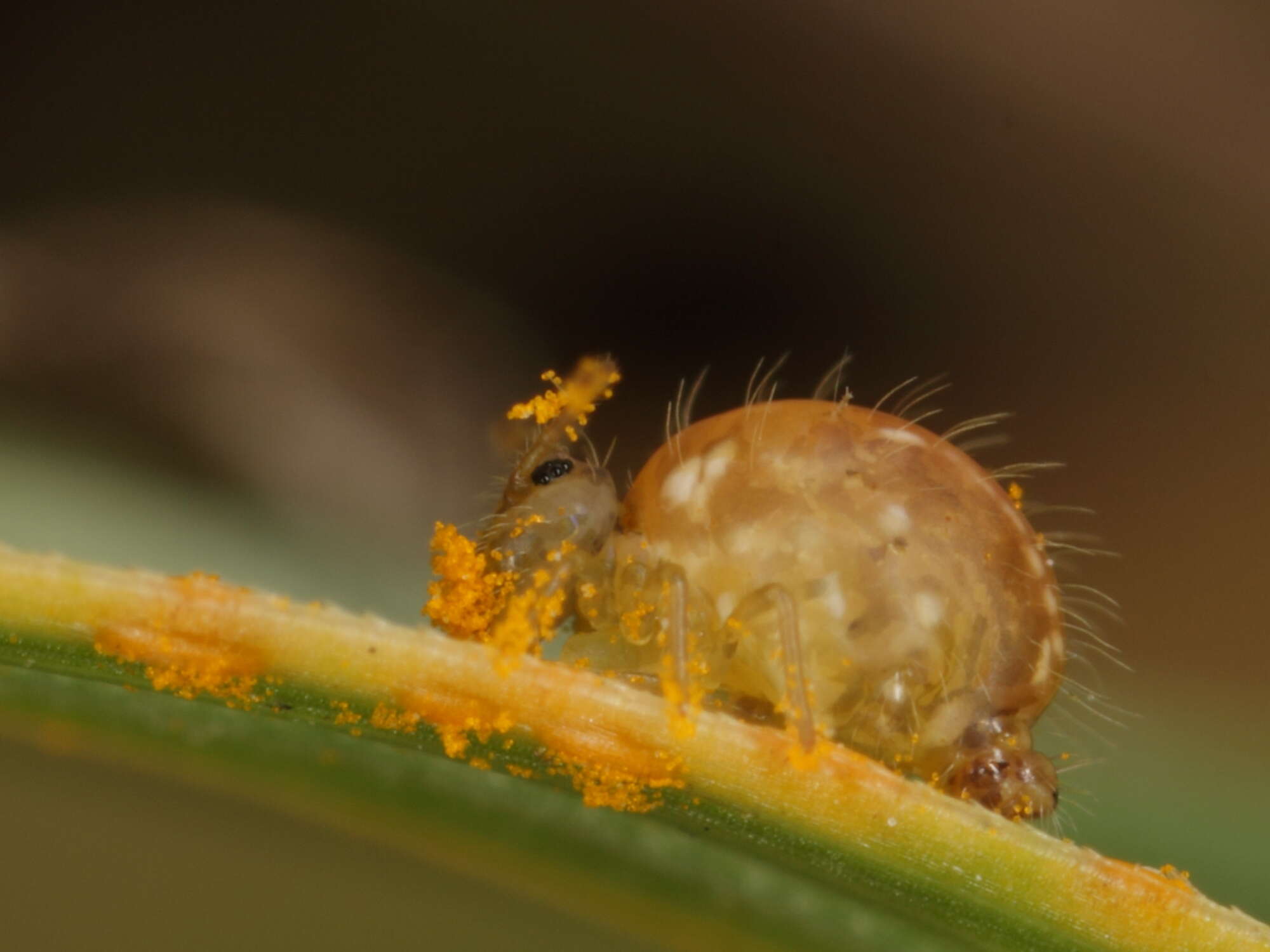
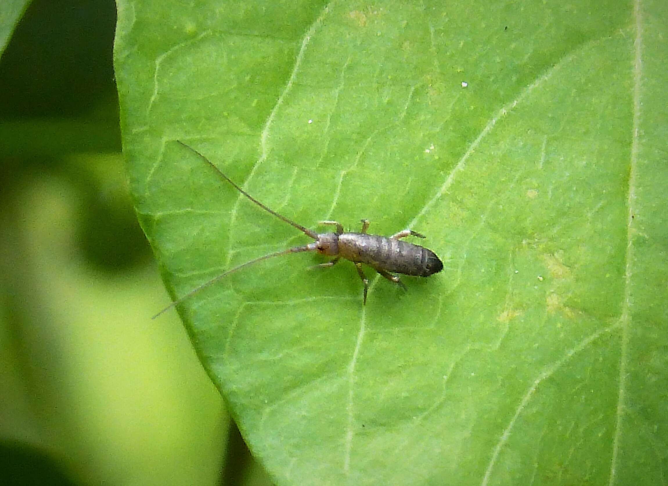
L: 2-3 mmCharacteristics: Bright yellow-green, antennae yellow, reddish towards the tip mouth orange, bristles (macrosetae) are distinctly present on the abdomen (if absent it is probably
Sminthurides). Compound eyes composed of eight ommatidia (single eyes).Habitat: Soil surface on a variety of habitats( including grass- and woodland).food
Sminthurus viridis is one of the few globular springtails that may be actually damaging leguminous (Fabaceae) plants; they rasp/abrade the plant surface and give way to secondary infections.Distribution: widespread, introduced pest species in AustraliaPhylum: Arthropoda LATREILLE, 1829 (arthropods, Gliederfer)Subphylum:Hexapoda BLAINVILLE, 1816Class: Entognatha STUMMER-TRAUNFELS, 1891 (Sackkiefler)Order: Collembola LUBBOCK, 1870 (collembolans or springtails, Springschwmze)Suborder: Symphypleona BRNER, 1901 Superfamily: Sminthuroidea BRETFELD, 1994Family: Sminthuridae LUBBOCK, 1862 (globular springtails, Kugelspringer)Subfamily: Sminthurinae LUBBOCK, 1862 [sensu DEHARVENG, L, 2004]Genus:
Sminthurus LATREILLE, 1804 [sensu BRNER, 1906]
Sminthurus viridis LINNAEUS, 1758 (Clover Springtail or Lucerne Flea, Luzernefloh)[det. Frans Janssens, 2013, based on photos]taxonomical info:
www.biolib.cz/en/taxonposition/id80646/some info:
www.naturespot.org.uk/species/clover-springtailmore info:
bugguide.net/node/view/86837more info:
www.nhm.ac.uk/nature-online/species-of-the-day/biodiversi...more info (German):
www.naturspaziergang.de/Andere/Collembola/Sminthurus_viri...Germany, Berlin: Tempelhof Flugfeld, 09.10.2012_______________________________________________MP-E 65mm f/2.8 1x-5x ( Macro Magnification - 2.7x), 1/250s, f/9.0, ISO100, 0EV, hand-held, ring-flash (diffusor)IMG_2625
-
Ipswich, England, United Kingdom
-
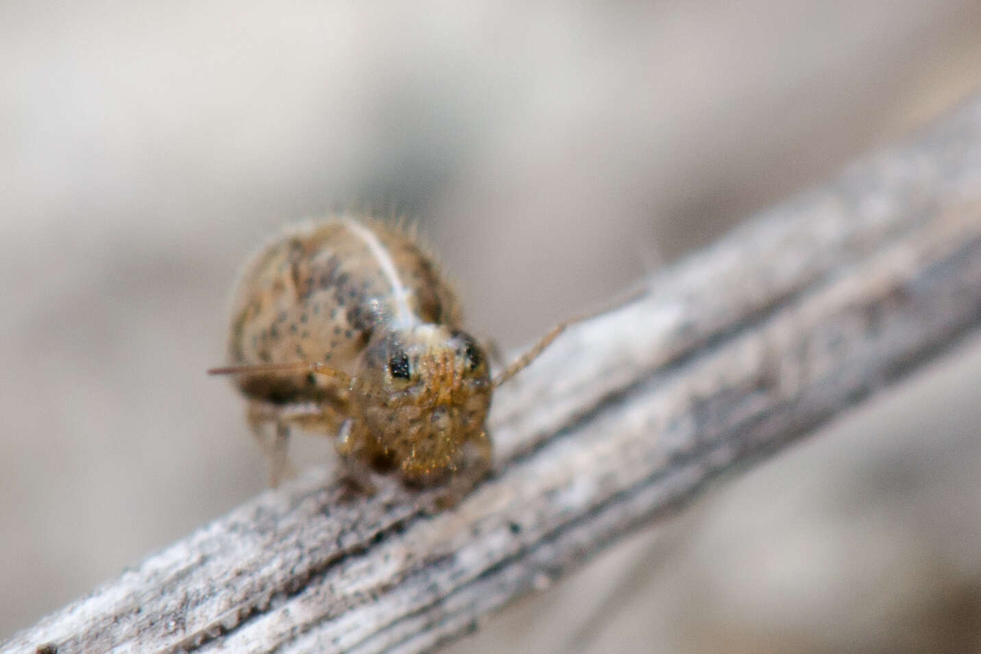
WWT Knapp Reserve, Worcs. SO748520
-
Some small black flies attracted my attention by having a tussle on the track in front of us. I stopped to see what flies they were, but they disappeared. I noticed these tiny Springtails jumping and was very lucky that this little one stayed on a stick for a while.
-
José G. Palacios-Vargas, Hugo H. Mejía-Madrid
Zookeys
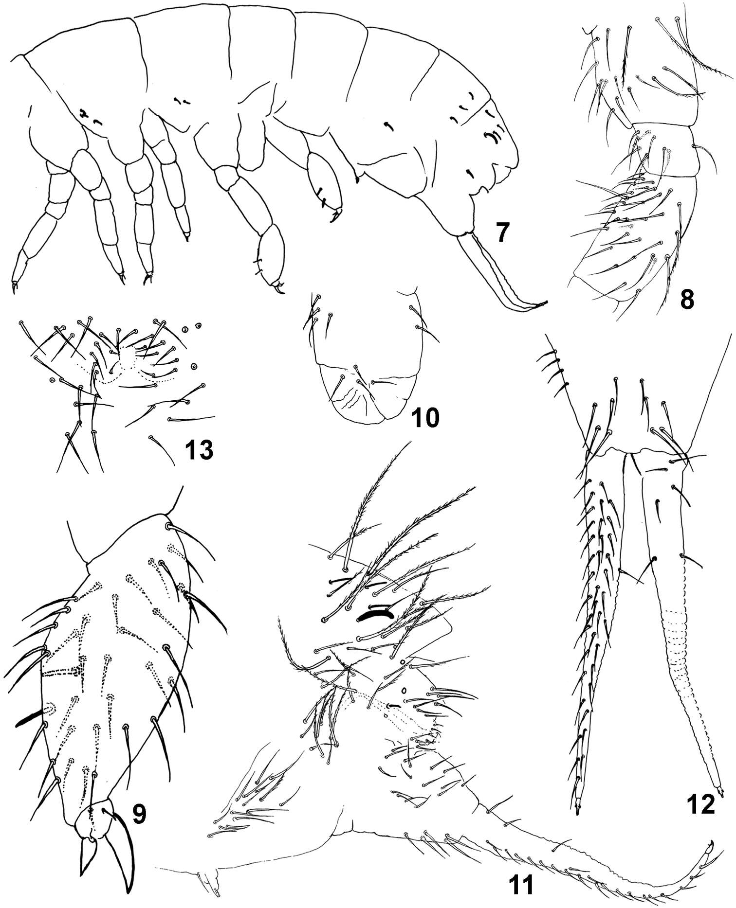
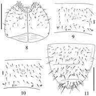
Figures 7–10.Pseudachorutes nica sp. n. 7 tibiotarsus III and unguis in ventral view 8 dorsal chaetotaxy of Th. II and III and Abd. I–VI (drawing style represents granulation close to setae) 9 furcula 10 female genital plate 11 male genital plate.
-
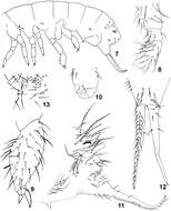
Maria Cleide de Mendonça, Eduardo A. Abrantes, Ana Carolina R. Neves
Zookeys
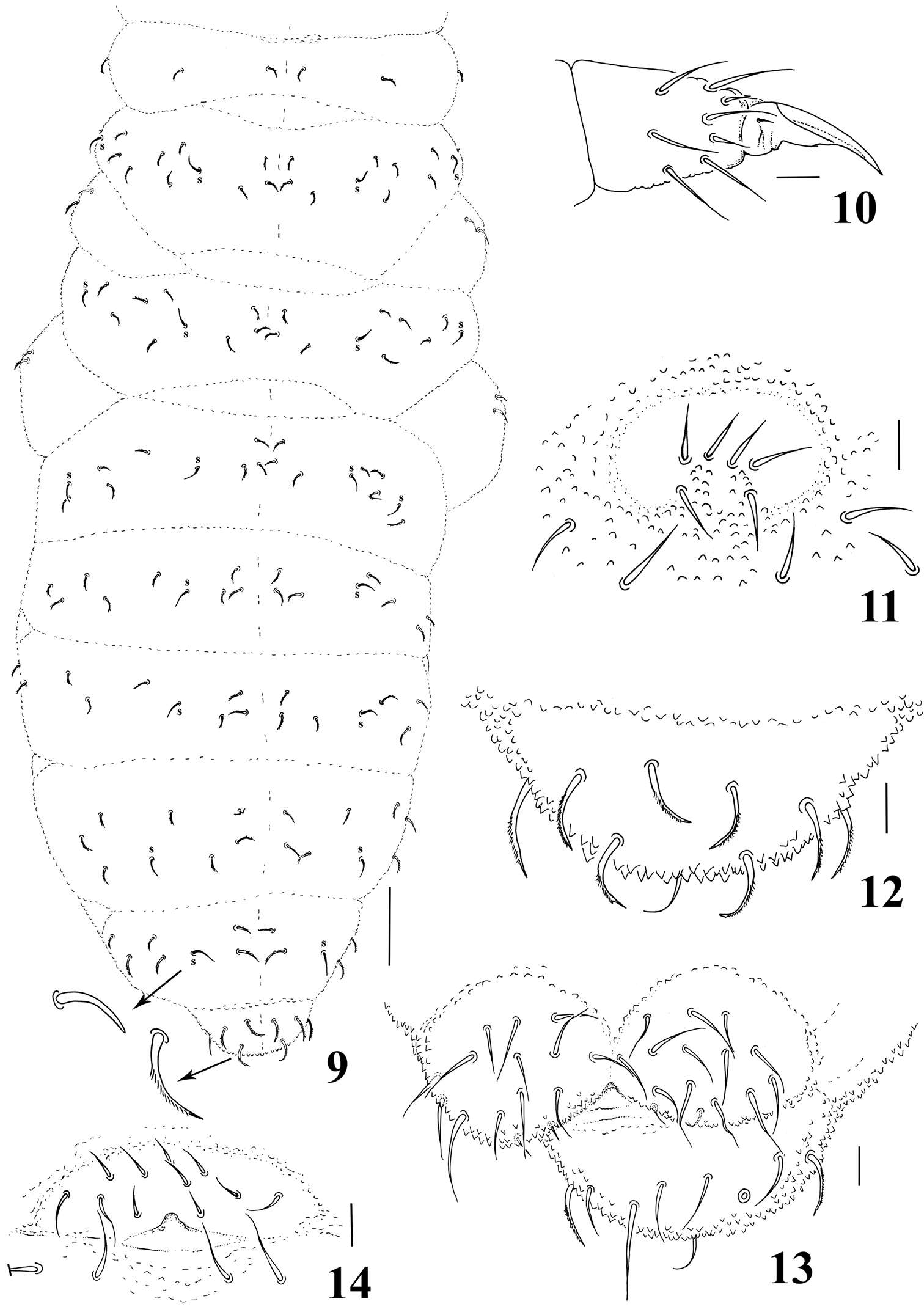
Figure 7–13.Isotomiella macedoi sp.n. 7 Sensillary pattern of the body 8 Subcoxa and femur of leg III 9 Tibiotarsus and unguis of leg III 10 Lateral view of ventral tube 11 Lateral view of abd. V-VI, subcoxa furcal, furca and tenaculum 12 Furca 13 Male genital plate.
-
Gabriel C. Queiroz, Maria Cleide de Mendonça
Zookeys
Figures 9–14.Micronella itacaman sp. n. 9. Dorsal body chaetotaxy with details of sensilla and chaetae 10 Tita of leg I 11 Furcal area 12 Dorsal view of Abd VI 13 Anal valves and ventral view of Abd VI 14 Female genital plate. Scale bars: 10μm (10–14); 50μm (9).
-
Xiang-Qun Yuan, Zhi-Xiang Pan
Zookeys
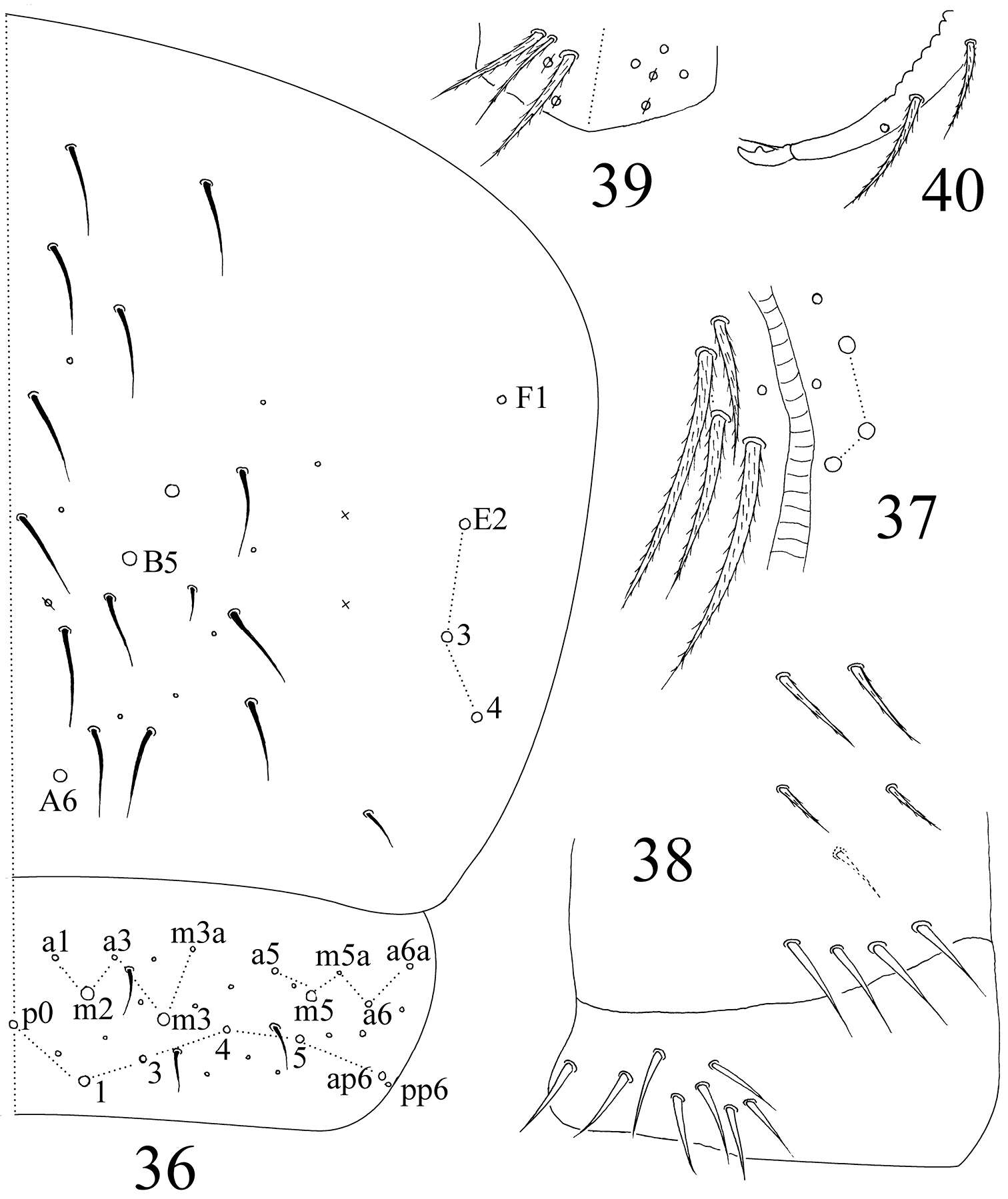
Figures 36–40.Sinella triseta sp. n. 36 dorsal chaetotaxy of Abd. IV–V 37 anterior face of VT 38 posterior face and lateral flap of VT 39 manubrial plaque 40 apical dentes and mucro.
-
Xin Sun, Louis Deharveng, Donghui Wu
Zookeys
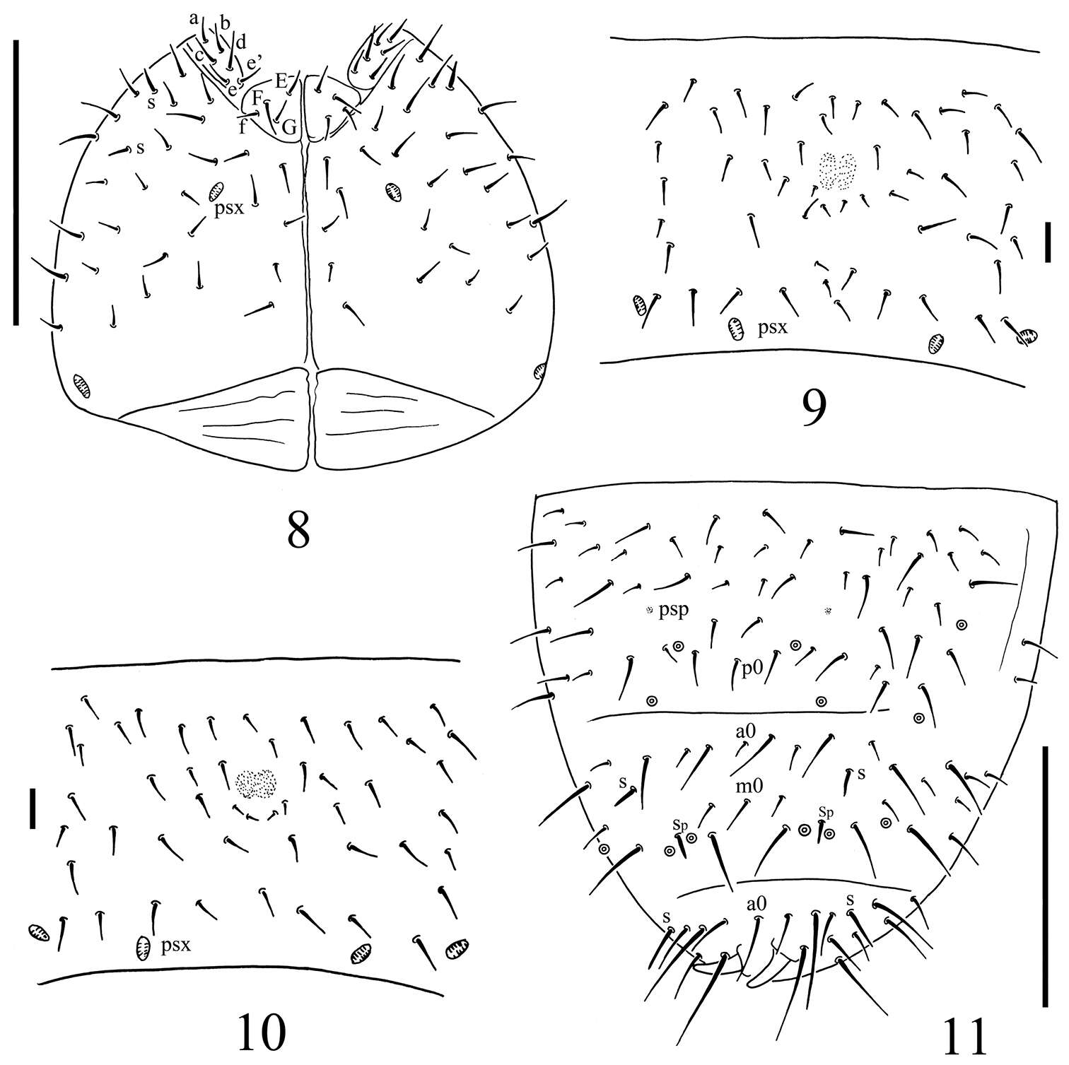
Figure 2.Thalassaphorura problematica sp. n. A dorsal side of head B ventral side of head C Abd. IV–VI terga D ventral tube (showing male ventral organ) E distal part of leg III F furca G anal valves. Scales: 0.1 mm (A–C and F–G), 0.01 mm (D–E)
-
Daoyuan Yu, Feng Zhang, Louis Deharveng
Zookeys
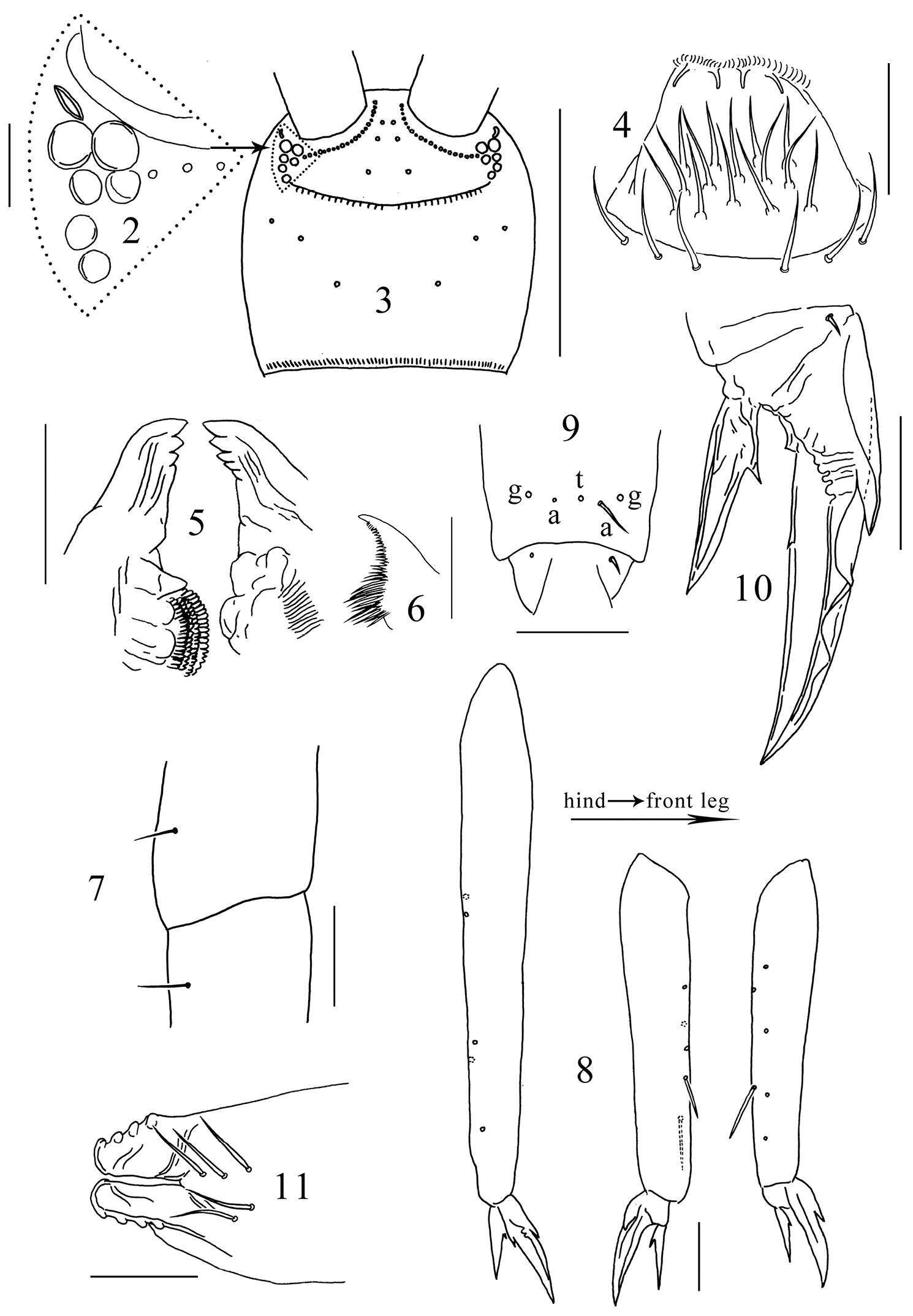
Figures 2–11.Tomocerus postantennalis sp. n. 2 PAO and ocelli 3 cephalic dorsal chaetotaxy 4 labrum 5 mandible 6 maxillary lamella five 7 trochanteral-femoral organ 8 tibiotarsus 9 anterior view of distal tibiotarsal chaetae (t: tenent hair, a: accessory chaetae, g: guard chaetae) 10 claw 11 tenaculum. Scale bars: 2, 7, 9, 10, 11 = 50 μm; 3 = 500 μm; 4, 5, 8 = 100 μm; 6= 20 μm.
-
José G. Palacios-Vargas, Ana E. Salazar Martínez
Zookeys
Figures 5–8.Tullbergia alcirae sp. n. 5 dorsal head and thoracic chaetotaxy 6 male genital plate 7 right half of labial and postlabial quetotaxy 8 dorsal abdominal cheatotaxy.
-
Figure 4.Onychiurus heilongjiangensis sp. n. A ventral side of head B anal valves C Abd. IV sternum. Scale bars: 0.1 mm (A–C).
-
Figure 2.Spinonychiurus sinensis sp. n. A ventral chaetotaxy of head B–C central part of abdominal sternum IV D dorsal chaetotaxy of Abd. IV–VI. Scale bars: 0.1 mm (A, D), 0.01 mm (B–C).