-
Marko Lukić, Céline Houssin, Louis Deharveng
Zookeys
Figures 1–3. Tritomurus veles sp. n. (optical stereomicroscope). 1, 2 Habitus (scale 1 mm) 3 Head (scale 0.2 mm).
-
Maria Cleide de Mendonça, Eduardo A. Abrantes, Ana Carolina R. Neves
Zookeys
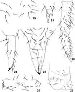
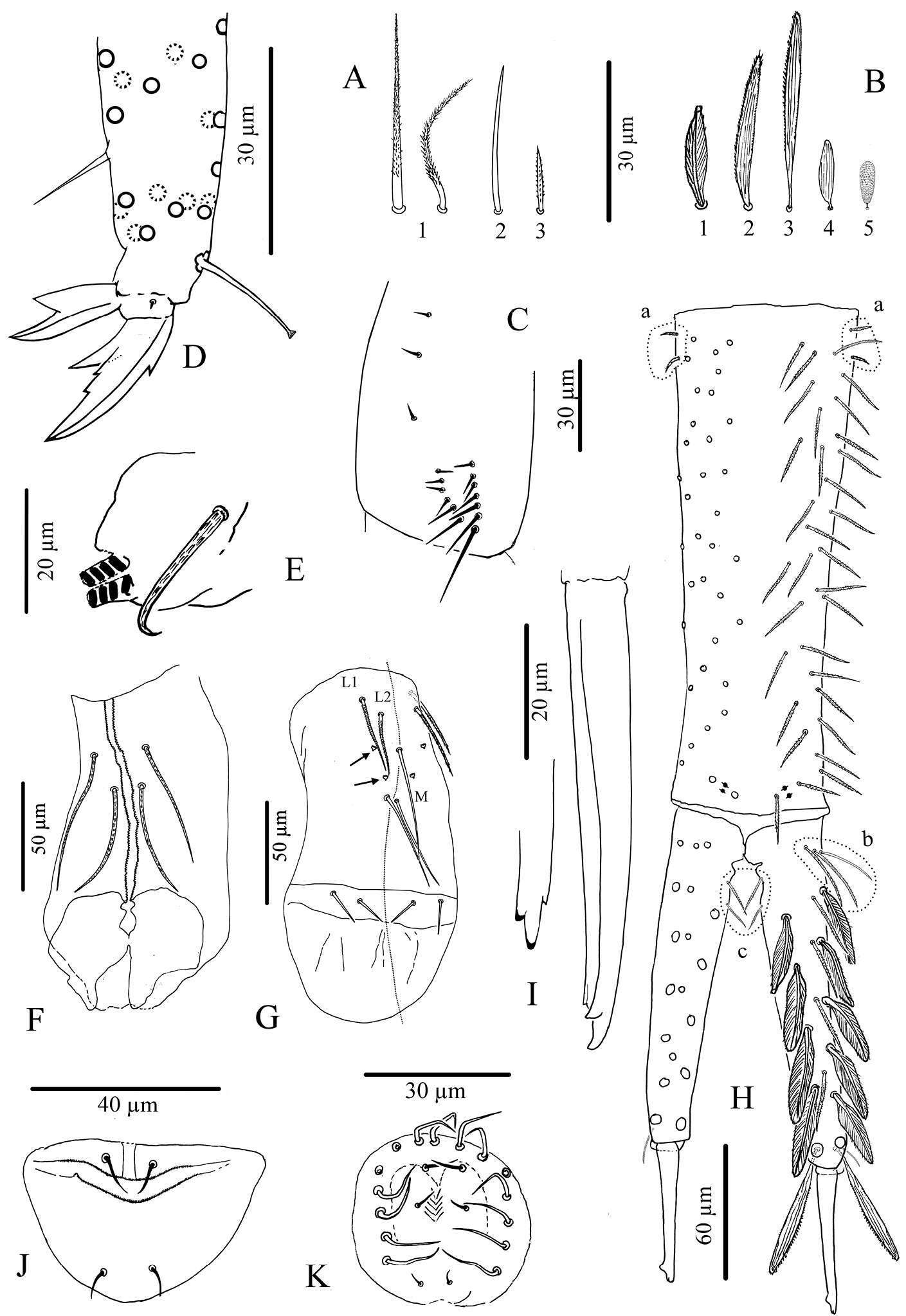
Figure 19–25.Isotomiella uai sp.n. 19 Detail of chaetotaxy of Abd II 20 Leg III 21 Unguis of leg III 22 Ventral tube 23 Furca 24 Lateral view of dens and mucro 25 Female genital opening.
-
Gabriel C. Queiroz, Maria Cleide de Mendonça
Zookeys
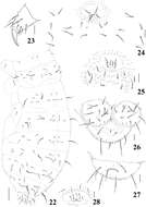
Figures 22–28.Micronella longisensilla sp. n. 22 Dorsolateral body chaetotaxy 23 Tita of leg I 24 Furcal area and its surrounding chaetae (adult) 25 Furcal area and its surrounding chaetae (juvenile) 26 Anal valves and ventral view of Abd VI 27 Dorsal view of Abd VI 28 Female genital plate. Scale bars: 10μm (23–28); 50μm (22). x represents missing chaeta.
-
Xiang-Qun Yuan, Zhi-Xiang Pan
Zookeys
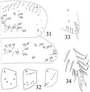
Figures 31–34.Sinella triseta sp. n. 31 dorsal chaetotaxy of Th. II–III 32 coxal mac formula (A fore leg; B mid leg; C hind leg) 33 trochanteral organ 34 tip tibiotarsus and claw of hind leg.
-
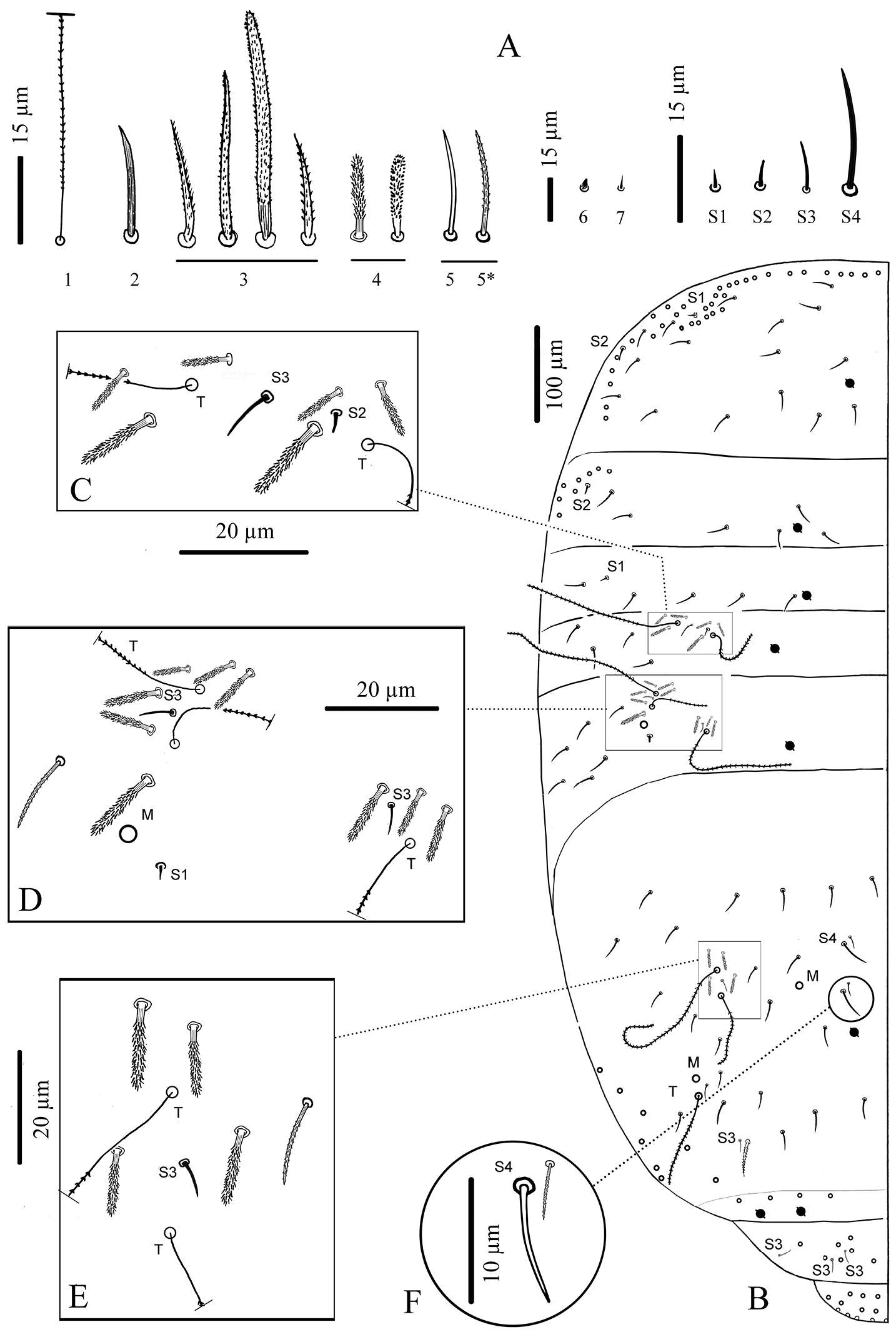
Sopark Jantarit, Chutamas Satasook, Louis Deharveng
Zookeys
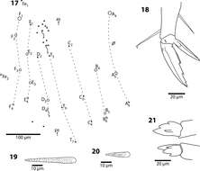
Figure 4.Cyphoderus songkhlaensis sp. n. continued A chaetae of tergites drawn from optical microscope, except 5* derived from SEM image B chaetotaxy of tergites with types of S-chaetae S1 to S4 C trichobothrial complexes of Abd.II D trichobothrial complexes of Abd.III E anterior trichobothrial complexes of Abd.IV F tandem of chaetae on Abd.IV; the smallest is a short type-5 mes and the largest a S4 sens.
-
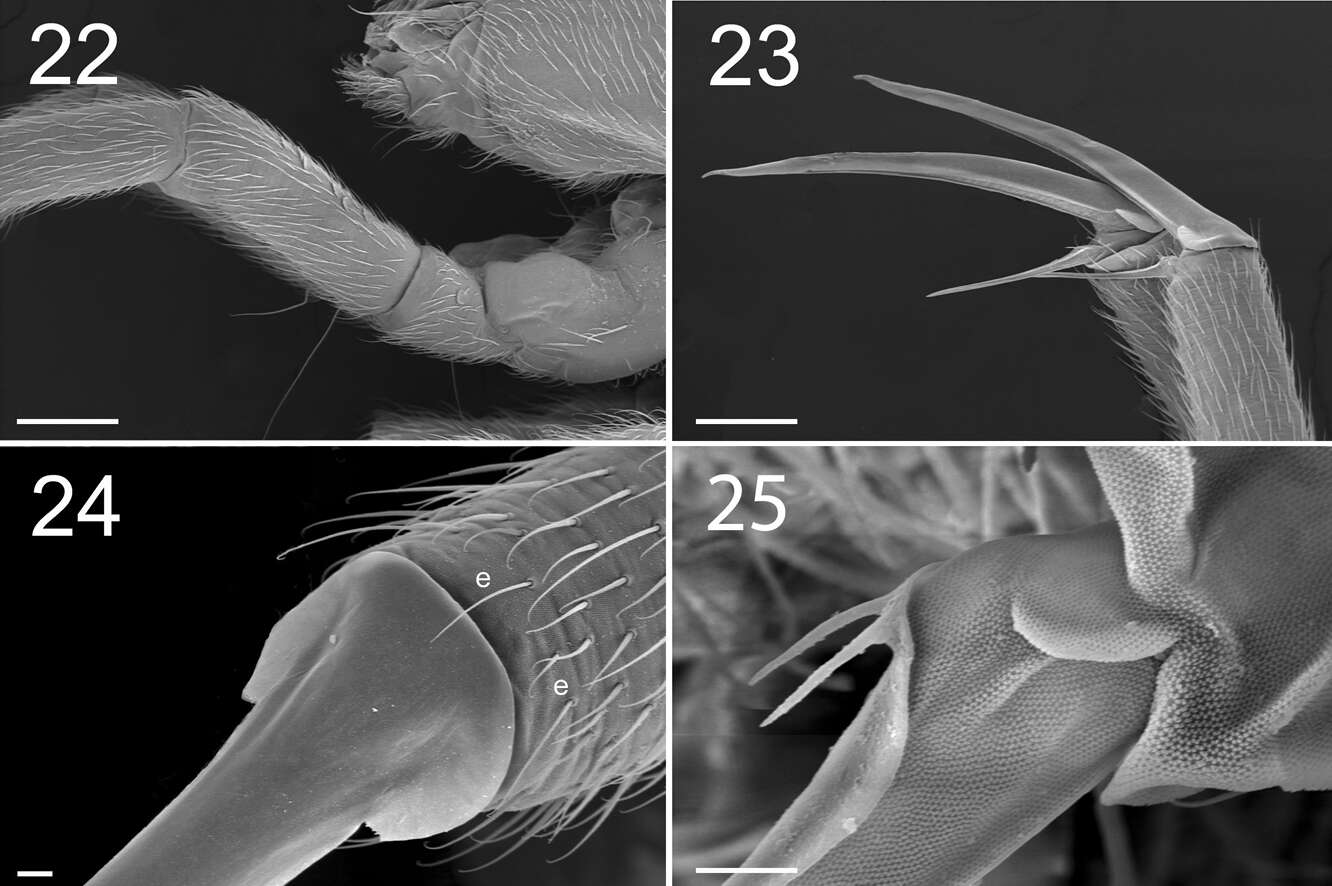
Marko Lukić, Céline Houssin, Louis Deharveng
Zookeys
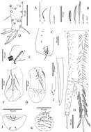
Figures 22–25. Tritomurus veles sp. n. (SEM). 22 Leg I, with ventro-basal macrochaetae of femur and ventral macrochaetae of trochanter (scale 100 μm); the second visible macrochaetae of femur belongs to other leg 23 Claws of legs I (scale 100 µm) 24 Claw of leg I, basal part in dorsal view (scale 10µm); e, thin distal tenent hairs 25 Bifurcate empodial appendage of leg II (scale 10 μm).
-
Gabriel C. Queiroz, Maria Cleide de Mendonça
Zookeys
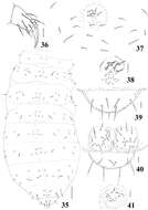
Figures 29–34.Micronella porcus (Denis, 1933). 29. Dorsal view of Ant I–IV 30 Ventral view of Ant I–IV 31 Detail of Ant III organ 32 Maxilla 33 Labium 34 Head chaetotaxy. Scale bars: 10μm (29–33); 20μm (34).
-
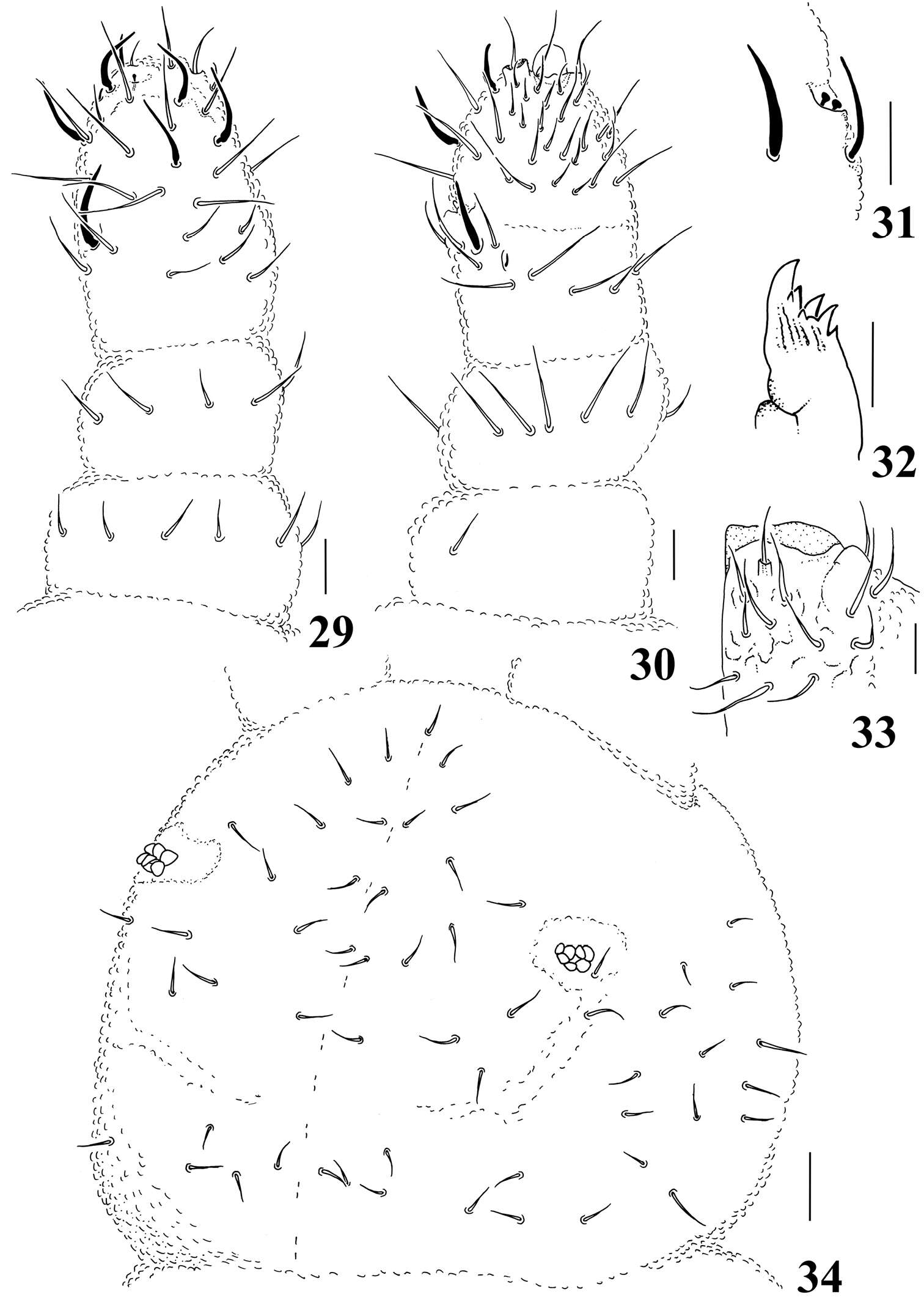
Xiang-Qun Yuan, Zhi-Xiang Pan
Zookeys
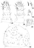
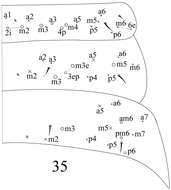
Figure 35.dorsal chaetotaxy of Abd. I–III of Sinella triseta sp. n.
-
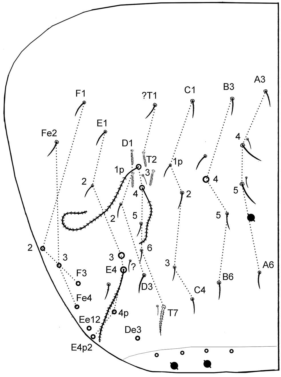
Sopark Jantarit, Chutamas Satasook, Louis Deharveng
Zookeys
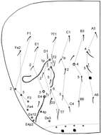
Figure 6.Cyphoderus songkhlaensis sp. n. continued, Szeptycki’s notation of tergal chaetae on Abd.IV (Szeptycki 1979).
-
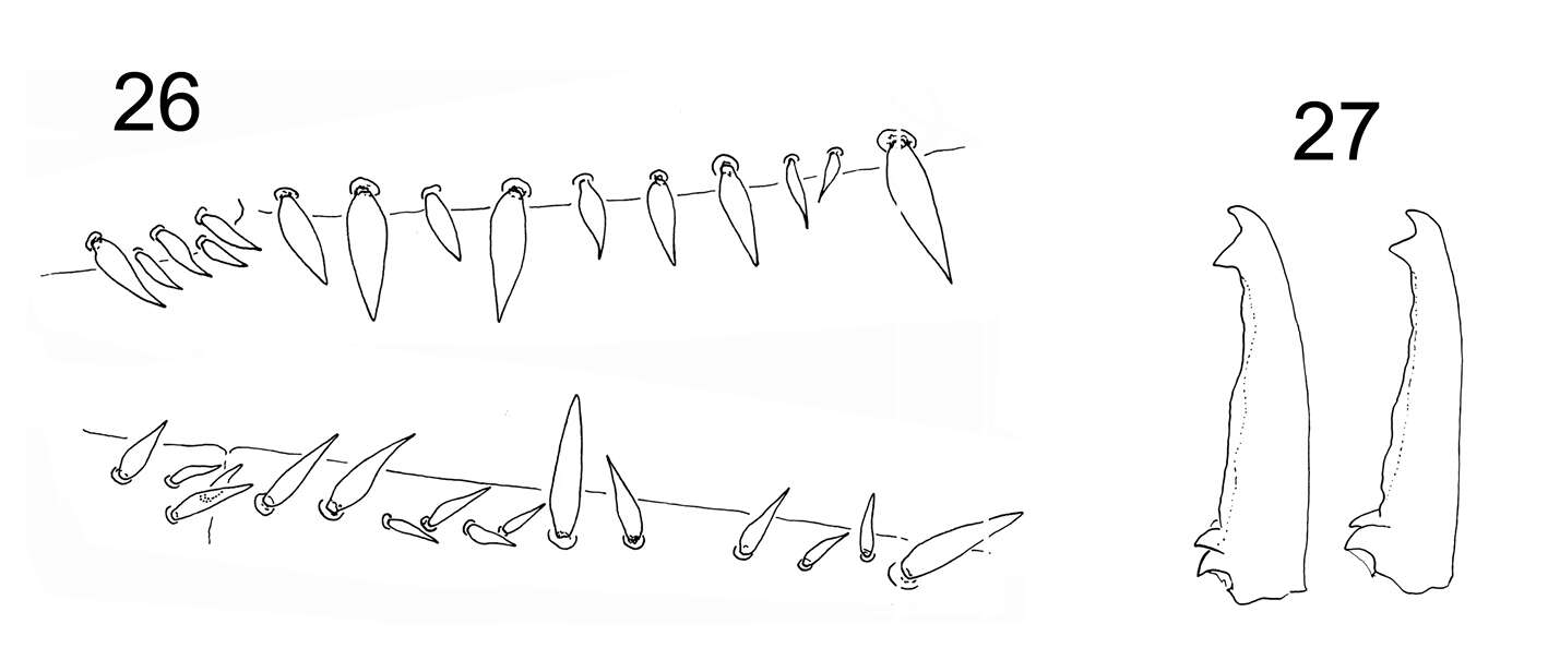
Marko Lukić, Céline Houssin, Louis Deharveng
Zookeys
Figures 26–27. Tritomurus veles sp. n. 26 Dental spines formula in a female specimen: 4/2,4,1,4,1 (lower, right dens) and 5/1,1,1,1, 2,1,2,1 (upper, left dens) 27 Mucro in two different specimens.
-
Gabriel C. Queiroz, Maria Cleide de Mendonça
Zookeys
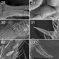
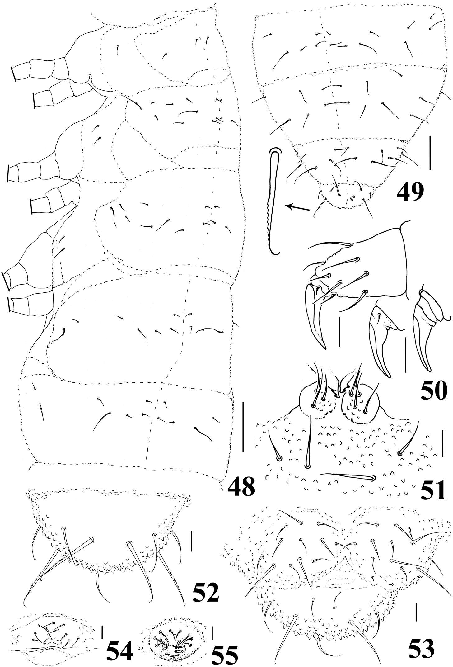
Figures 35–41.Micronella porcus (Denis, 1933). 35 Dorsal body chaetotaxy 36 Tita of leg I 37 Furcal area and its surrounding chaetae 38 Detail of furcal area 39 Dorsal view of Abd VI 40 Anal valves and ventral view of Abd VI 41 Female genital plate. Scale bars: 10μm (36–41); 50μm (35).
-
Sopark Jantarit, Chutamas Satasook, Louis Deharveng
Zookeys
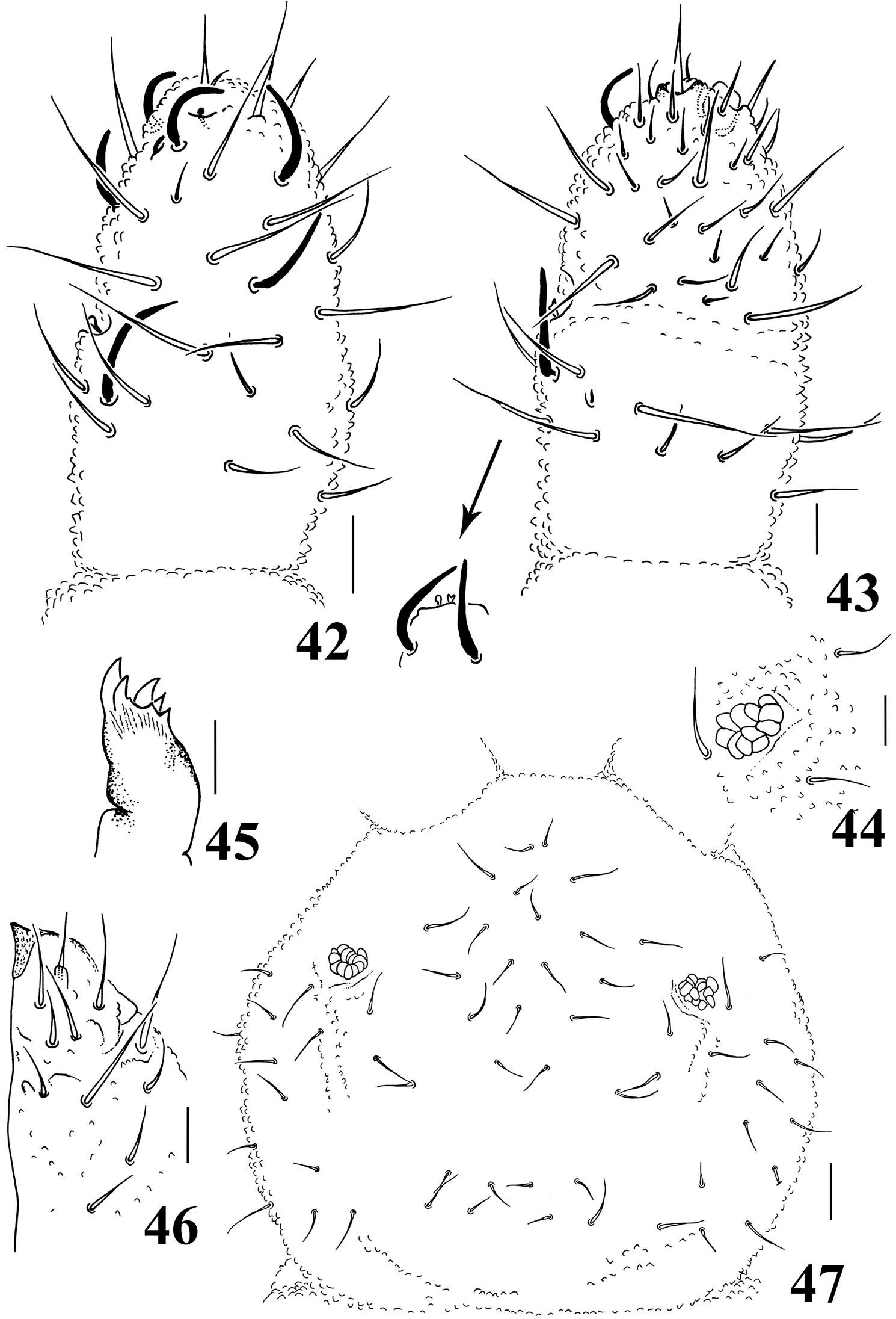
Figure 7.Cyphoderus songkhlaensis sp. n. continued A chaetae of furca B scales of furca C trochanteral organ D claw and distal part of tibiotarsus III E tenaculum F anterior face of the ventral tube G posterior face of the ventral tube; the peg-like setulae are indicated by arrows H furca; encircled by dotted lines are the 2+2 latero-basal mesochaetae of manubrium (a) the 3 outer basal mesochaetae of dens (b) and the 2+2 inner basal mesochaetae of dens (c) (I) mucro in lateral view (right) and in dorsal view (left) showing a third minute external tooth J female genital plate K male genital plate.
-
Marko Lukić, Céline Houssin, Louis Deharveng
Zookeys
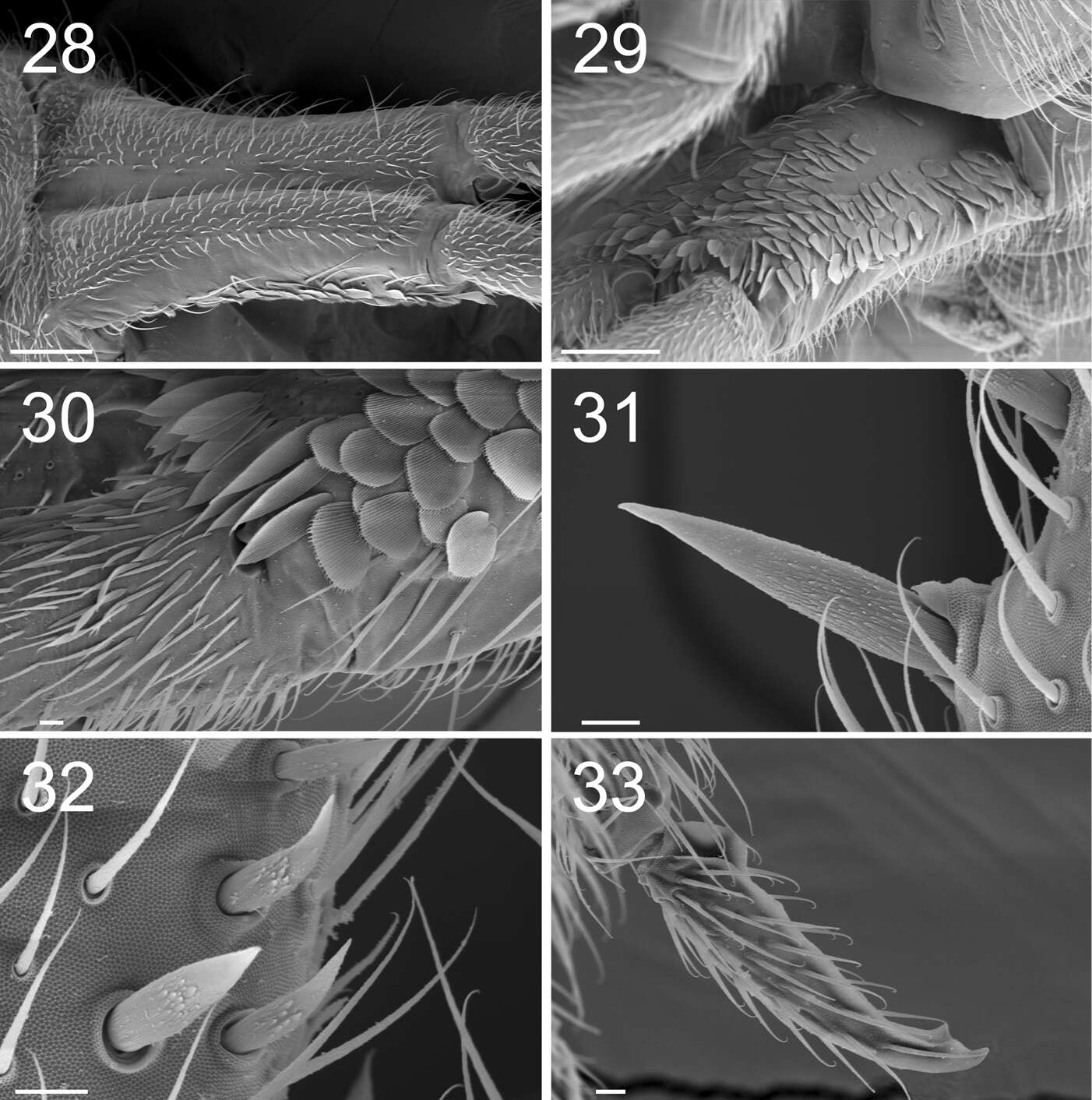
Figures 28–33. Tritomurus veles sp. n. (SEM). 28 Manubrium in dorsal view (scale 100 μm) 29 Manubrium in ventral view (scale 100 μm) 30 Manubrium ventro-distally and dens ventro-basally (scale 10 μm) 31, 32 Dental spines (scale 10 μm) 33 Mucro (scale 10 μm).
-
Gabriel C. Queiroz, Maria Cleide de Mendonça
Zookeys
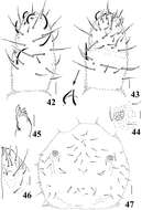
Figures 42–47.Neorganella rotundatae sp. n. 42 Dorsal view of Ant II–IV 43 Ventral view of Ant III–IV with detail of Ant III organ 44 Detail of PAO 45 Maxilla 46 Labium 47 Head chaetotaxy. Scale bars: 10μm (42–46); 20μm (47).
-
Sopark Jantarit, Chutamas Satasook, Louis Deharveng
Zookeys
Figure 8.Cyphoderus khaochakanus sp. n. A trochanteral organ B claw and distal part of tibiotarsus III C posterior face of the ventral tube D furca; feathered chaetae in lateral view, only one of the two vanes attached to the rachis is visible E mucro.
-
Marko Lukić, Céline Houssin, Louis Deharveng
Zookeys
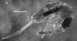
Figures 34–35. Tritomurus veles sp. n. (34, SEM; 35, optical microscope). 34 Sternite of Abd.V with genital plate (scale 100μm); arrow points to minute lateral S-microchaeta 35 Internal parasitic larva (Nematomorpha) (scale 20 µm).
-
Gabriel C. Queiroz, Maria Cleide de Mendonça
Zookeys
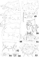
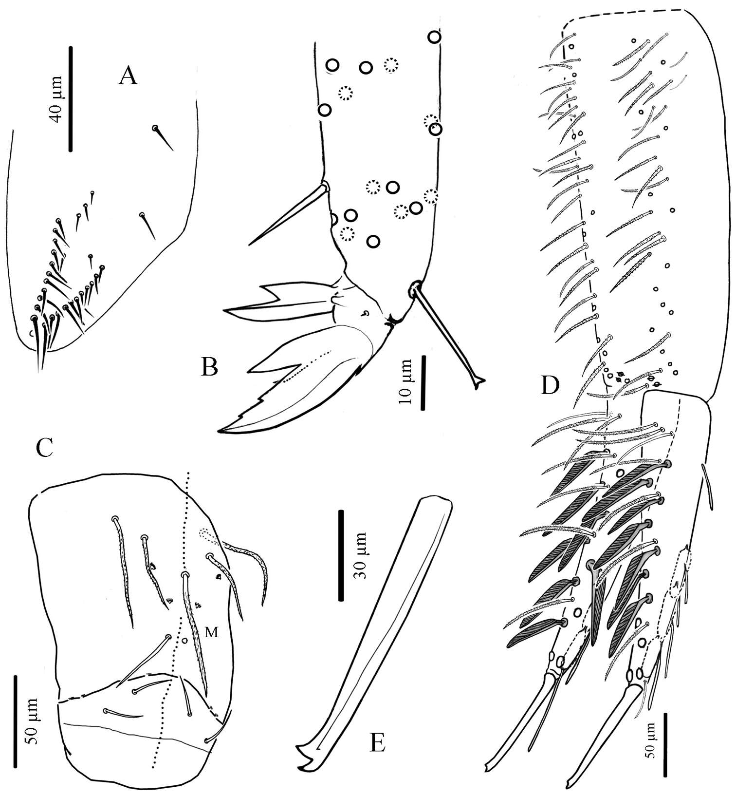
Figures 48–55.Neorganella rotundatae sp. n. 48 Dorsolateral chaetotaxy of Th I–Abd II 49 Dorsolateral chaetotaxy of Abd III–VI with detail of chaetae 50 Tita of leg II with detail of two unguis (left: unguis III; right: unguis II) 51 Tenaculum and reduced furca 52 Dorsal view of Abd VI 53 Anal valves and ventral view of Abd VI 54 Female genital plate 55 Male genital plate. Scale bars: 10μm (50–55); 50μm (48–49).
-
Sopark Jantarit, Chutamas Satasook, Louis Deharveng
Zookeys
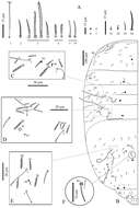
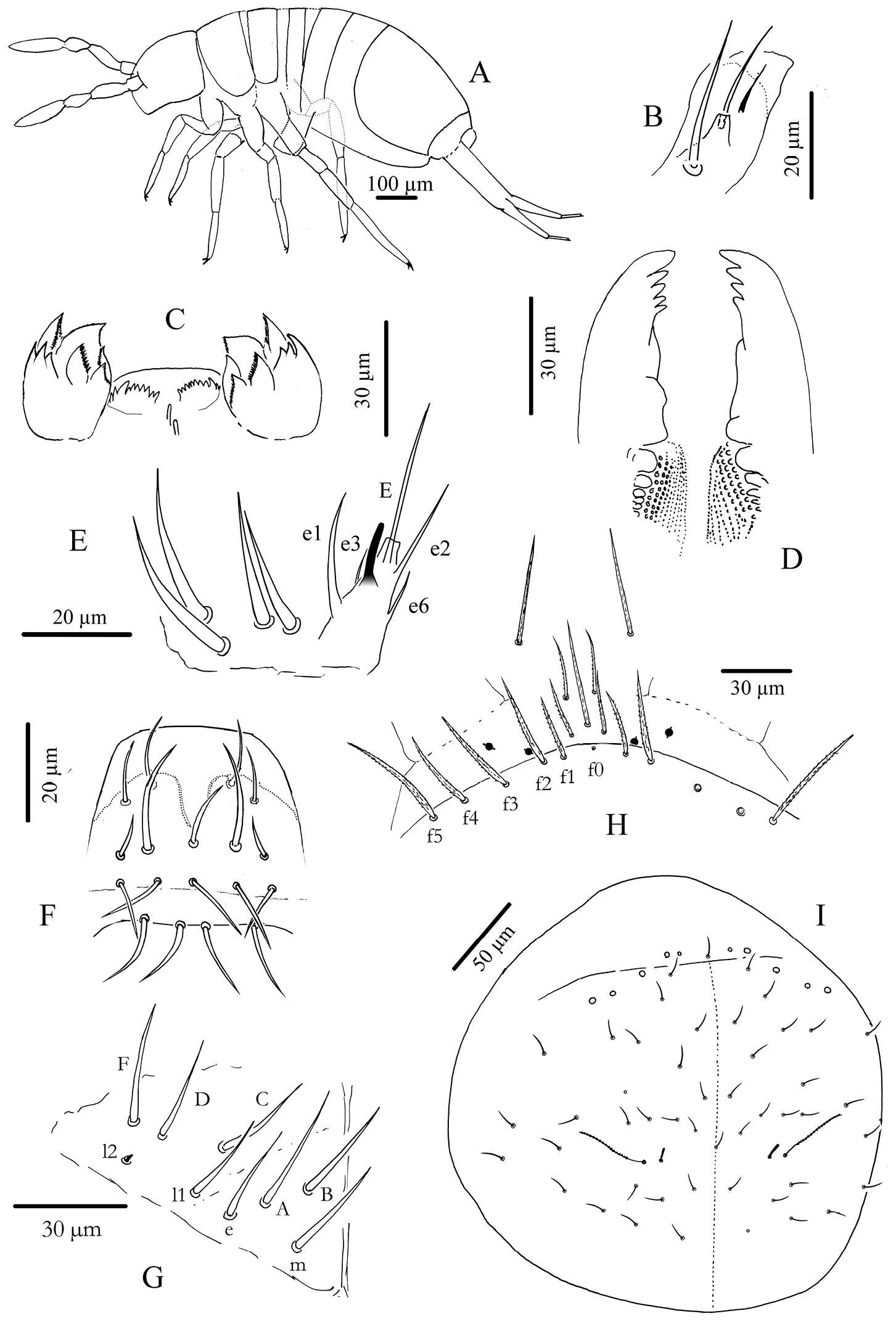
Figure 2.Cyphoderus songkhlaensis sp. n. A habitus B outer maxillary lobe C maxilla head and ventral complex of the labrum D mandible E labial palp: proximal chaetae and external papilla E F labrum, dorsal view G chaetotaxy of labial basis; frontal chaetae H frontal chaetae and pseudopores of head I dorsal chaetotaxy of head.
-
Marko Lukić, Céline Houssin, Louis Deharveng
Zookeys
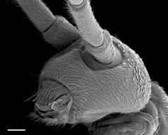
Figure 4. Tritomurus veles sp. n., head in lateral view (SEM, scale 100 μm).
-
Felipe N. Soto-Adames, Steven J. Taylor
Zookeys
Figure 1.Trogolaphysa giordanoae sp. n. habitus, scale=0.5 mm.
-
Marko Lukić, Céline Houssin, Louis Deharveng
Zookeys
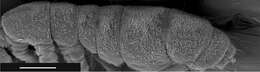
Figure 5. Tritomurus veles sp. n., body (SEM, scale 400 μm).
-
Felipe N. Soto-Adames, Steven J. Taylor
Zookeys
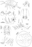
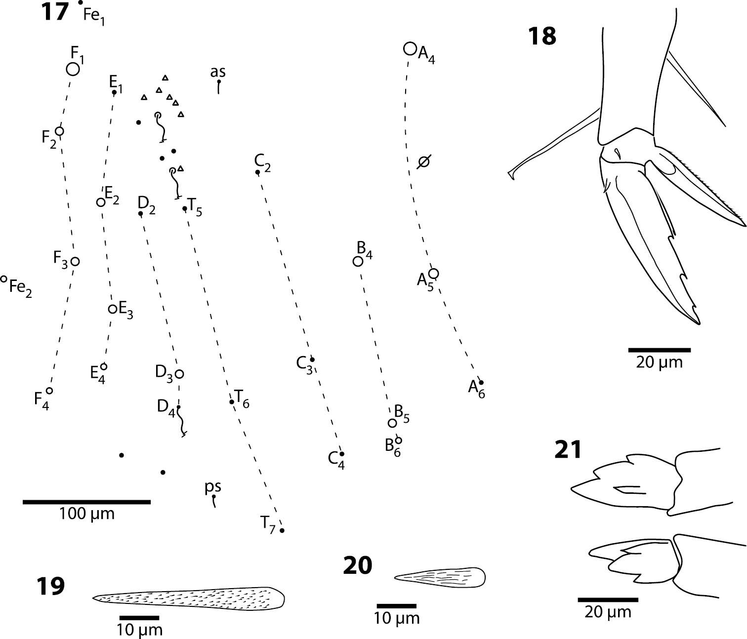
Figures 17–21.Trogolaphysa giordanoae sp. n. 17 Fourth abdominal segment dorsal chaetotaxy, diameter of circle is approximately proportional to size of macrochaeta 18 Metathoracic claw complex 19 Dens basal spine, outer row 20 Dens basal spine, inner row 21 Mucro.
-
Marko Lukić, Céline Houssin, Louis Deharveng
Zookeys
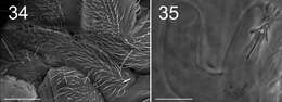
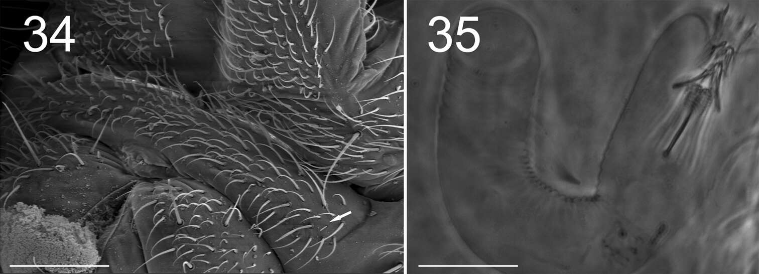
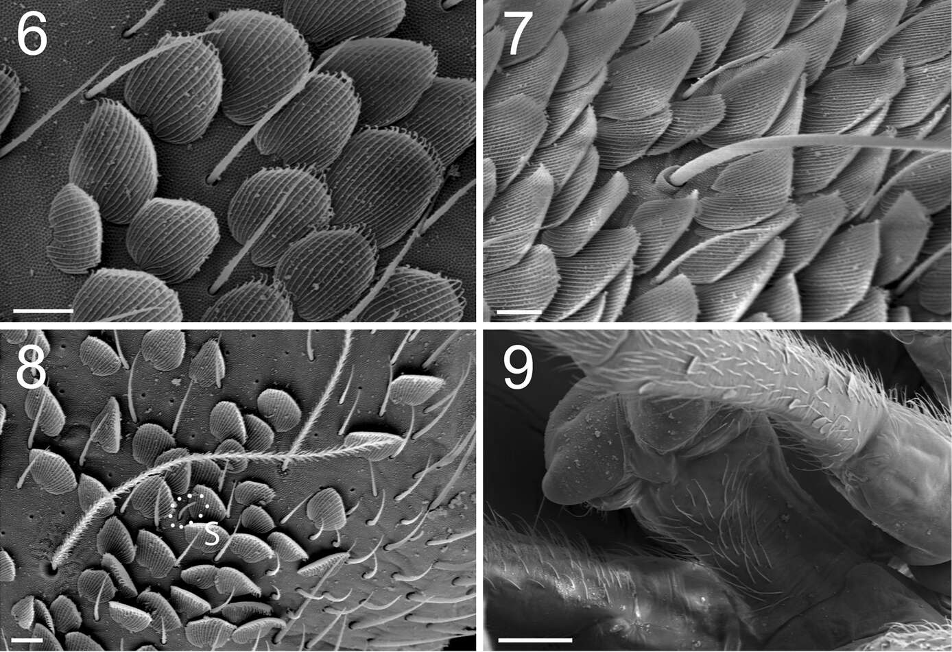
Figures 6–9. Tritomurus veles sp. n. (SEM). 6 Scales and ordinary mesochaetae on Th.II (scale 10 μm) 7 Scales, mesochaetae and macrochaeta on Abd.III (scale 10 µm) 8 Bothriotrichal area of Th.II, illustrating the presence of the four main chaetal types: bothriotricha, mesochaetae, S-chaetae (S) and scales (scale 10 µm) 9 Ventral tube in anterior view (scale 100 µm).
-
Felipe N. Soto-Adames, Steven J. Taylor
Zookeys
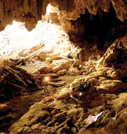
Figure 22.Trogolaphysa giordanoae sp. n. paratype habitat Okebal Ha entrance/twilight zone. Specimens were collected from a small pile of fruit bat guano near the researchers in the foreground, below a bat roost site. Sample site was much darker than it appears in this enhanced image. Photo courtesy of MES.