-
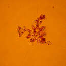
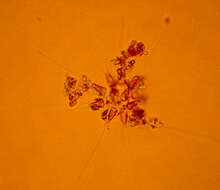
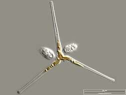
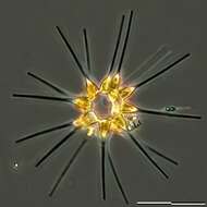
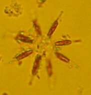
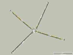
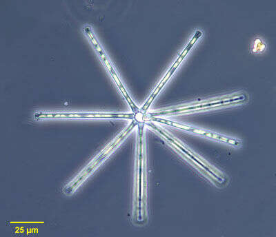
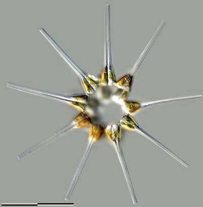
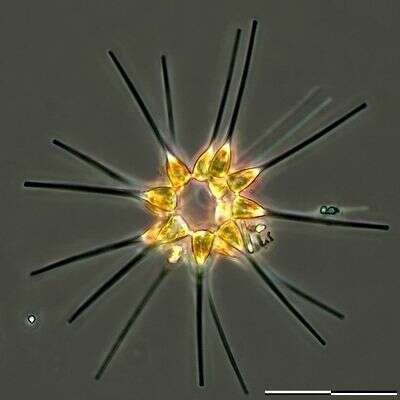
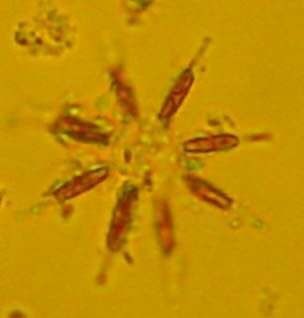
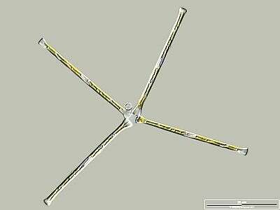
Cells are heteropolar with a triangular foot pole which narrows into a thin extension. Cells are connected by the valve faces of the foot poles into chains which are often star-shaped. A. glacialis can form extensive blooms. A. glacialis is a cosmpolitan species.
-
-
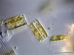
The cells in this diatom are attached side by side to form long curving belts or ribbons. The chloroplasts adhere to the walls where the cells join together. Differential interference contrast.
-
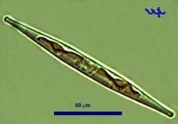
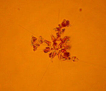
of the colonial diatom, Asterionella formosa (Hassall, 1850). The linear frustules have expanded ends. The frustules of the colony are connected by gelatinous cushions at the larger of their two ends in a radial array all in more or less the same plane The yellow chloroplasts are seen here. Blooms of this diatom may impart a disagreeable fishy taste to fresh water. Collected from freshwater pond near Boise, Idaho January 2003. Phase contrast illumination.
-
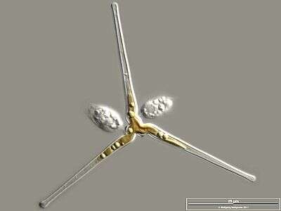
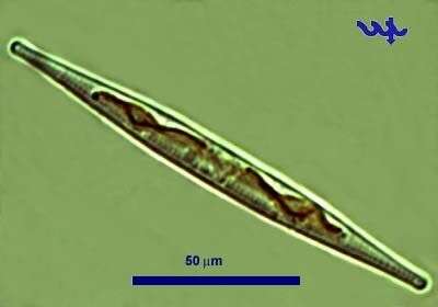
Cell accompanied by epibiotic flagellates. Scale bar indicates 50 µm. Sample from the Federsee near Lake Constance. The image was built up using several photomicrographic frames with manual stacking technique. Images were taken using Zeiss Universal with Olympus C7070 CCD camera.Image under Creative Commons License V 3.0 (CC BY-NC-SA).
-
Helical arranged cell colony. Differential interference contrast.Scale bar indicates 50 µm. Sample from North Sea near Heligoland (spring diatom bloom). The image was built up using several photomicrographic frames with manual stacking technique. Images were taken using Zeiss Universal with Olympus C7070 CCD camera.
-
-
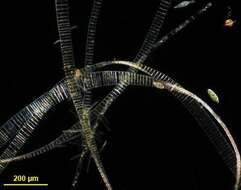
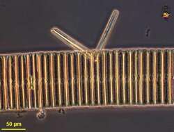
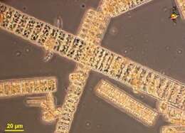
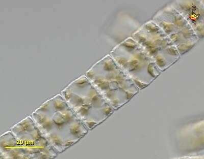
Filaments of Fragilaria, a pennate diatom in which the valves of adjacent cells are joined by small hooks to form long tough filaments. Dark ground illumination.
-
Helical arranged cell colony. Phase contrast.Scale bar indicates 50 µm. Sample from North Sea near Heligoland (spring diatom bloom). The image was built up using several photomicrographic frames with manual stacking technique. Images were taken using Zeiss Universal with Olympus C7070 CCD camera.
-
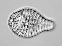
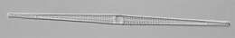
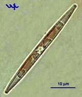
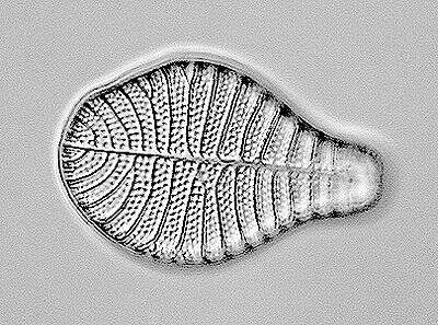
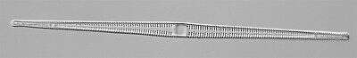
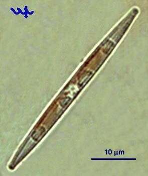
A very common pennate diatom in the summer plankton of Lake Kinneret in recent years. Identification uncertain.
-
Filaments of Fragilaria, a pennate diatom in which the valves of adjacent cells are joined by small hooks to form long tough filaments. Differential interference contrast image emphasizing plastids.
-
This is a northern cold water to temperate species. Like A. glacialis it can form star shaped chains.
-
A very common pennate diatom in the summer plankton of Lake Kinneret in recent years.
-
Filaments of Fragilaria, a pennate diatom in which the valves of adjacent cells are joined by small hooks to form long tough filaments. Differential interference contrast image emphasizing plastids, nuclei and nucleoli.
-
-
Filaments of Fragilaria, a pennate diatom in which the valves of adjacent cells are joined by small hooks to form long tough filaments. Phase contrast image with attached Synedra.
-
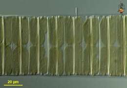
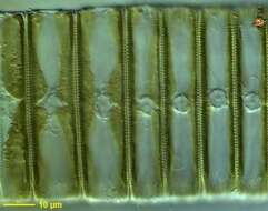
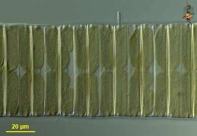
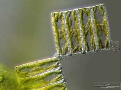
Colony of Diatoma vulgare. The mucilaginous connection material is shown. Scale bar indicates 25 µm. Sample from Lake Constance (Bodensee, Southern Germany) near Bodman. Images were taken using Zeiss Universal with Olympus C7070 CCD camera.
-
Scale bar indicates 25 µm. Sample from the Lake Constance (vicinity of Bodman). The image was built up using several photomicrographic frames with manual stacking technique. Images were taken using Zeiss Universal with Olympus C7070 CCD camera.Image under Creative Commons License V 3.0 (CC BY-NC-SA).
-
Colony of Diatoma vulgare. The mucilaginous connection material is shown. Scale bar indicates 50 µm. Sample from Lake Constance near Bodman. Images were taken using Zeiss Universal with Olympus C7070 CCD camera.Image under Creative Commons License V 3.0 (CC BY-NC-SA).
-
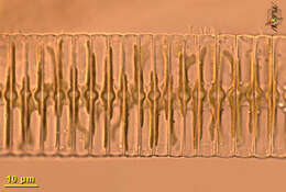
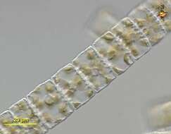
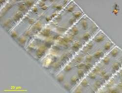
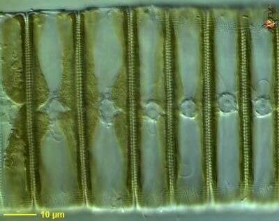
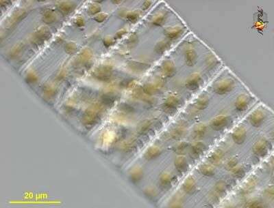
Diatoma (die-at-home-a ... ish), filamentous centric diatom. Cells are not circular in cross section but compressed. Cell with internal strengthening ridges. Many small peripheral chloroplasts and a central nucleus. May form very long filaments, this sample collected from a large (easily visible with the naked eye) clump. Differential interference contrast.
-
Diatoma (die-at-home-a ... ish), filamentous centric diatom. Cells are not circular in cross section by compressed - as can be seen in the cell to the left. Cell with internal strengthening ridges. Many small peripheral chloroplasts and a central nucleus. May form very long filaments, this sample collected from a large (easily visible with the naked eye) clump. Differential interference contrast.
-
Diatoma (die-at-home-a ... ish), filamentous centric diatom. Cells are not circular in cross section but compressed. Cell with internal strengthening ridges. Many small peripheral chloroplasts and a central nucleus. May form very long filaments, this sample collected from a large (easily visible with the naked eye) clump. Differential interference contrast.
-
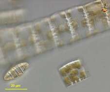
Diatoma (die-at-home-a ... ish), filamentous centric diatom. Cells are not circular in cross section but compressed. Cell with internal strengthening ridges. May form very long filaments, this sample collected from a large (easily visible with the naked eye) clump. Phase contrast.