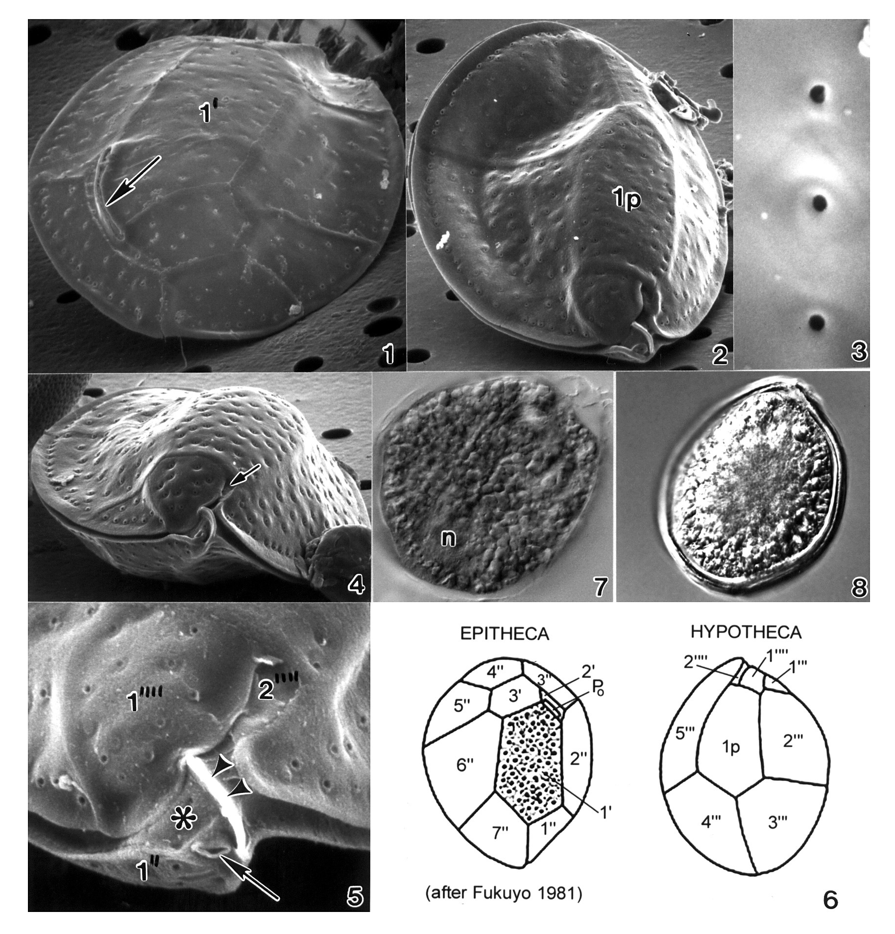
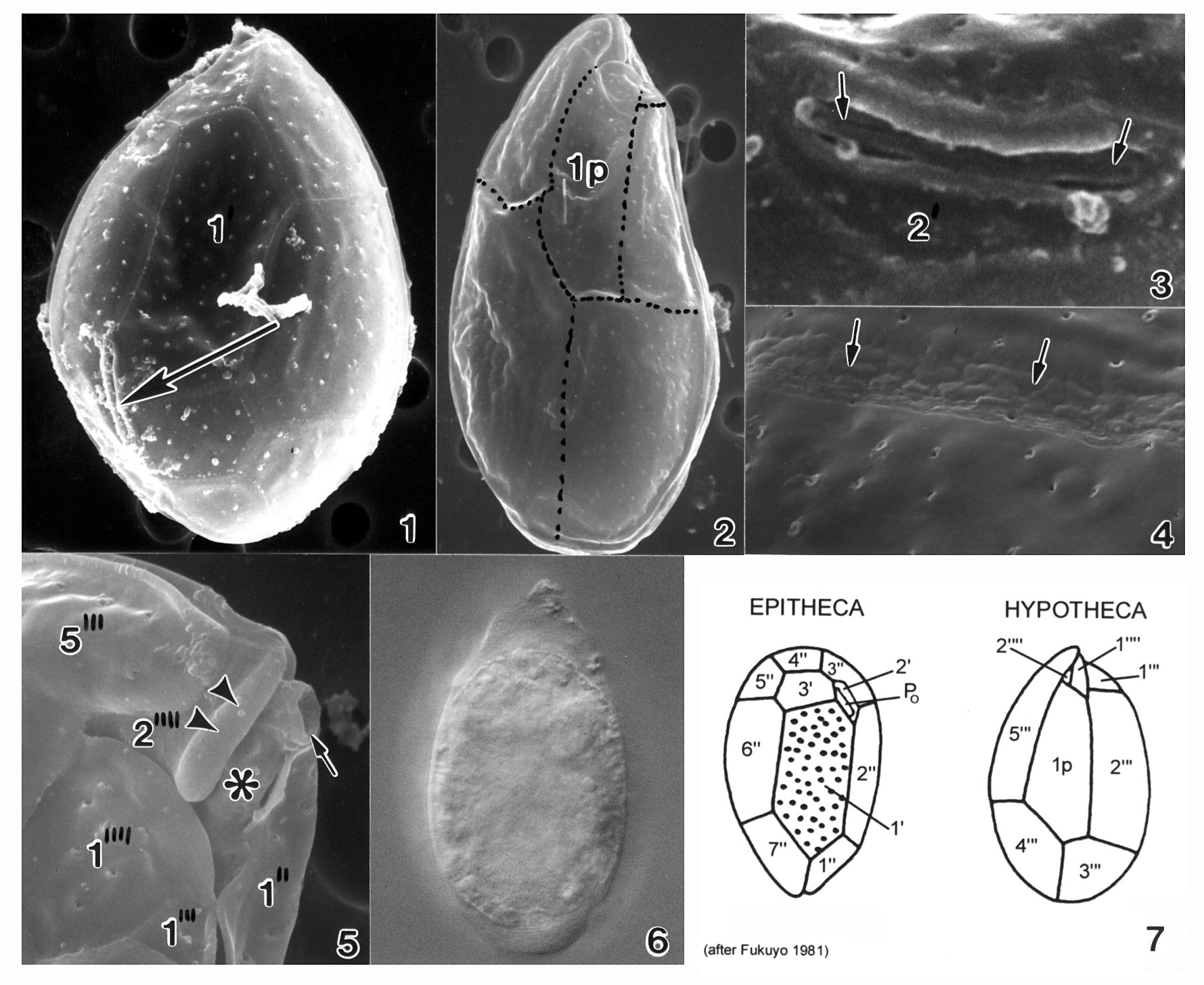
Figs 1-5. Cells of Ostreopsis marinus sp. nov. Figs 1-4. Scanning electron microscopy. Fig. 1. Morphology of epithecal plates and position of apical pore plate are shown (Po). Fig. 2. Hypothecal plates. Fig. 3. Antapical plate 1" is larger; plate 2" is tiny. Thecal surface is smooth with small, evenly distributed pores (arrows). Fig. 4. The ventral opening (Vo) is situated in the cingulum adjacent to a ridged plate (Rp). Fig. 5. Epifluorescence light microscopy of hypothecal plates; Ip plate is in the center.
EMu: HOLOTYPE SEM NEGATIVE # 212055; SEM STUB # 212; FIELD # 96/10; ACCESSION # 2002799; CATALOG #1537; FIGURE # 1.
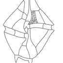
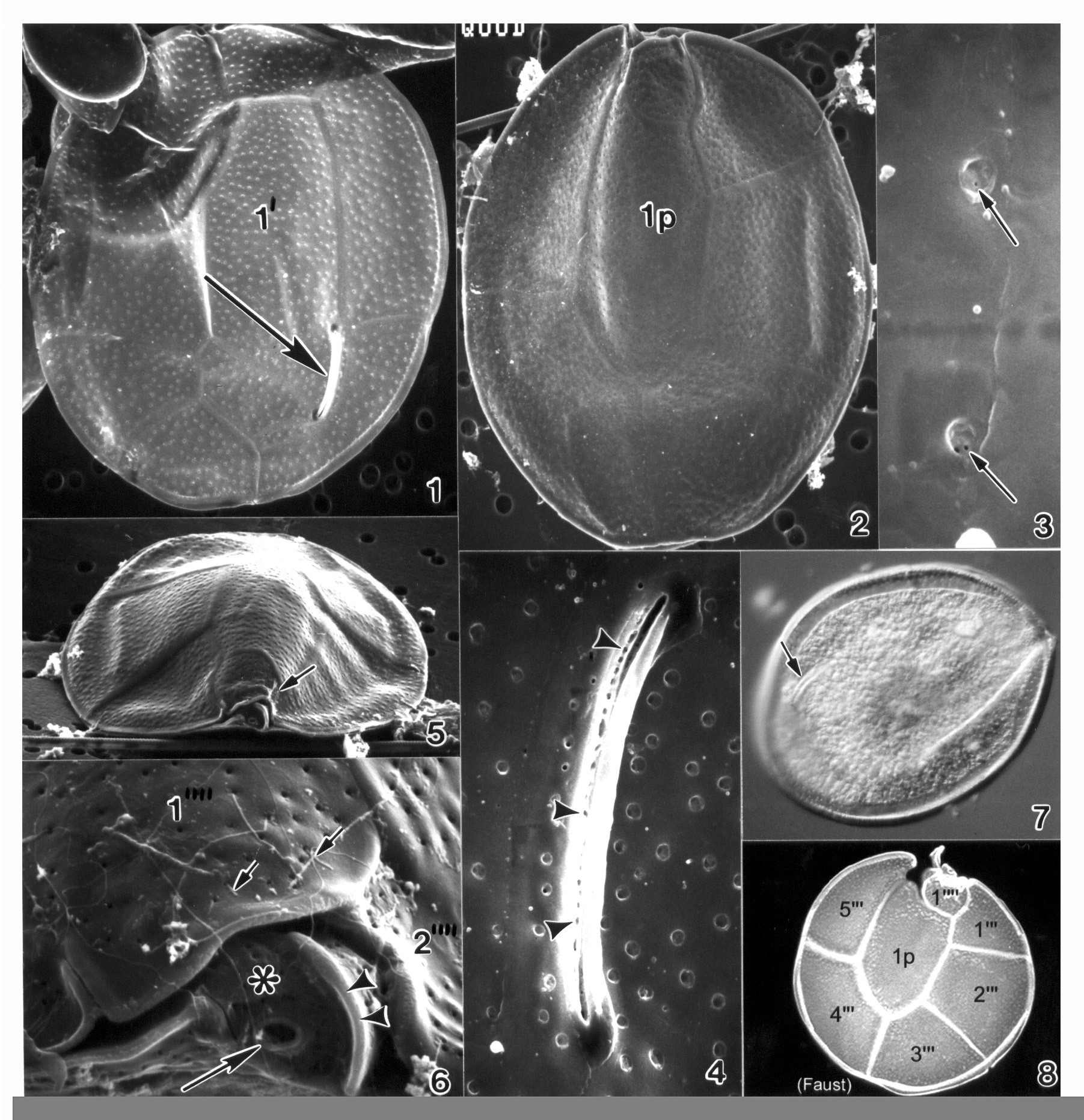
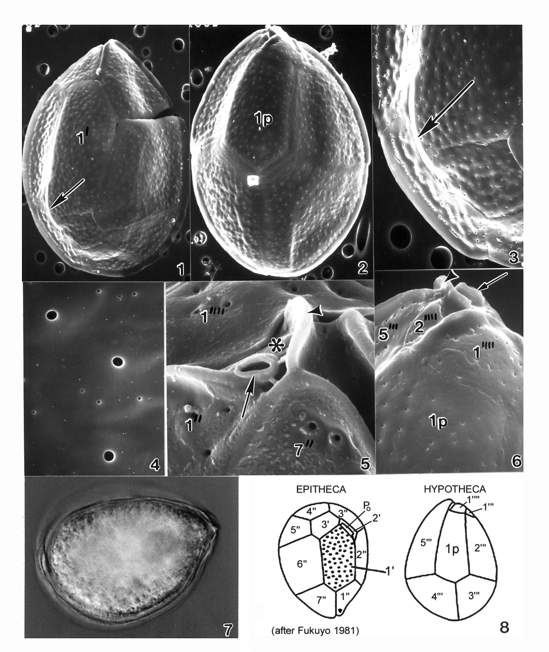
Plate 33. Ostreopsis mascarenensis. Figs. 1-5. SEM. Fig. 1. Epitheca: inner thecal surface. Cell very large, broadly ovate, large plates. Plate 1' elongate and hexagonal. Apical pore plate (Po) nearly straight. Fig. 2. Hypotheca: plate 1p long and wide. Fig. 3. Smooth cell surface with round pores; pores with two small openings (arrows). Fig. 4. Po with long narrow apical pore; small pores line the opening (arrowheads). Figs. 5-6. Ventral view of epitheca. Fig. 5. Cell compressed anterio-posteriorly; cingulum narrow with smooth edge. Small sulcus hidden (arrow). Fig. 6. Location of ventral opening (large arrow), ventral plate (asterisk), and rigid plate (arrowheads) within cingulum. Pores with ejected trichocysts (small arrows). Fig. 7. LM. Epitheca: Po (arrow) and cingulum in focus. Fig. 8. Line drawing: hypotheca plate arrangement.