-
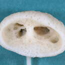
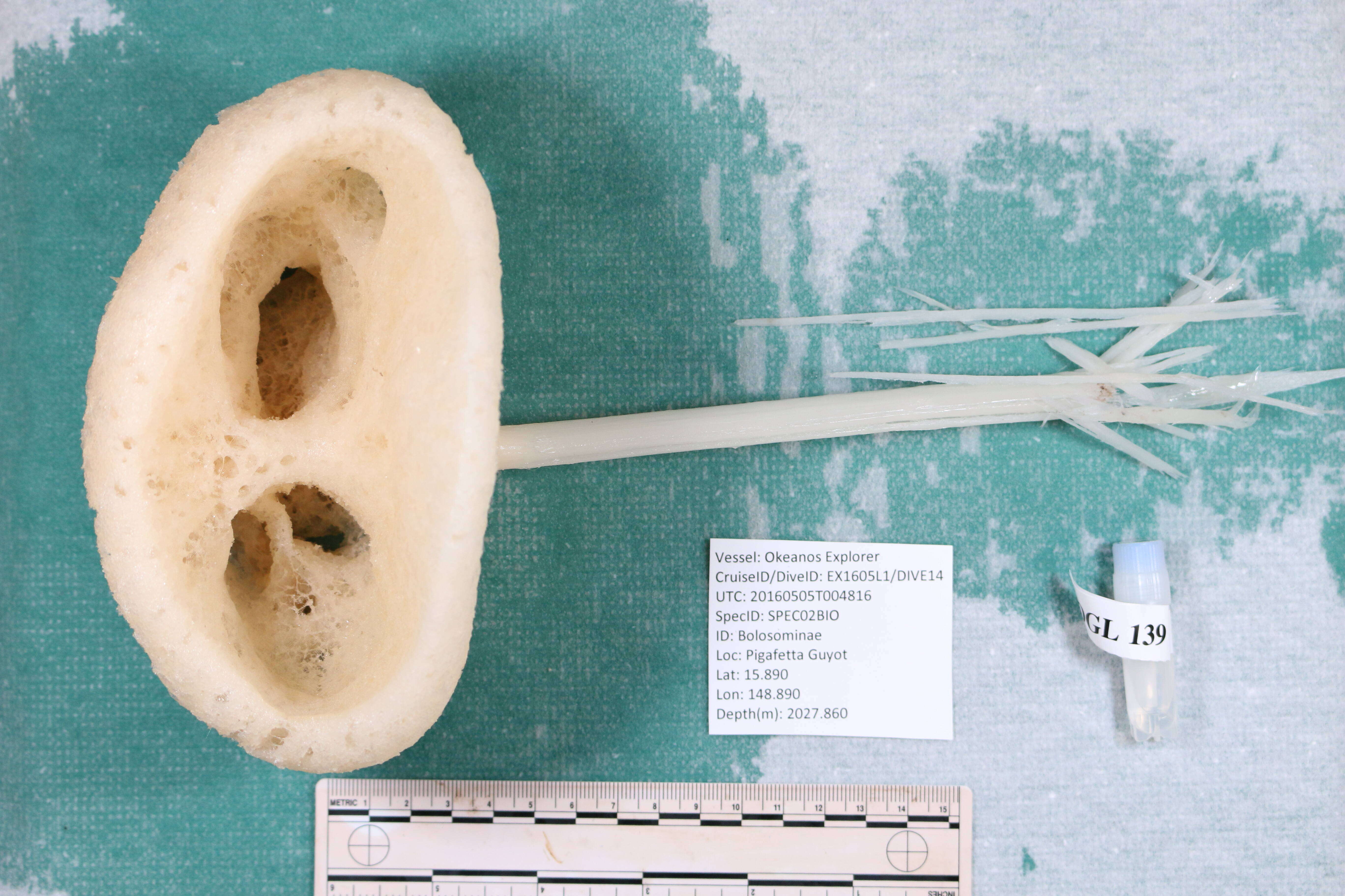
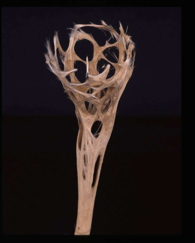
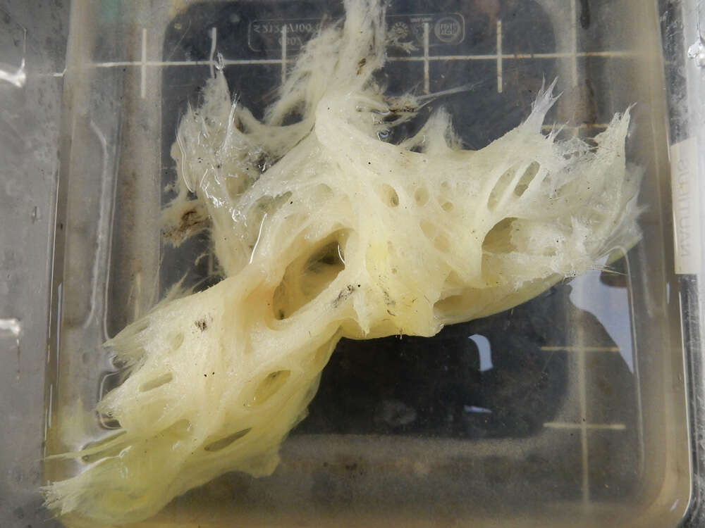
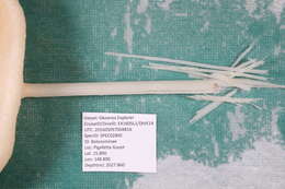
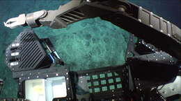
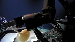
USNM 1424107 Lab Image 2; EX1605L1_20160505T004816_D2_DIVE14_SPEC02BIO_L06.JPG
-
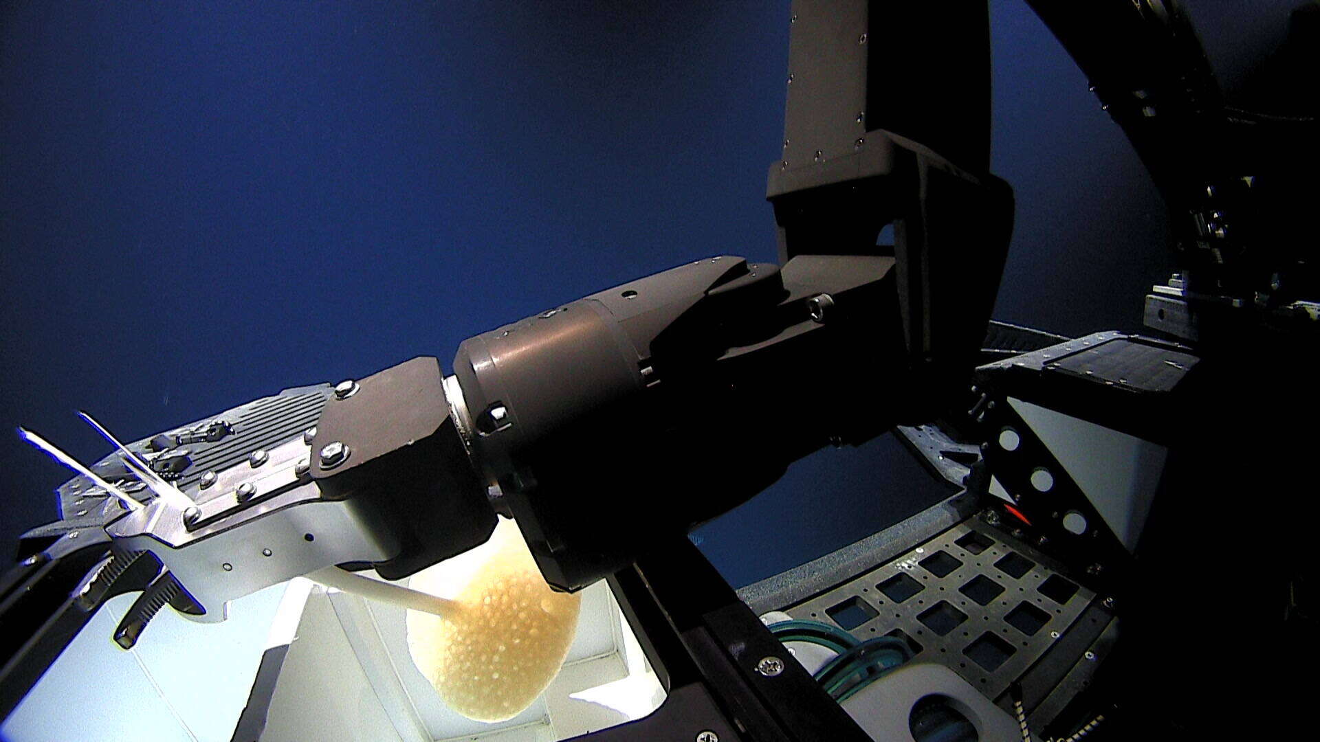
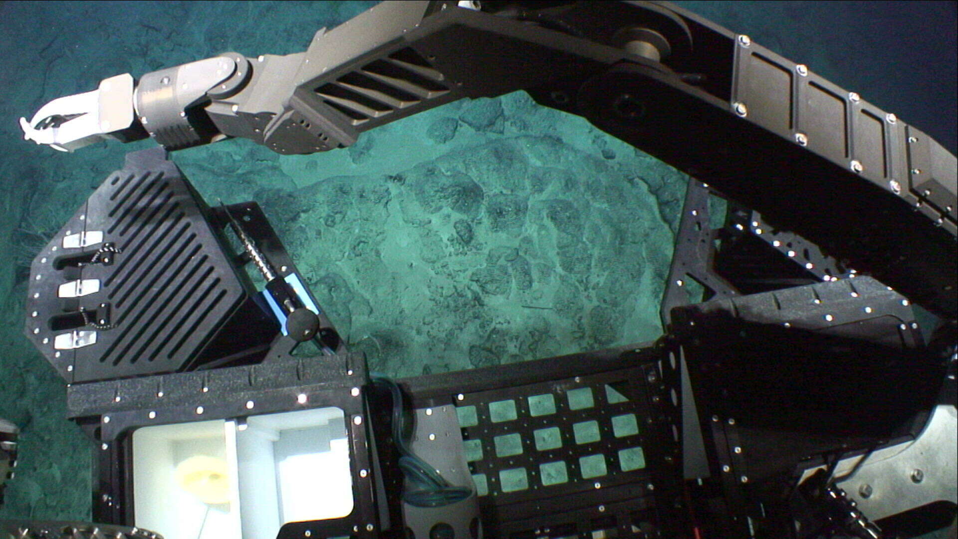
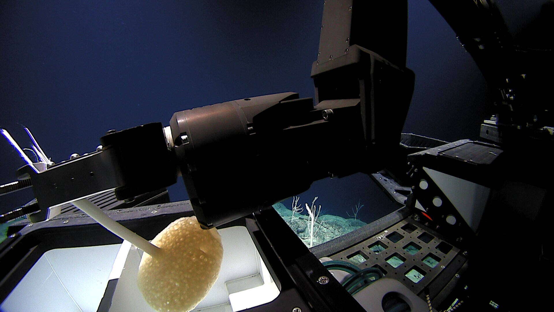
USNM 1424107 Frame Grab 7; EX1605L1_IMG_20160505T004555Z_D2_DIVE14_SPEC02BIO_04.jpg
-
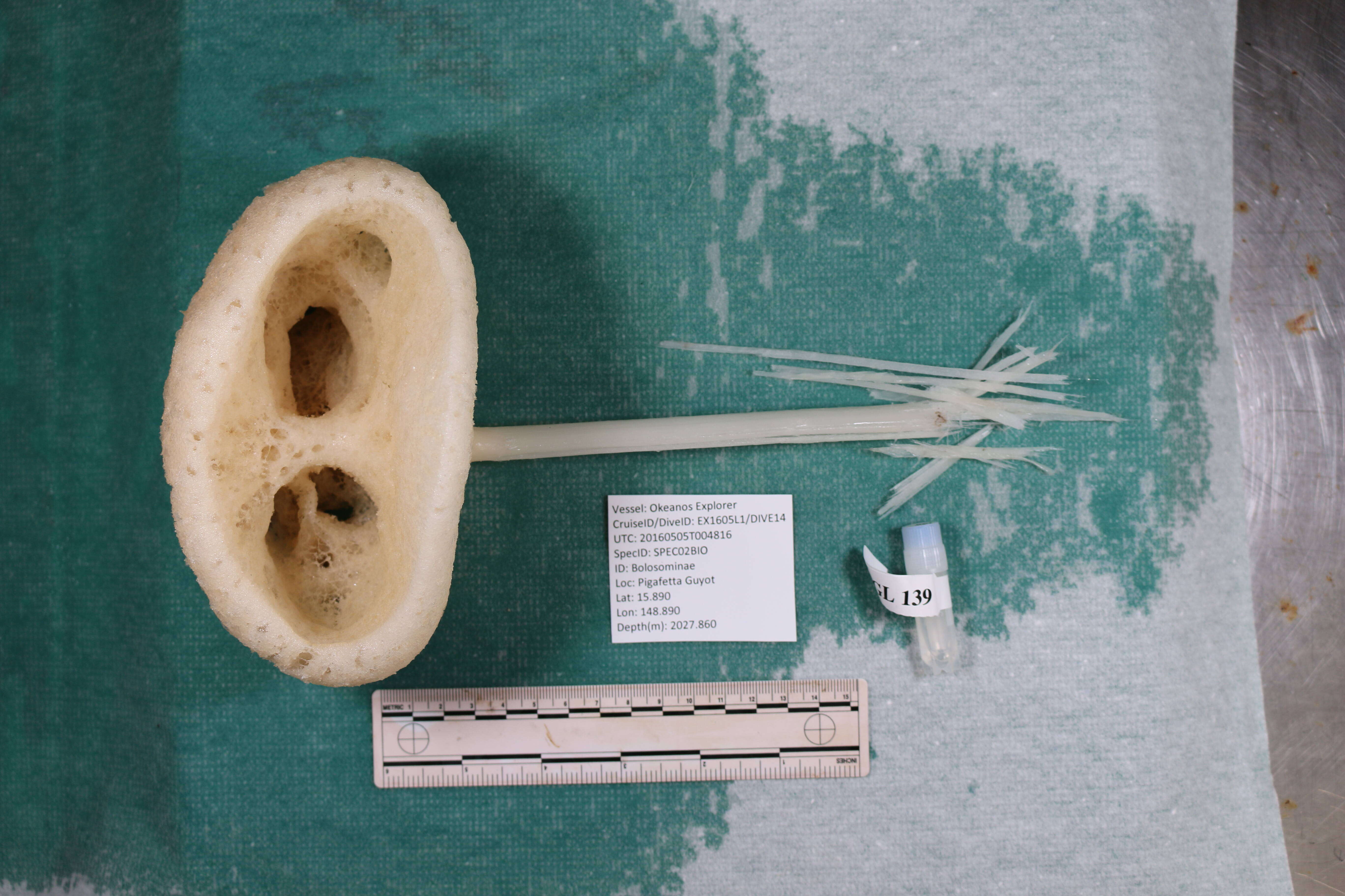
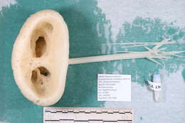
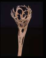
USNM 1424107 Lab Image 3; EX1605L1_20160505T004816_D2_DIVE14_SPEC02BIO_L07.JPG
-
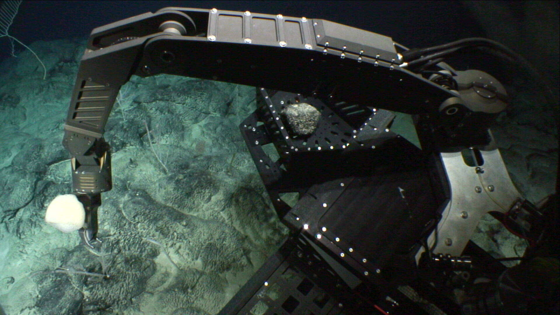
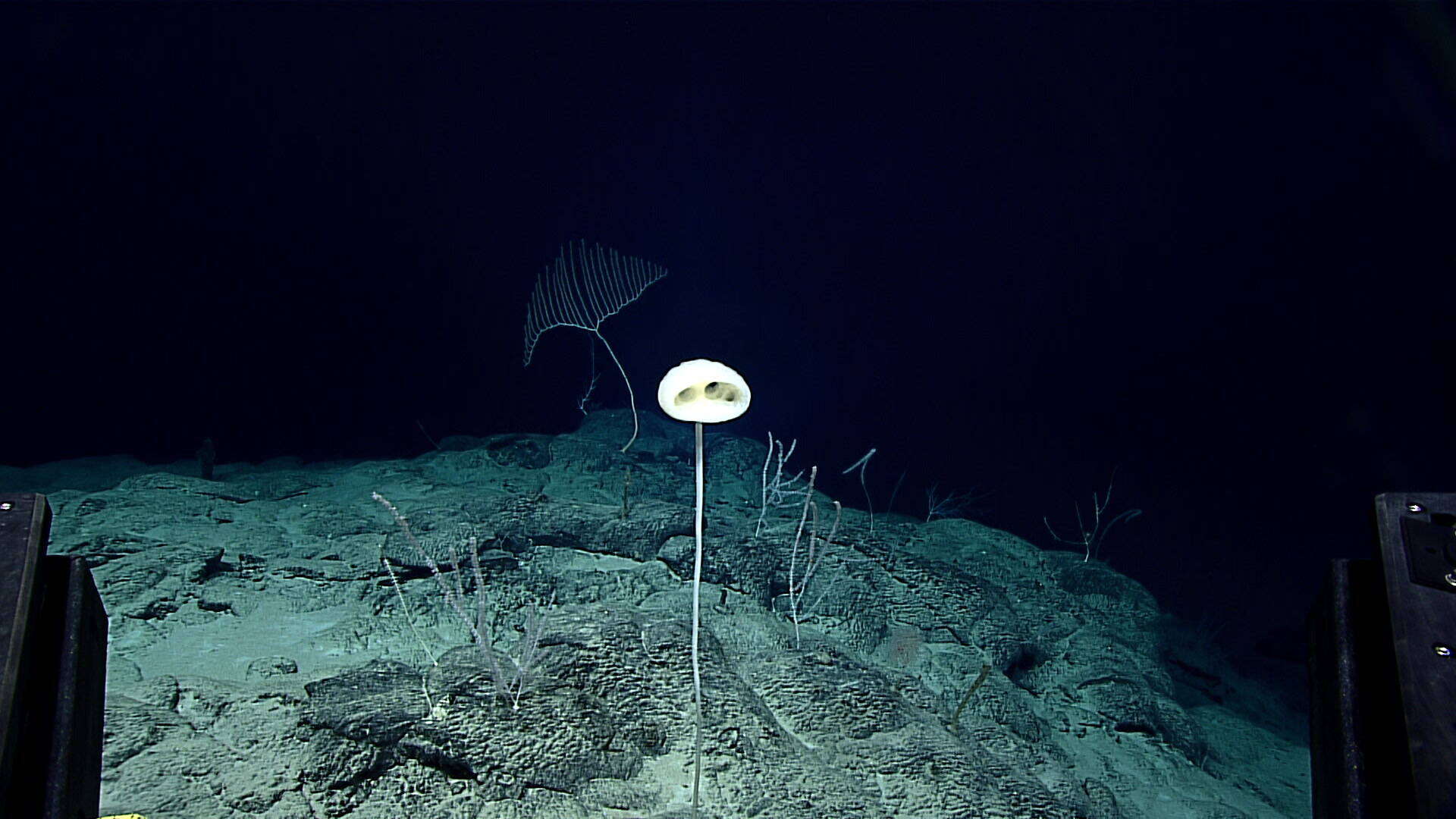
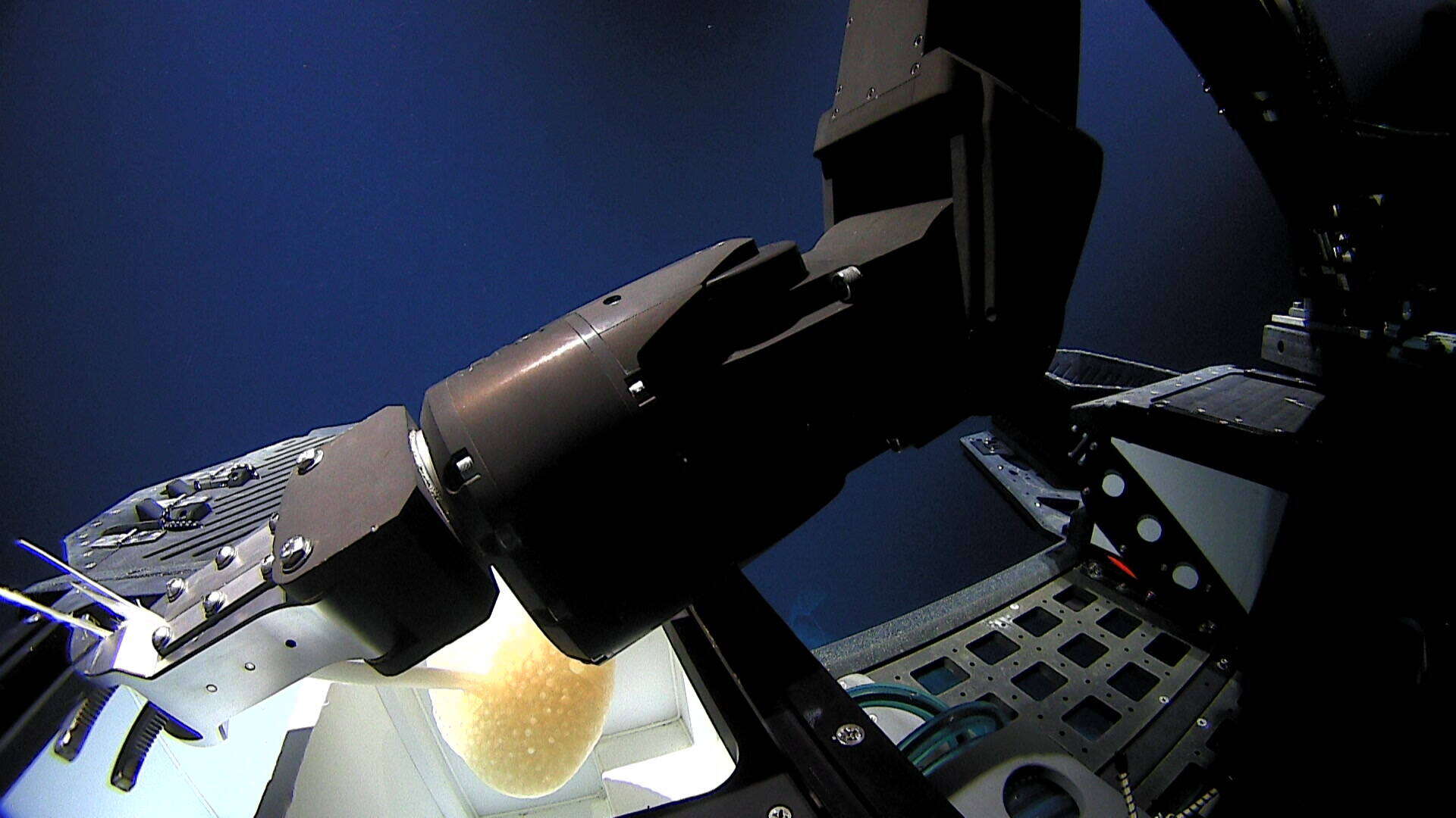
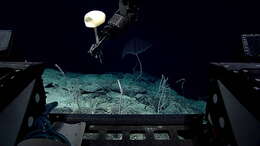
USNM 1424107 Frame Grab 5; EX1605L1_IMG_20160505T004158Z_D2_DIVE14_SPEC02BIO_02.jpg
-
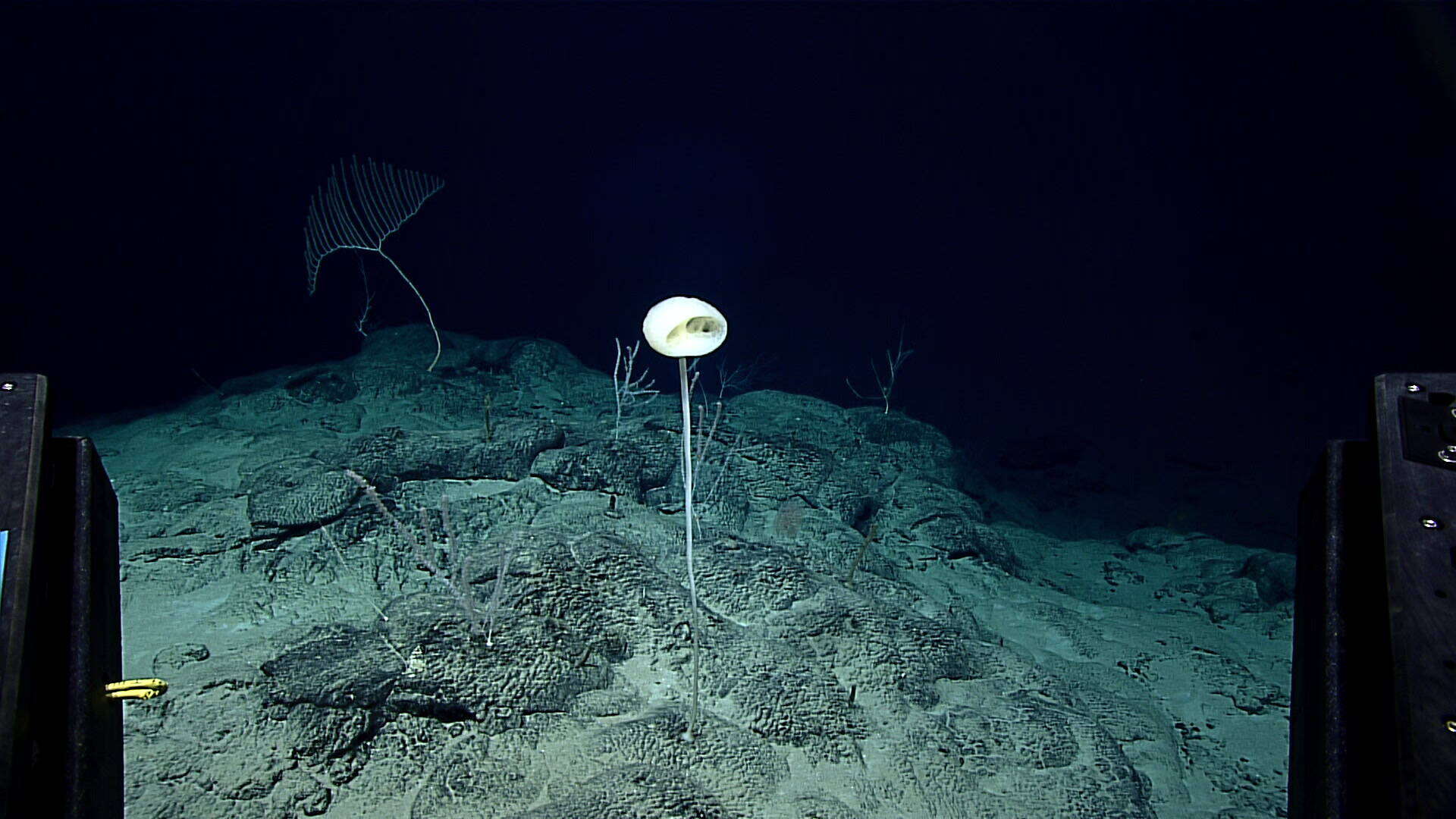
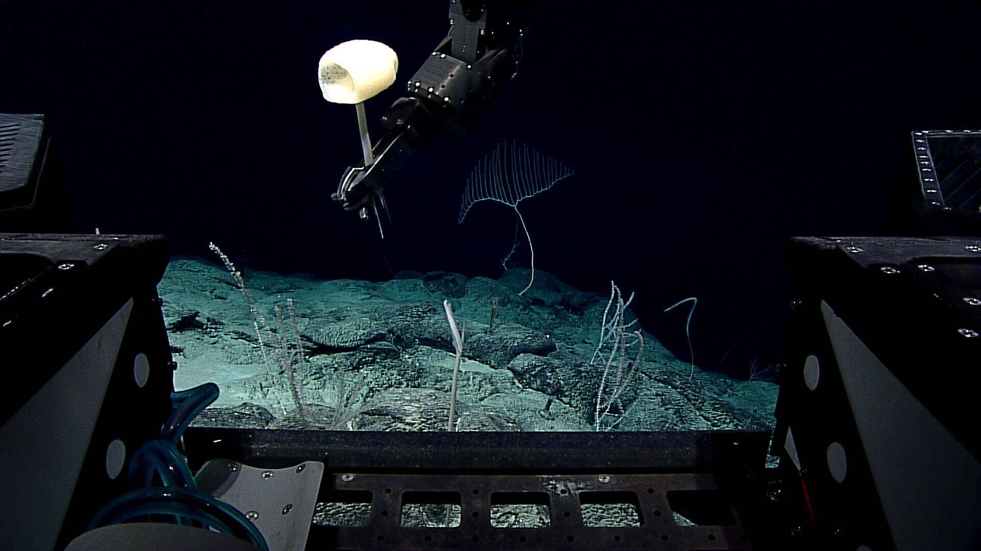
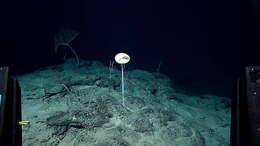
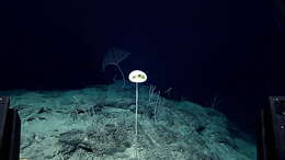
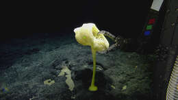
USNM 1424107 Frame Grab 1; EX1605L1_IMG_20160505T003042Z_D2_DIVE14_SPEC02BIO_01.jpg
-
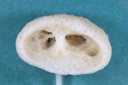
USNM 1424107 Lab Image 1; EX1605L1_20160505T004816_D2_DIVE14_SPEC02BIO_L04.JPG
-
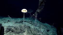
USNM 1424107 Frame Grab 9; EX1605L1_IMG_20160505T004759Z_D2_DIVE14_SPEC02BIO_02.jpg
-
USNM 1424107 Lab Image 4; EX1605L1_20160505T004816_D2_DIVE14_SPEC02BIO_L10.JPG
-
USNM 1424107 Frame Grab 2; EX1605L1_IMG_20160505T003310Z_D2_DIVE14_SPEC02BIO_01.jpg
-
USNM 1424107 Frame Grab 4; EX1605L1_IMG_20160505T004153Z_D2_DIVE14_SPEC02BIO_01.jpg
-
USNM 1424107 Frame Grab 8; EX1605L1_IMG_20160505T004642Z_D2_DIVE14_SPEC02BIO_04.jpg
-
USNM 1424107 Frame Grab 3; EX1605L1_IMG_20160505T004048Z_D2_DIVE14_SPEC02BIO_01.jpg
-
USNM 1424107 Frame Grab 6; EX1605L1_IMG_20160505T004407Z_D2_DIVE14_SPEC02BIO_04.jpg
-
published in Rarest of the Rare
-
-
-
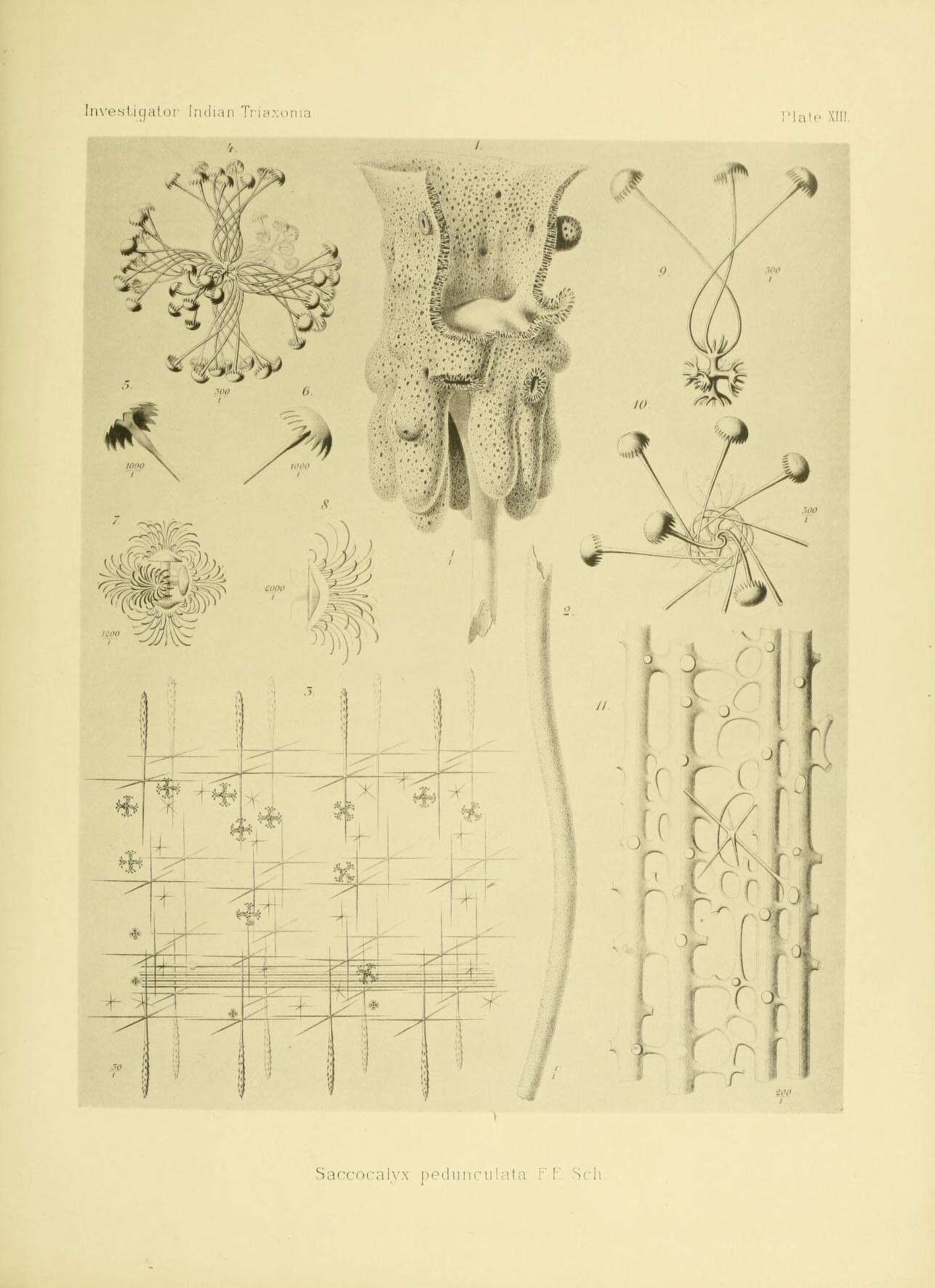
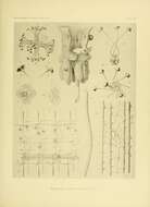
An account of the Indian Triaxonia collected by the Royal Indian Marine Survey Ship Investigator /.
Calcutta :Printed by order of the Trustees of the Indian Museum,1902..
biodiversitylibrary.org/page/9628654
-
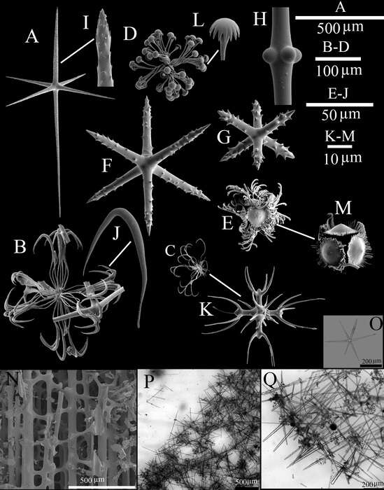
Image courtesy of the NOAA Office of Ocean Exploration and Research, 2016 Deepwater Exploration of the Marianas. Image is part of Figure 8 from the following publication:(https://marinespecies.org/aphia.php?p=sourcedetails&id=382781)
-
-
A, dermal or atrial hexactin; B, drepanocome I; C, drepanocome II; D, spirodiscohexaster; E, plumicome; F, microhexactin I; G, microhexactin II; H, detail of tubercles in the middle of a diactine; I, detail of the pinular ray of a hexactin; J, detail of the hook-like secondary ray of a drepanocome I; K, detail of the middle part of a drepanocome II; L, detail of the tooth disc of a spirodiscohexaster; M, detail of the whorl of a plumicome; N, spicules of the peduncle; O–Q, LM images of spicule and skeleton; O, choanosomal hexactin; P, tangential view of choanosomal structure; Q, transversal view of choanosomal structure.