-
Asker, Akershus, Norge
-
Asker, Akershus, Norge
-
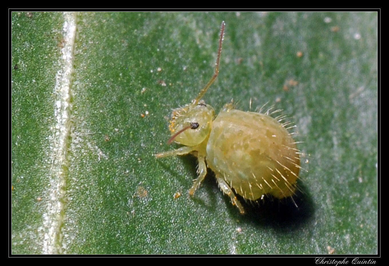
Maria Cleide de Mendonça, Eduardo A. Abrantes, Ana Carolina R. Neves
Zookeys
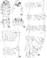
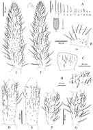
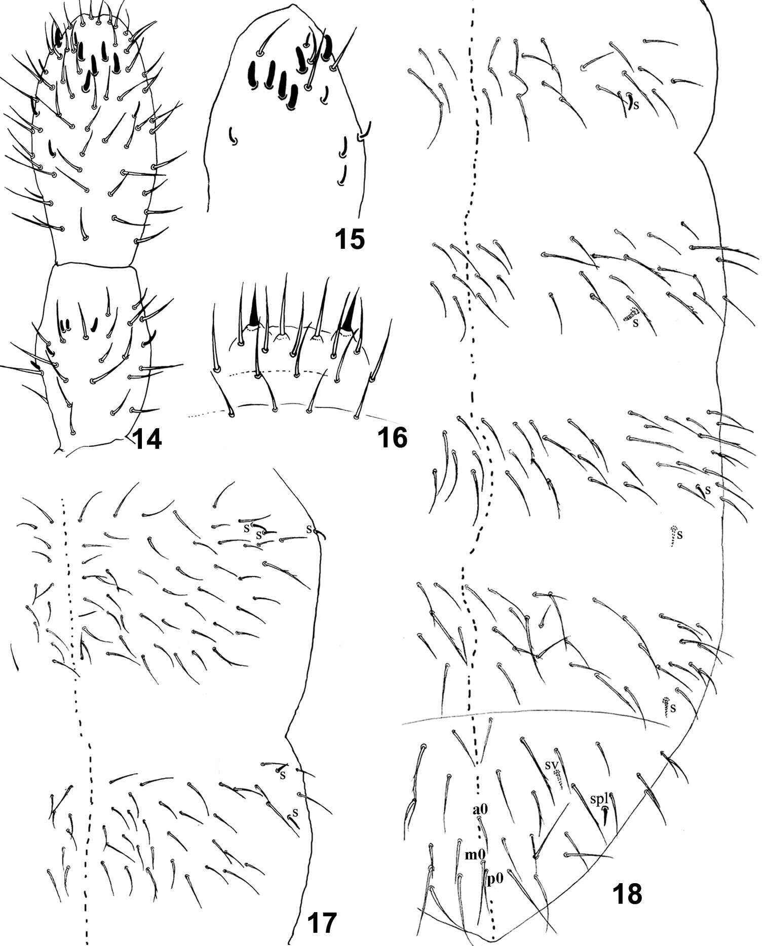
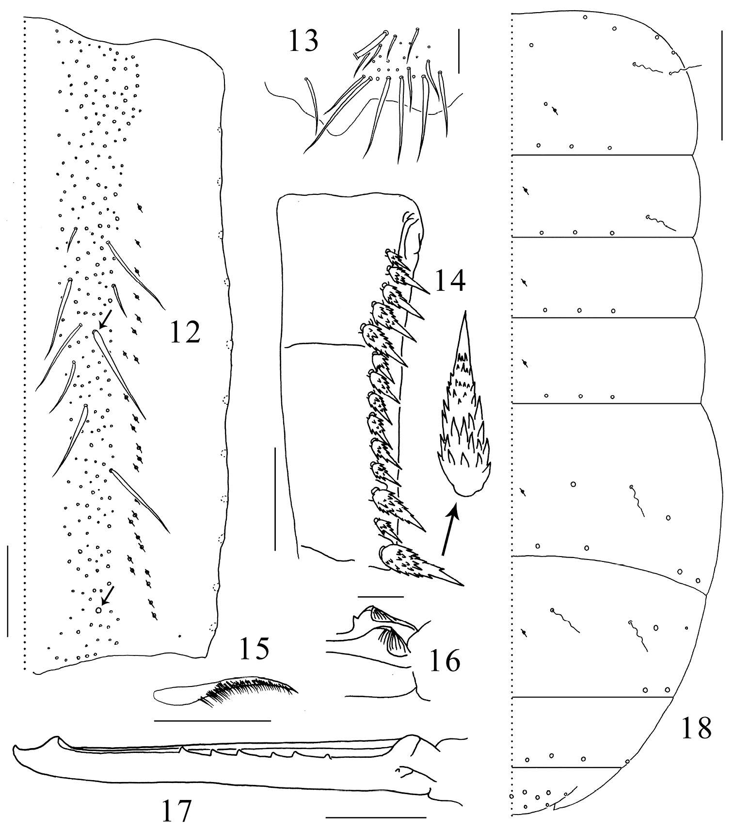
Figure 14–18.Isotomiella uaisp.n. 14 Ant III-IV Dorsal view 15 Sensillary pattern of Ant IV 16 Labral chaetae 17 Dorsal chaetotaxy of Th II-III 18 Dorsal chaetotaxy of Abd I-VI.
-
Gabriel C. Queiroz, Maria Cleide de Mendonça
Zookeys
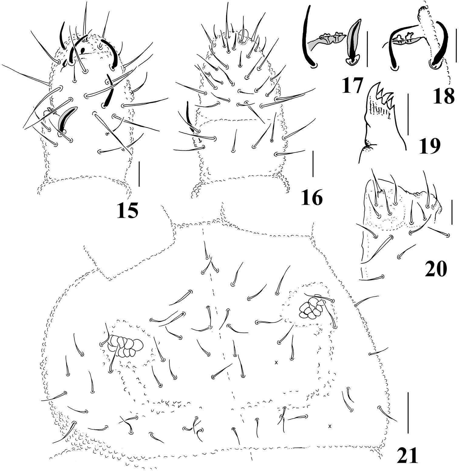
Figures 15–21.Micronella longisensilla sp. n. 15 Dorsal view of Ant III–IV 16 Ventral view of Ant III–IV 17 Detail of Ant III organ 18 Detail of Ant III organ (same specimen of Fig. 17, right antennae) 19 Maxilla 20 Labium 21 Head. Scale bars: 10μm (15–20); 20μm (21). x represents missing chaeta.
-
Xiang-Qun Yuan, Zhi-Xiang Pan
Zookeys
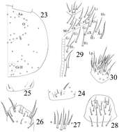
Figures 23–29.Sinella triseta sp. n. 23 dorsal cephalic chaetotaxy 24 basal chaetae of Ant. I 25 basal chaetae of Ant. II 26 Ant. III organ 27 clypeus 28 labrum 29 labial base 30 labial palp.
-
Sopark Jantarit, Chutamas Satasook, Louis Deharveng
Zookeys
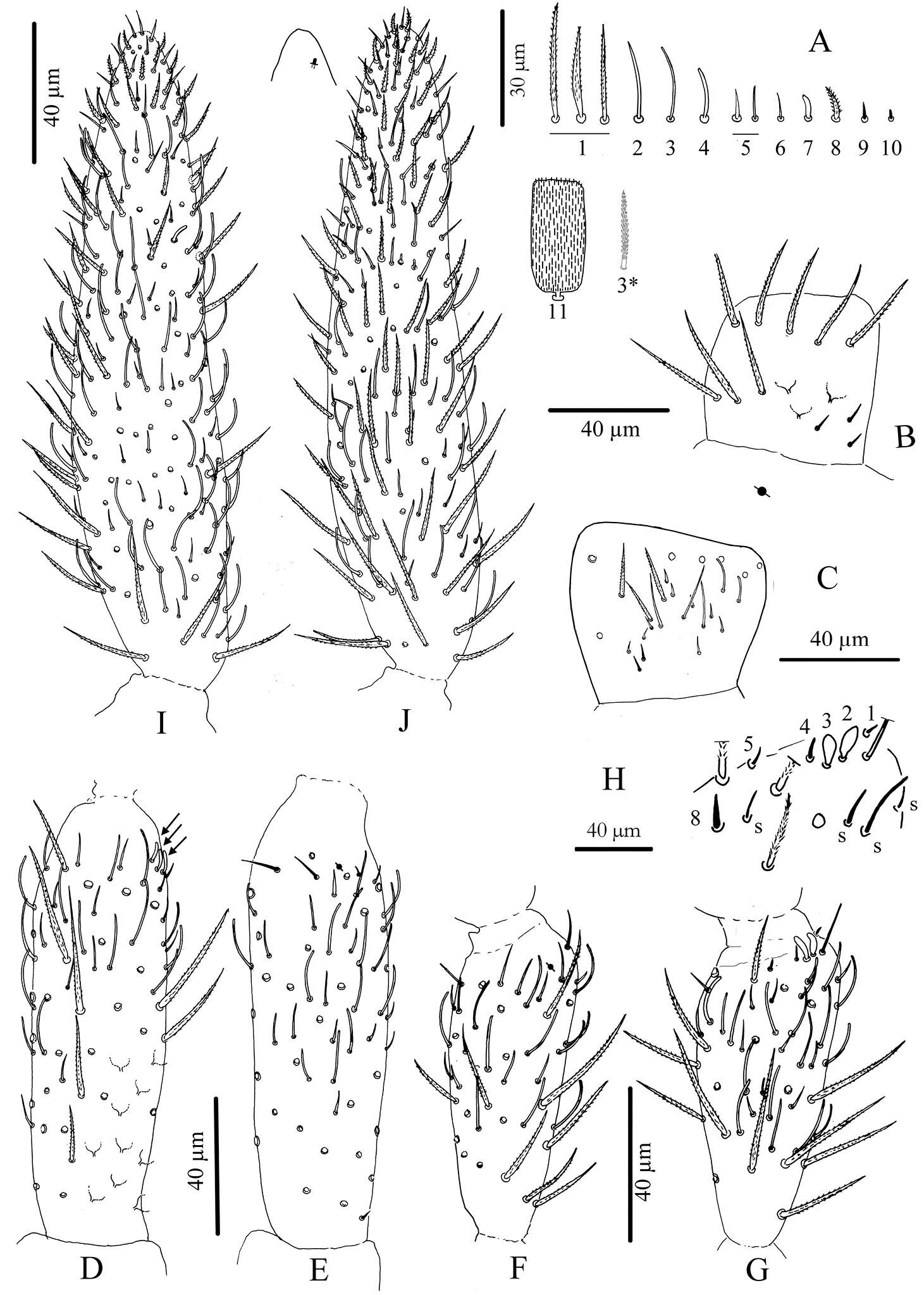
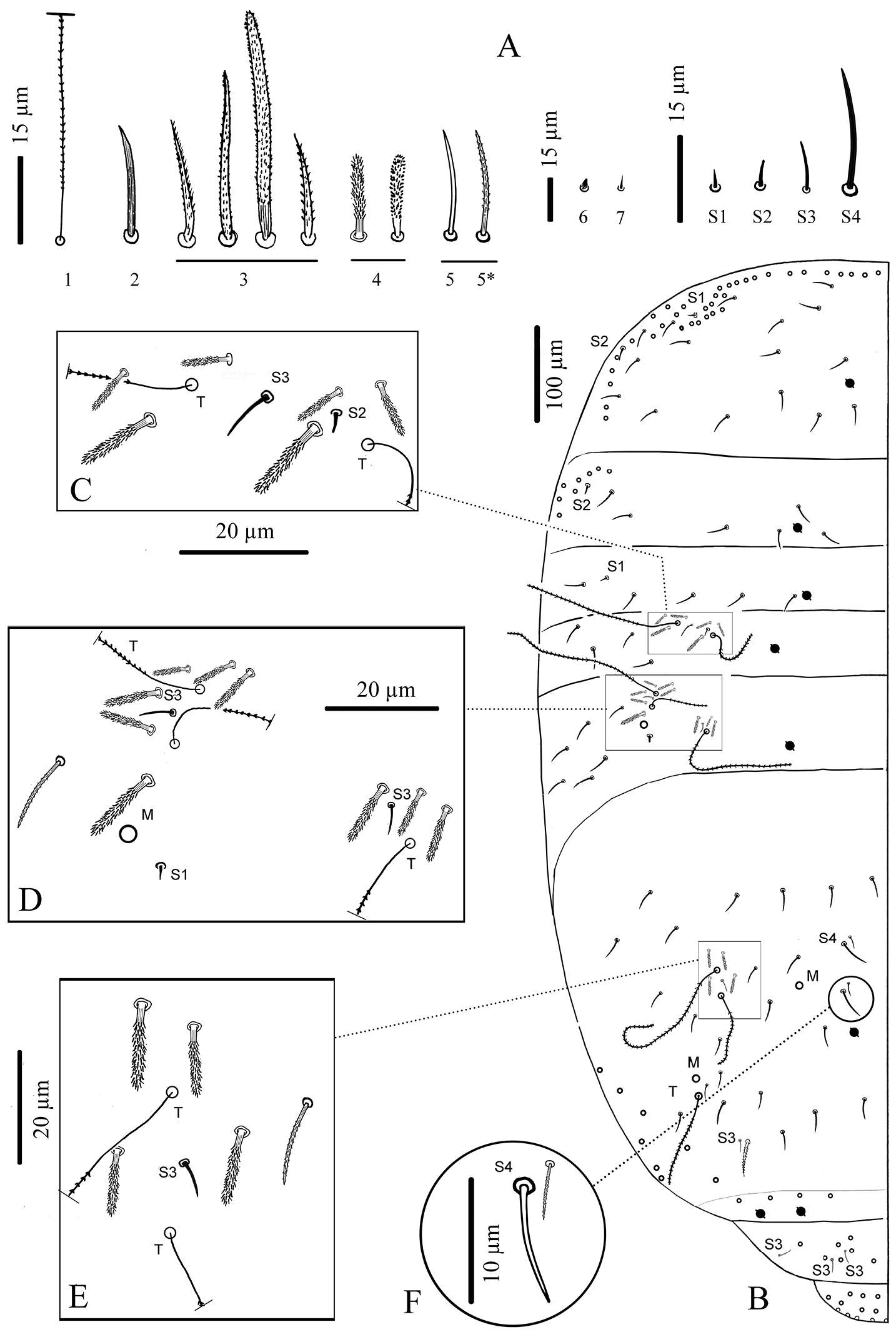
Figure 3.Cyphoderus songkhlaensis sp. n. continued A chaetae of antenna drawn from optical microscope, except 3* derived from SEM image B dorsal side of right Ant.I C ventral side of right Ant.I D dorsal side of right Ant.II; the apical swollen sens of type-7 are indicated by arrows E ventral side of right Ant.II with apical pseudopore F ventral side of right Ant.III with apical pseudopore G dorsal side of right Ant.III H distal organite of Ant.III I ventral side of Ant.IV J dorsal side of Ant.IV with separate view of the subapical organite (left).
-
Daoyuan Yu, Feng Zhang, Louis Deharveng
Zookeys
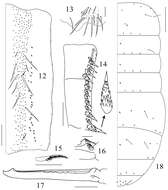
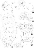
Figures 12–18.Tomocerus postantennalis sp. n. 12 dorsal face of manubrium (right side; prominent chaetae arrowed) 13 disto-dorsal chaetae on manubrium (left side) 14 dental spines (left side) 15 feathered chaeta on dens 16 basal teeth of left mucro 17 right mucro 18 body chaetotaxy. Scale bars: 12, 14 = 100μm; 13, 17 = 50 μm; 15, 16 = 21 μm; 18 = 400 μm. Large circles: macrochaetae; small circles: mesochaetae; wavy lines: bothriotricha; circles with a slash: pseudopores.
-
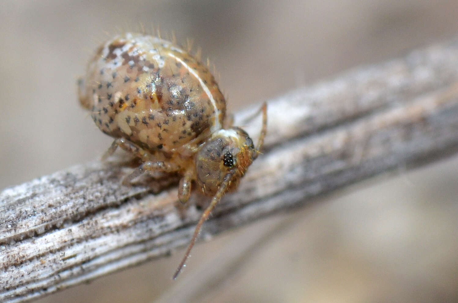
Marko Lukić, Céline Houssin, Louis Deharveng
Zookeys
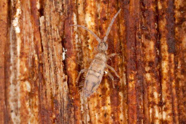
Figures 1–3. Tritomurus veles sp. n. (optical stereomicroscope). 1, 2 Habitus (scale 1 mm) 3 Head (scale 0.2 mm).
-
Melbourne, Victoria, Australia
-
Ballan, Victoria, Australia
-
Saint-Brieuc, Brittany, France
-
Wareham, England, United Kingdom
-
Perth, Western Australia, Australia
-
Camin, Veneto, Italy
-
Ballan, Victoria, Australia
-
Asker, Akershus, Norge
-
Asker, Akershus, Norge
-
Asker, Akershus, Norge
-
Maria Cleide de Mendonça, Eduardo A. Abrantes, Ana Carolina R. Neves
Zookeys
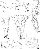
Figure 19–25.Isotomiella uai sp.n. 19 Detail of chaetotaxy of Abd II 20 Leg III 21 Unguis of leg III 22 Ventral tube 23 Furca 24 Lateral view of dens and mucro 25 Female genital opening.
-
Gabriel C. Queiroz, Maria Cleide de Mendonça
Zookeys
Figures 22–28.Micronella longisensilla sp. n. 22 Dorsolateral body chaetotaxy 23 Tita of leg I 24 Furcal area and its surrounding chaetae (adult) 25 Furcal area and its surrounding chaetae (juvenile) 26 Anal valves and ventral view of Abd VI 27 Dorsal view of Abd VI 28 Female genital plate. Scale bars: 10μm (23–28); 50μm (22). x represents missing chaeta.
-
Xiang-Qun Yuan, Zhi-Xiang Pan
Zookeys
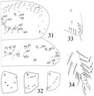
Figures 31–34.Sinella triseta sp. n. 31 dorsal chaetotaxy of Th. II–III 32 coxal mac formula (A fore leg; B mid leg; C hind leg) 33 trochanteral organ 34 tip tibiotarsus and claw of hind leg.
-
Sopark Jantarit, Chutamas Satasook, Louis Deharveng
Zookeys
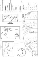
Figure 4.Cyphoderus songkhlaensis sp. n. continued A chaetae of tergites drawn from optical microscope, except 5* derived from SEM image B chaetotaxy of tergites with types of S-chaetae S1 to S4 C trichobothrial complexes of Abd.II D trichobothrial complexes of Abd.III E anterior trichobothrial complexes of Abd.IV F tandem of chaetae on Abd.IV; the smallest is a short type-5 mes and the largest a S4 sens.
-
Marko Lukić, Céline Houssin, Louis Deharveng
Zookeys
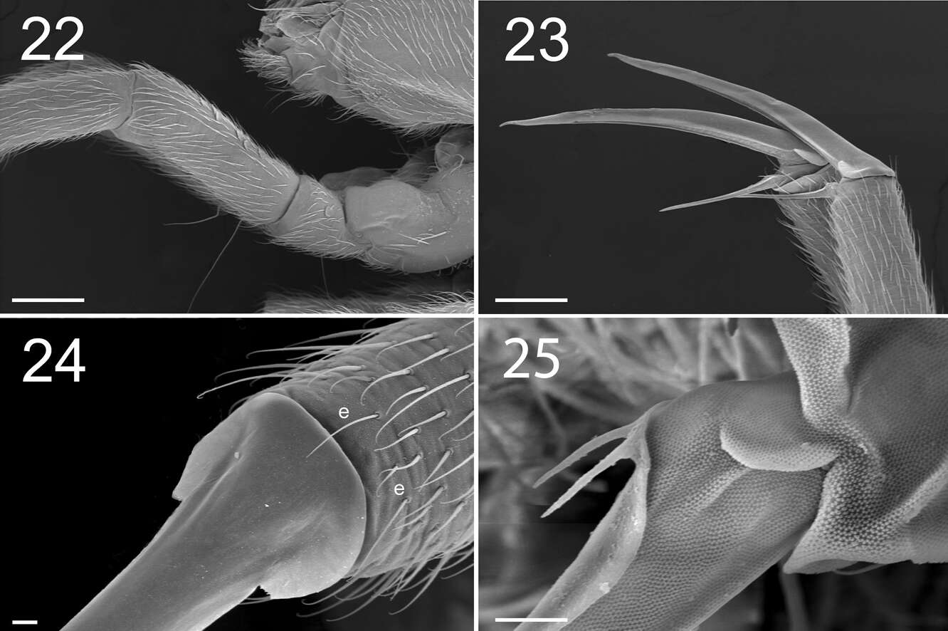
Figures 22–25. Tritomurus veles sp. n. (SEM). 22 Leg I, with ventro-basal macrochaetae of femur and ventral macrochaetae of trochanter (scale 100 μm); the second visible macrochaetae of femur belongs to other leg 23 Claws of legs I (scale 100 µm) 24 Claw of leg I, basal part in dorsal view (scale 10µm); e, thin distal tenent hairs 25 Bifurcate empodial appendage of leg II (scale 10 μm).
-
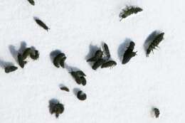
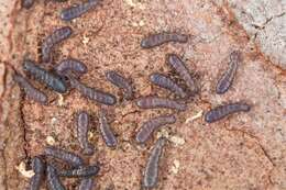
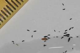
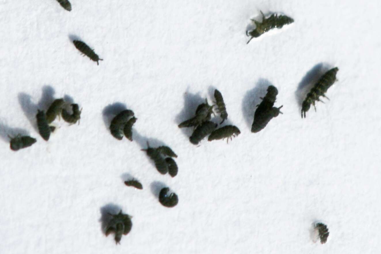
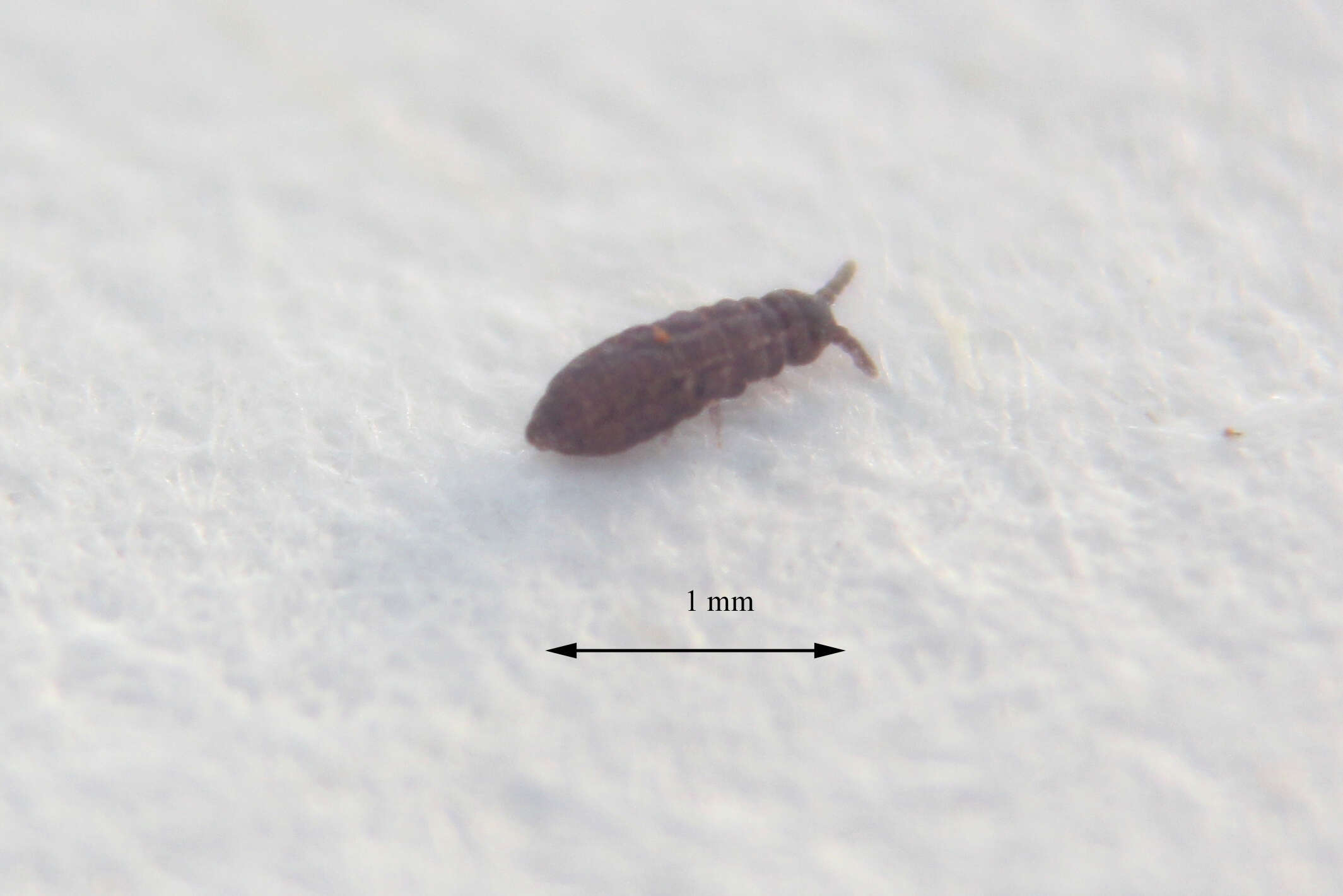
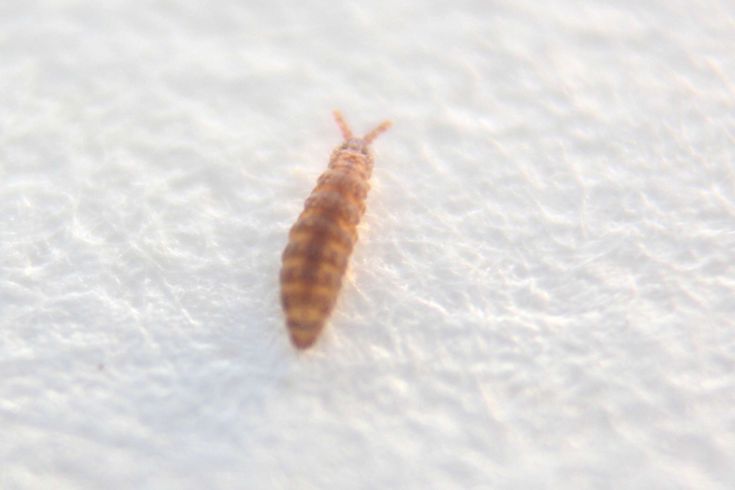
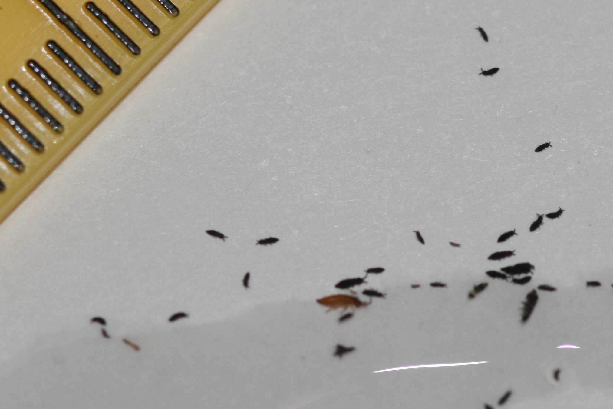
Purple-scum Springtails (Hypogastrura vernalis) - Collembola - congregating on swimming pool at Forest Hill, Melbourne, Victoria, Australia. Photographed on 28 May 2009.c. 1mm in lenth. They congregate in thousands on the surface of the pool.see Greenslade, P., et al. (2014). Biology and key to the Australian species of Hypogastrura and Ceratophysella (Collembola: Hypogastruridae).
Austral Entomolgy 53(1): 53-74.
www.inaturalist.org/observations/55659751