-
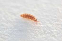
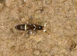
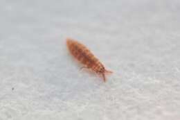
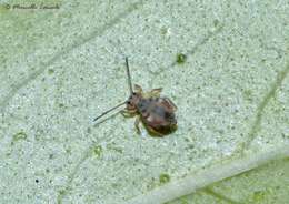
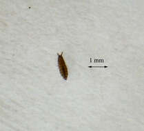
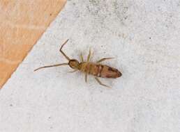
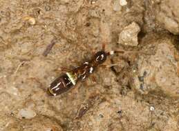
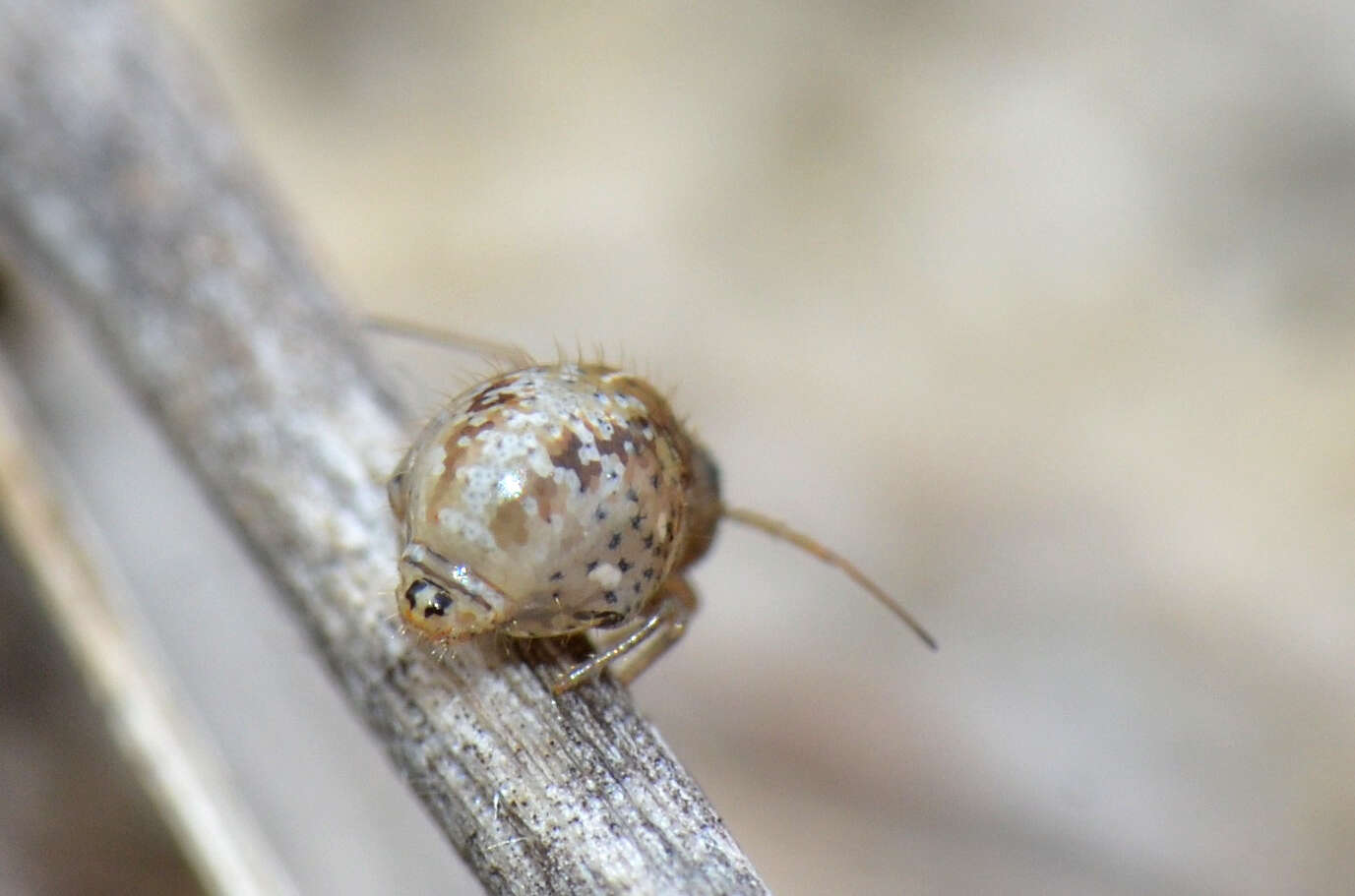
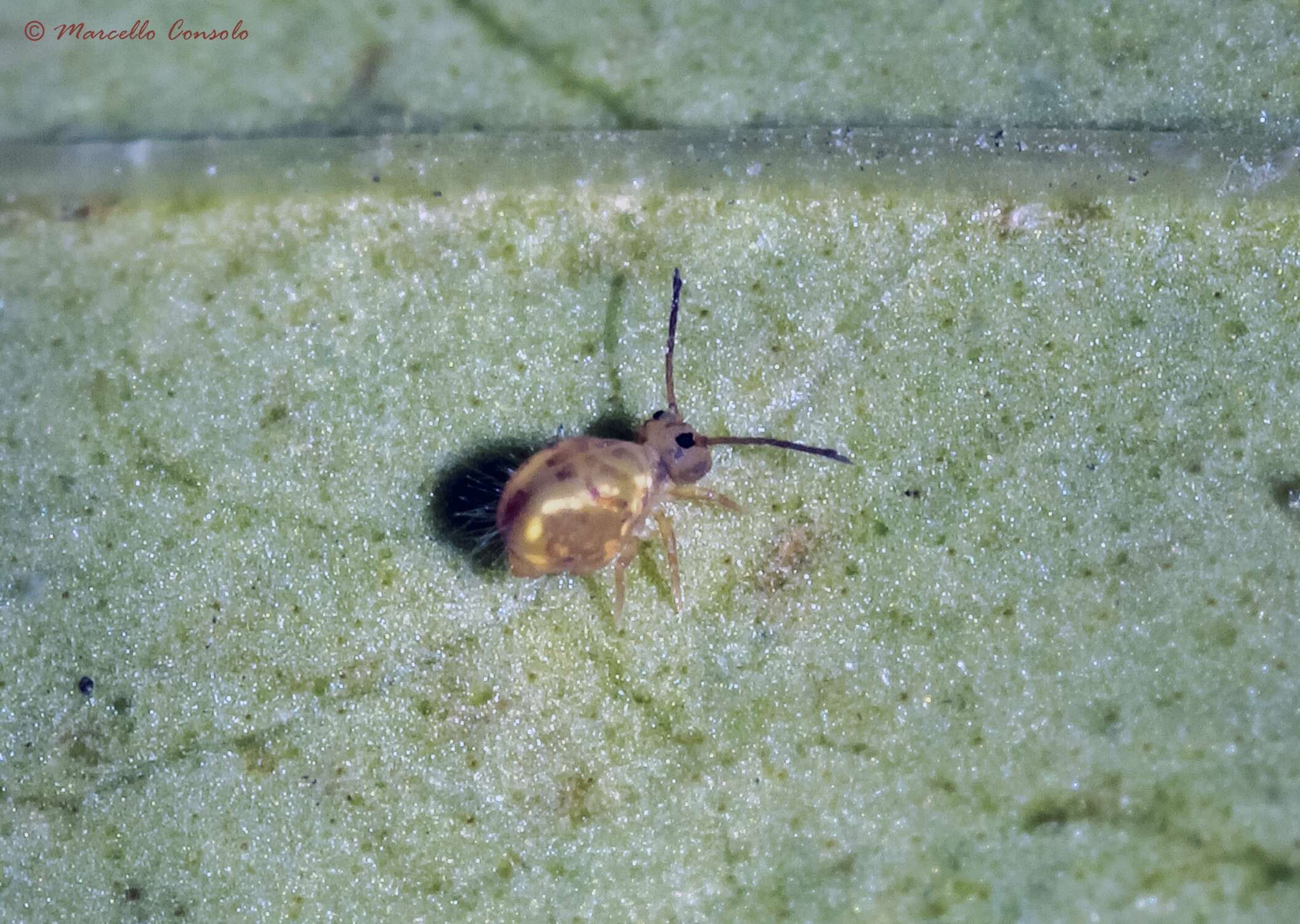
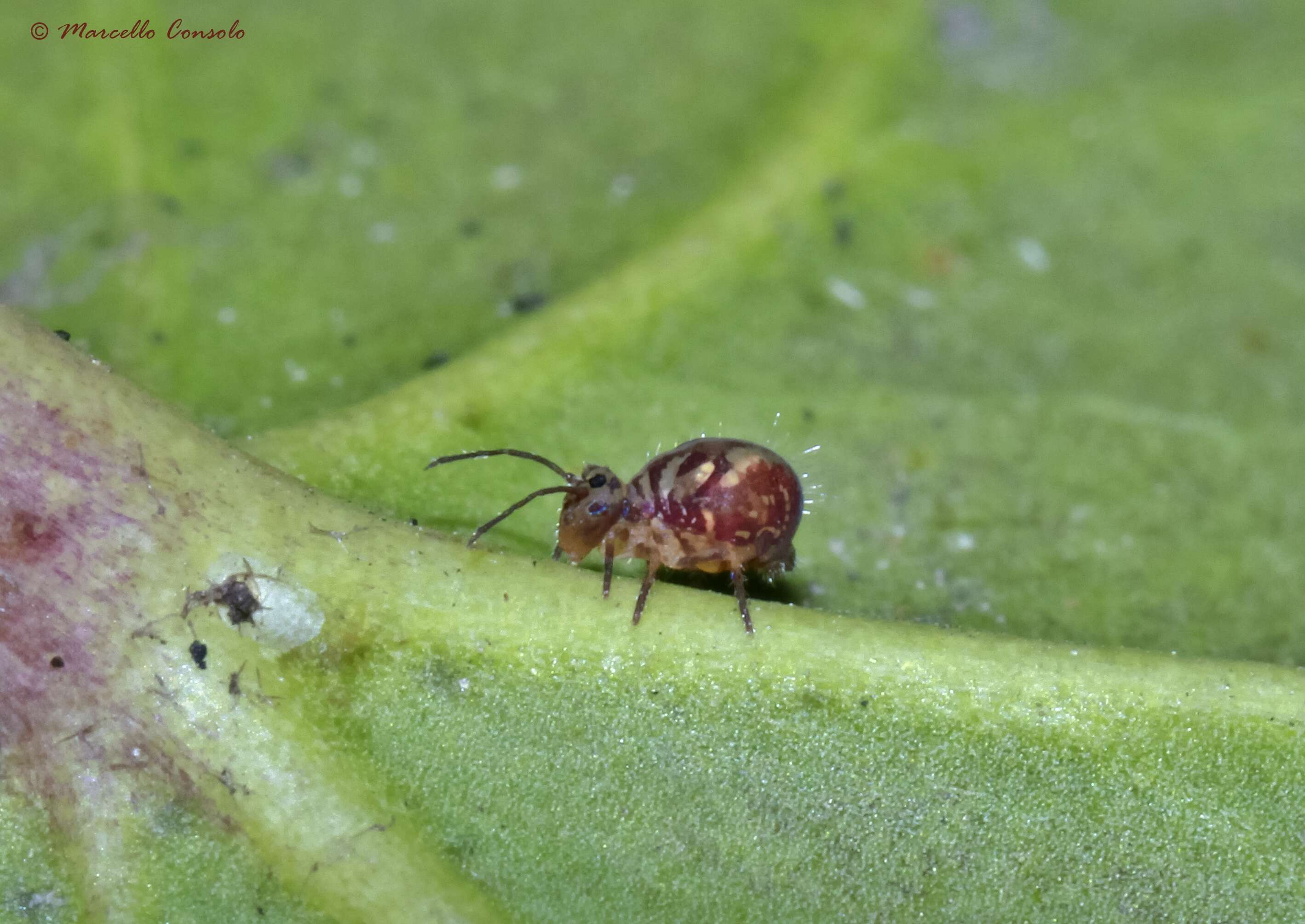
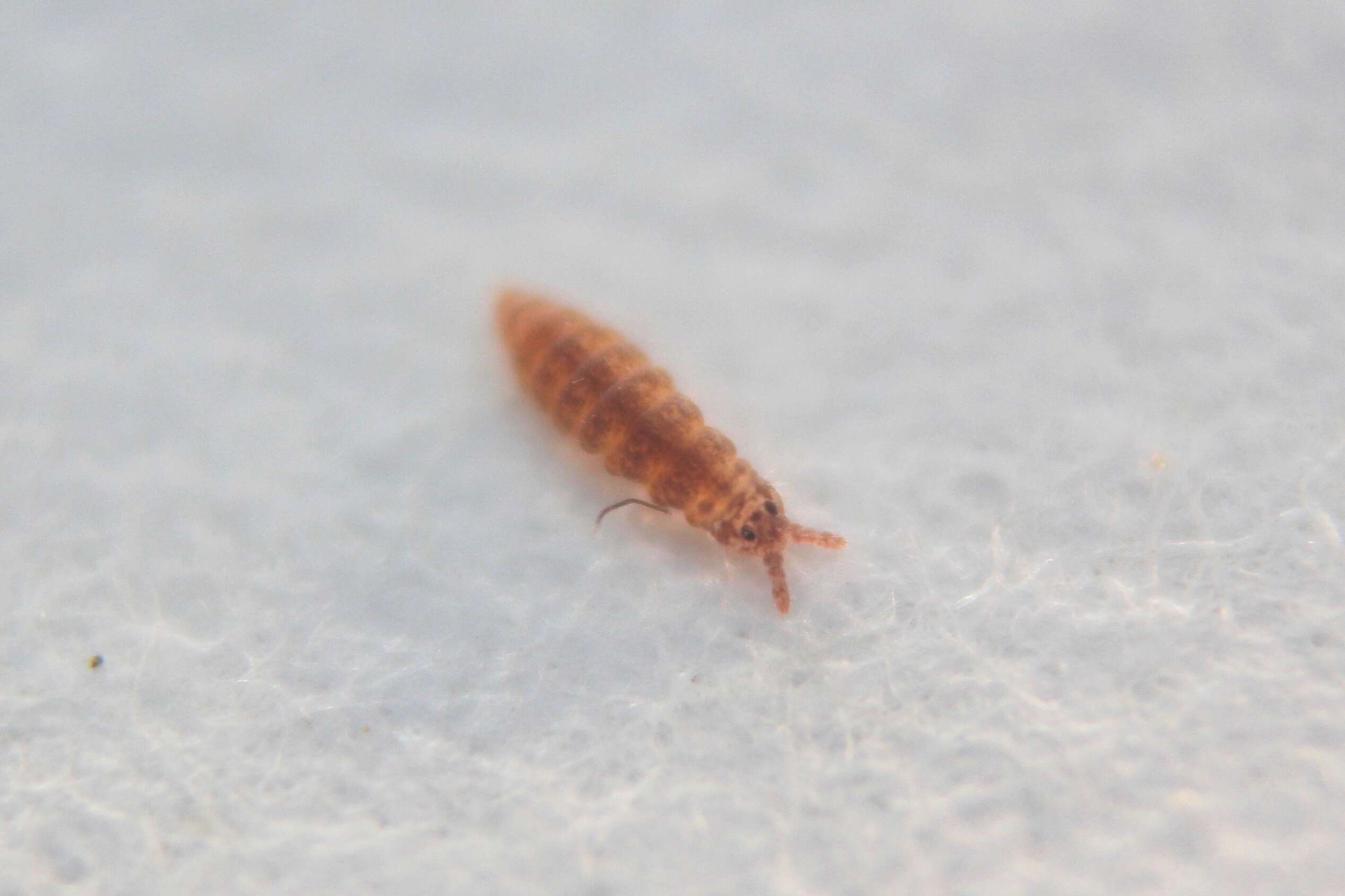
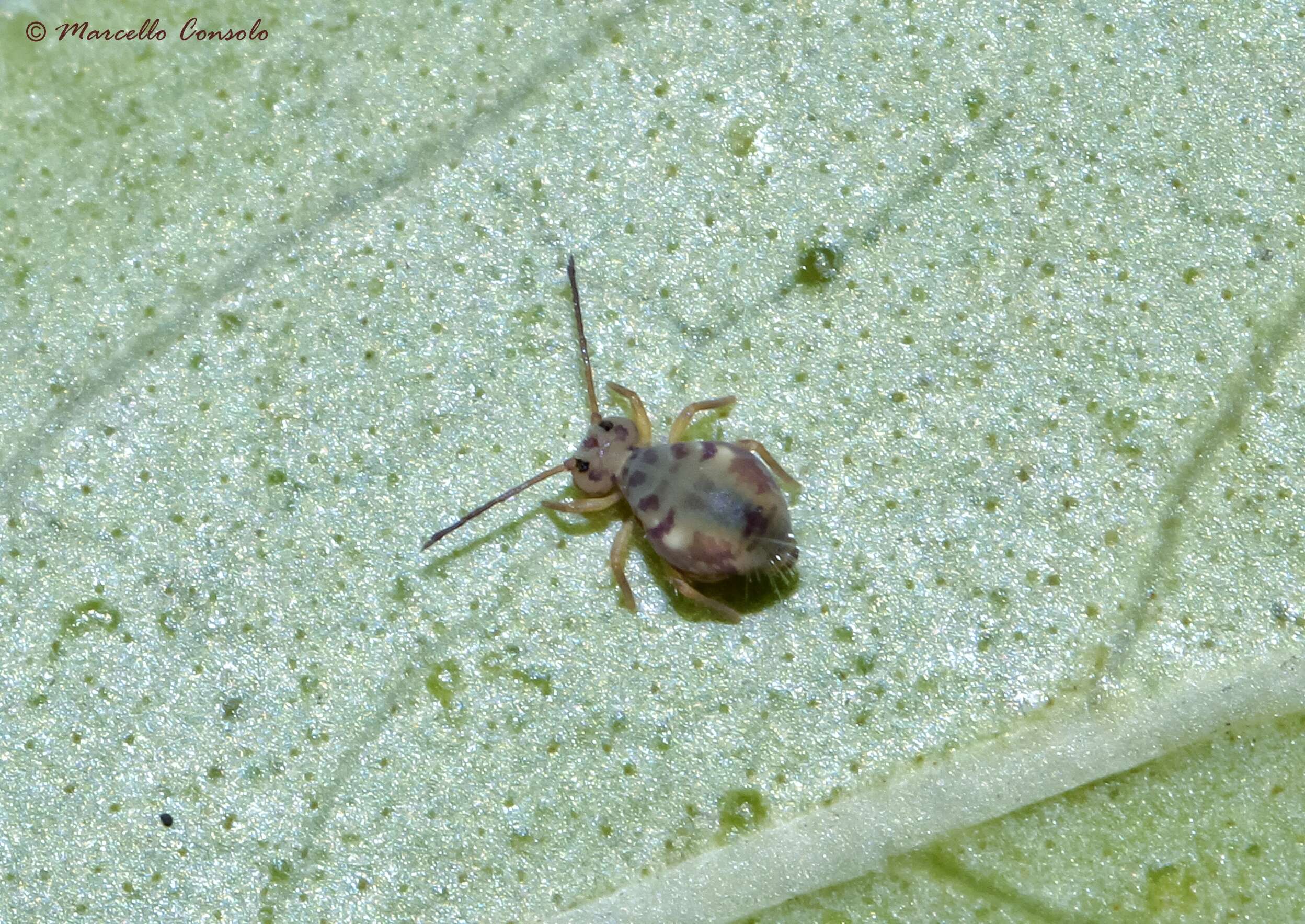
Some small black flies attracted my attention by having a tussle on the track in front of us. I stopped to see what flies they were, but they disappeared. I noticed these tiny Springtails jumping and was very lucky that this little one stayed on a stick for a while.
-
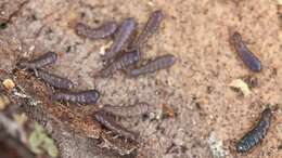
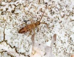
Camin, Veneto, Italy
-
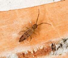
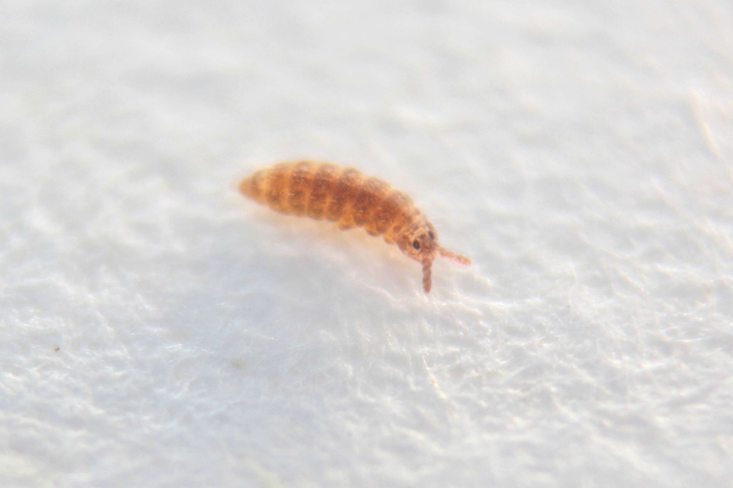
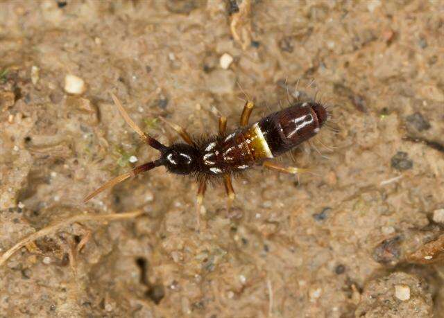
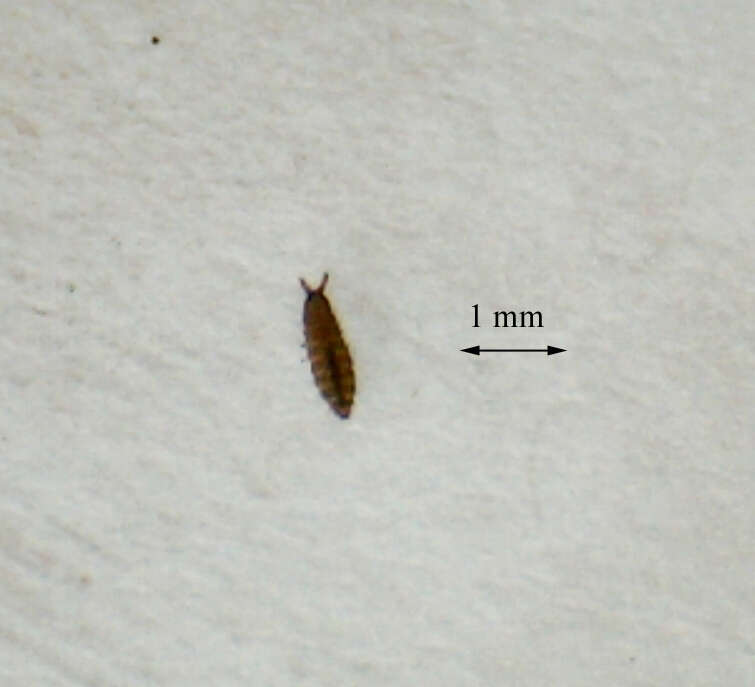
Ballan, Victoria, Australia
-
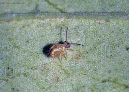
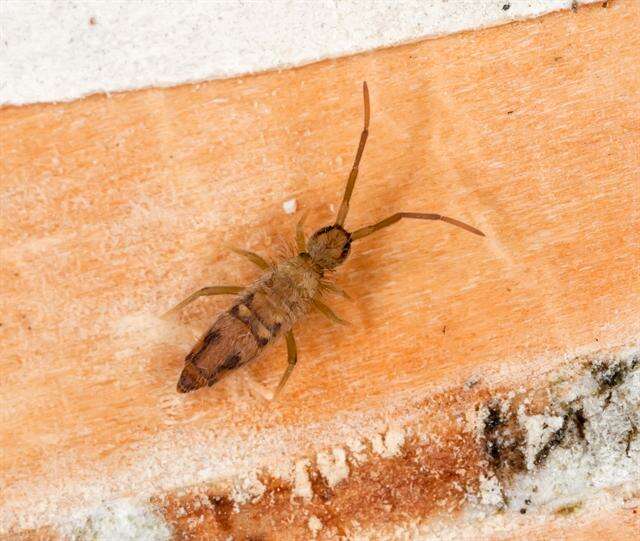
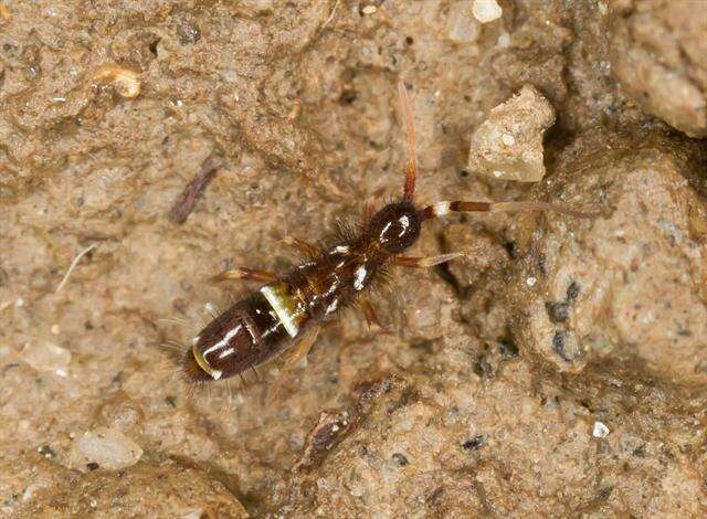
Asker, Akershus, Norge
-
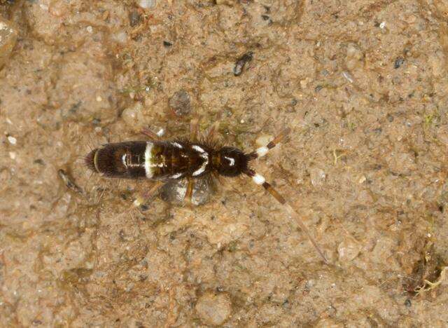
Asker, Akershus, Norge
-
Asker, Akershus, Norge
-
Gabriel C. Queiroz, Maria Cleide de Mendonça
Zookeys
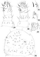
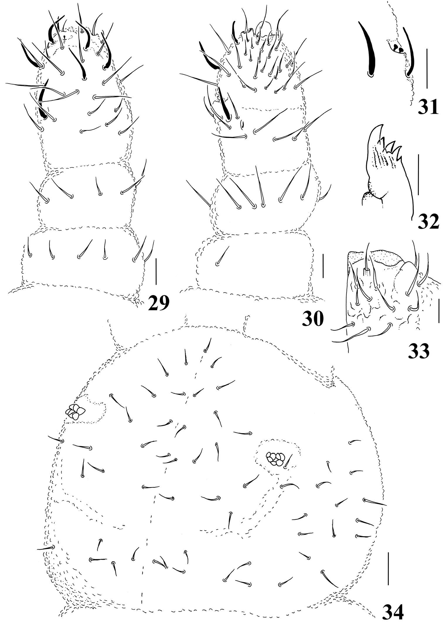
Figures 29–34.Micronella porcus (Denis, 1933). 29. Dorsal view of Ant I–IV 30 Ventral view of Ant I–IV 31 Detail of Ant III organ 32 Maxilla 33 Labium 34 Head chaetotaxy. Scale bars: 10μm (29–33); 20μm (34).
-
Xiang-Qun Yuan, Zhi-Xiang Pan
Zookeys
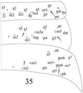
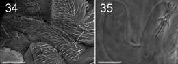
Figure 35.dorsal chaetotaxy of Abd. I–III of Sinella triseta sp. n.
-
Sopark Jantarit, Chutamas Satasook, Louis Deharveng
Zookeys
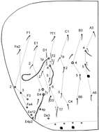
Figure 6.Cyphoderus songkhlaensis sp. n. continued, Szeptycki’s notation of tergal chaetae on Abd.IV (Szeptycki 1979).
-
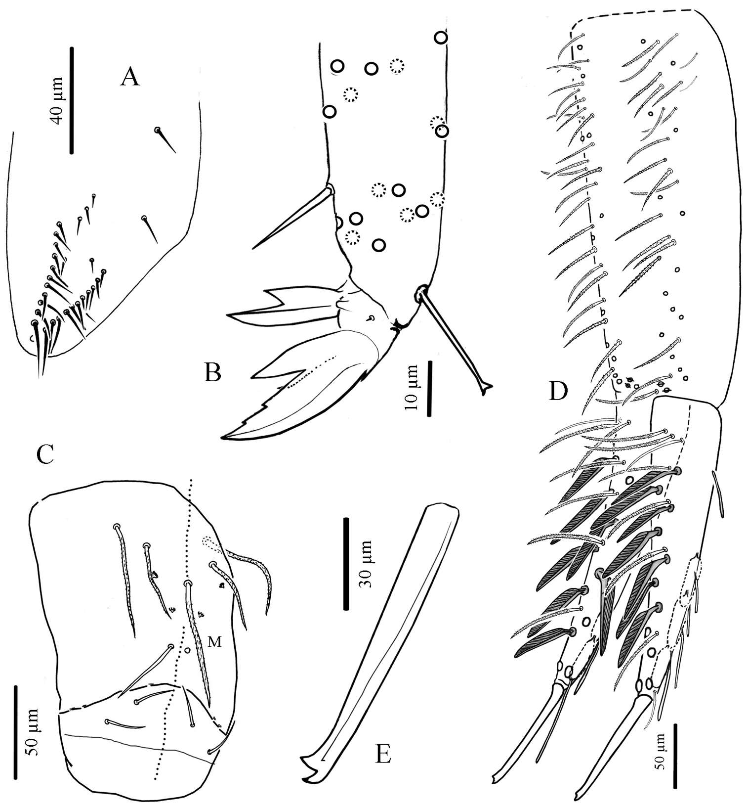
Marko Lukić, Céline Houssin, Louis Deharveng
Zookeys
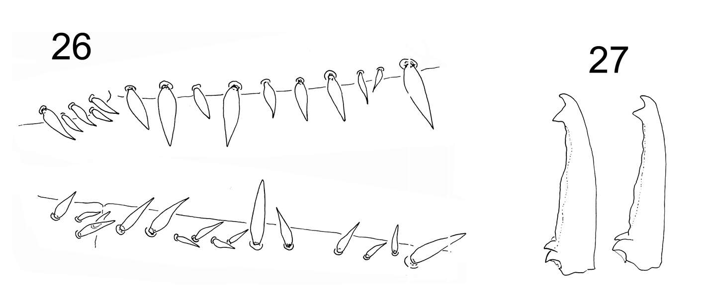
Figures 26–27. Tritomurus veles sp. n. 26 Dental spines formula in a female specimen: 4/2,4,1,4,1 (lower, right dens) and 5/1,1,1,1, 2,1,2,1 (upper, left dens) 27 Mucro in two different specimens.
-
Camin, Veneto, Italy
-
Ballan, Victoria, Australia
-
Asker, Akershus, Norge
-
Asker, Akershus, Norge
-
Gabriel C. Queiroz, Maria Cleide de Mendonça
Zookeys
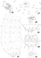
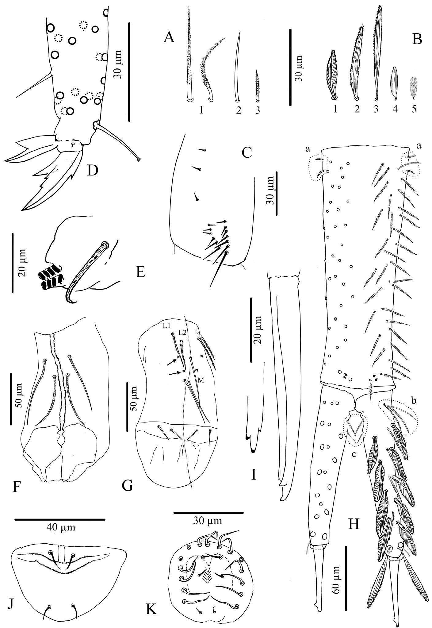
Figures 35–41.Micronella porcus (Denis, 1933). 35 Dorsal body chaetotaxy 36 Tita of leg I 37 Furcal area and its surrounding chaetae 38 Detail of furcal area 39 Dorsal view of Abd VI 40 Anal valves and ventral view of Abd VI 41 Female genital plate. Scale bars: 10μm (36–41); 50μm (35).
-
Sopark Jantarit, Chutamas Satasook, Louis Deharveng
Zookeys
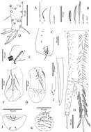
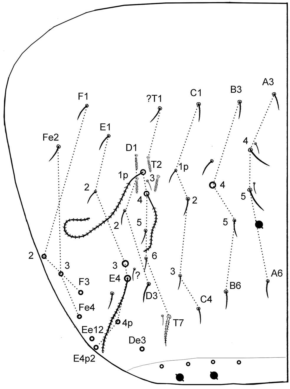
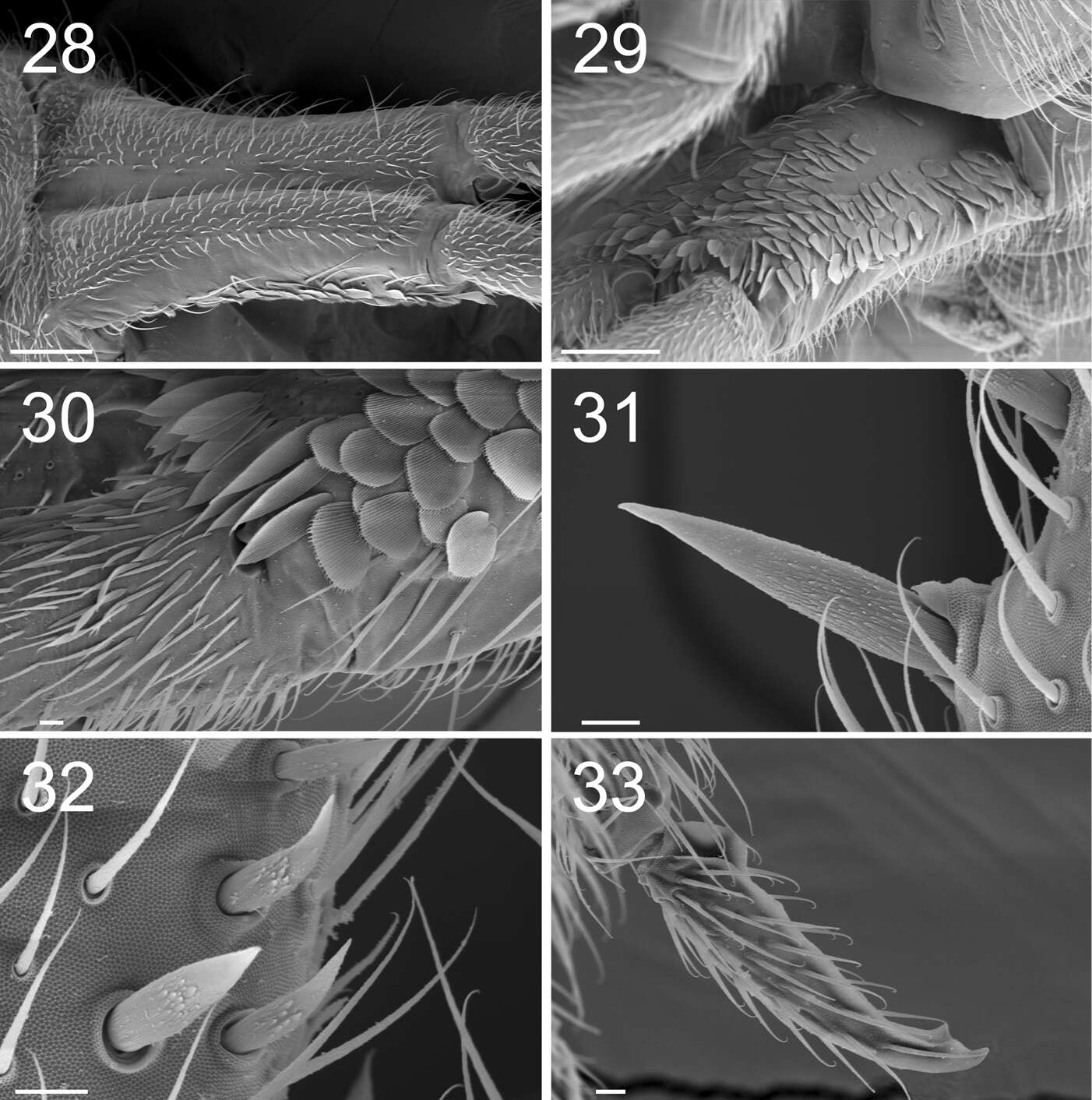
Figure 7.Cyphoderus songkhlaensis sp. n. continued A chaetae of furca B scales of furca C trochanteral organ D claw and distal part of tibiotarsus III E tenaculum F anterior face of the ventral tube G posterior face of the ventral tube; the peg-like setulae are indicated by arrows H furca; encircled by dotted lines are the 2+2 latero-basal mesochaetae of manubrium (a) the 3 outer basal mesochaetae of dens (b) and the 2+2 inner basal mesochaetae of dens (c) (I) mucro in lateral view (right) and in dorsal view (left) showing a third minute external tooth J female genital plate K male genital plate.
-
Marko Lukić, Céline Houssin, Louis Deharveng
Zookeys
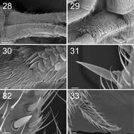
Figures 28–33. Tritomurus veles sp. n. (SEM). 28 Manubrium in dorsal view (scale 100 μm) 29 Manubrium in ventral view (scale 100 μm) 30 Manubrium ventro-distally and dens ventro-basally (scale 10 μm) 31, 32 Dental spines (scale 10 μm) 33 Mucro (scale 10 μm).
-
Camin, Veneto, Italy
-
Ballan, Victoria, Australia
-
Asker, Akershus, Norge
-
Asker, Akershus, Norge
-
Gabriel C. Queiroz, Maria Cleide de Mendonça
Zookeys
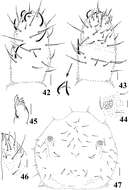
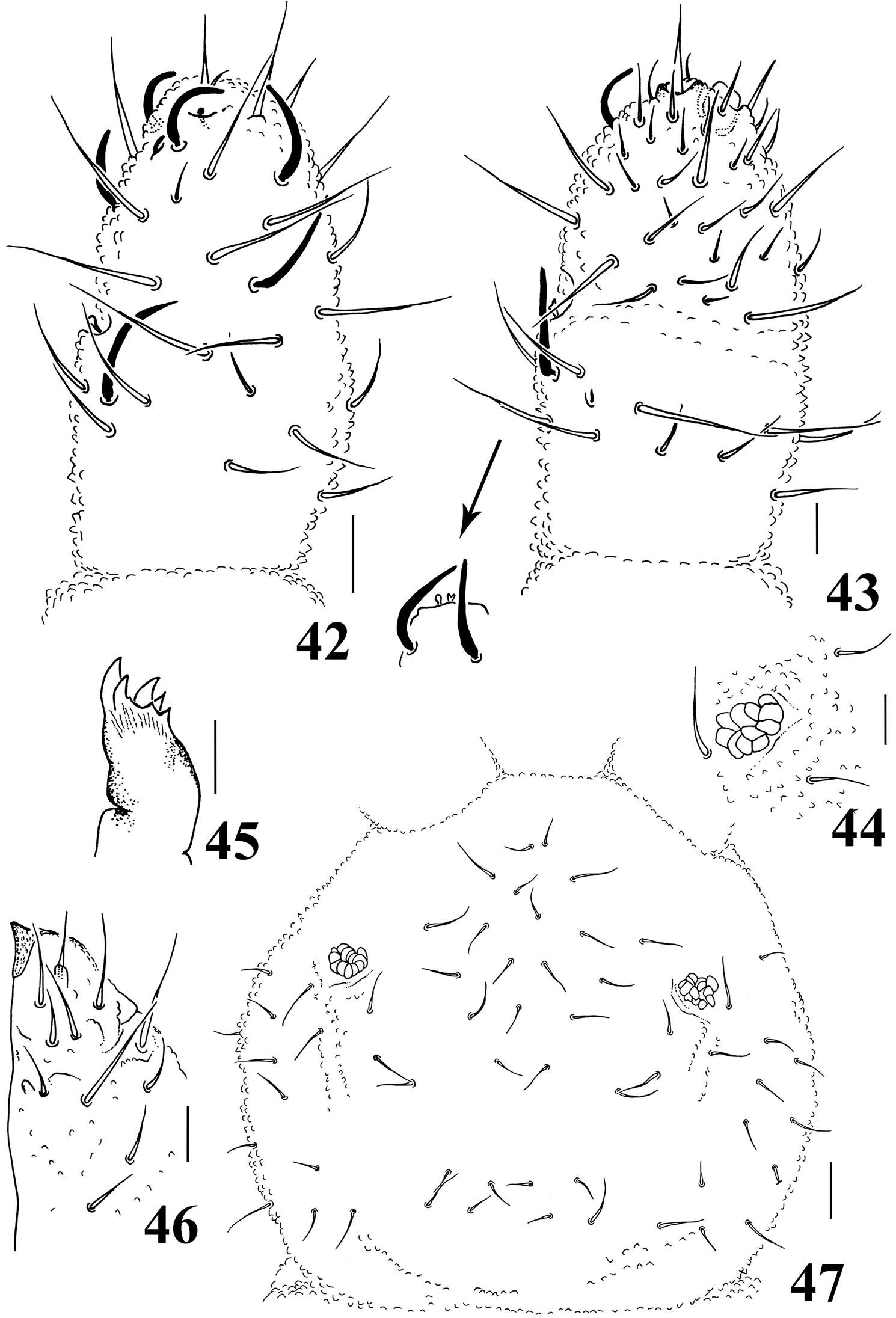
Figures 42–47.Neorganella rotundatae sp. n. 42 Dorsal view of Ant II–IV 43 Ventral view of Ant III–IV with detail of Ant III organ 44 Detail of PAO 45 Maxilla 46 Labium 47 Head chaetotaxy. Scale bars: 10μm (42–46); 20μm (47).
-
Sopark Jantarit, Chutamas Satasook, Louis Deharveng
Zookeys
Figure 8.Cyphoderus khaochakanus sp. n. A trochanteral organ B claw and distal part of tibiotarsus III C posterior face of the ventral tube D furca; feathered chaetae in lateral view, only one of the two vanes attached to the rachis is visible E mucro.
-
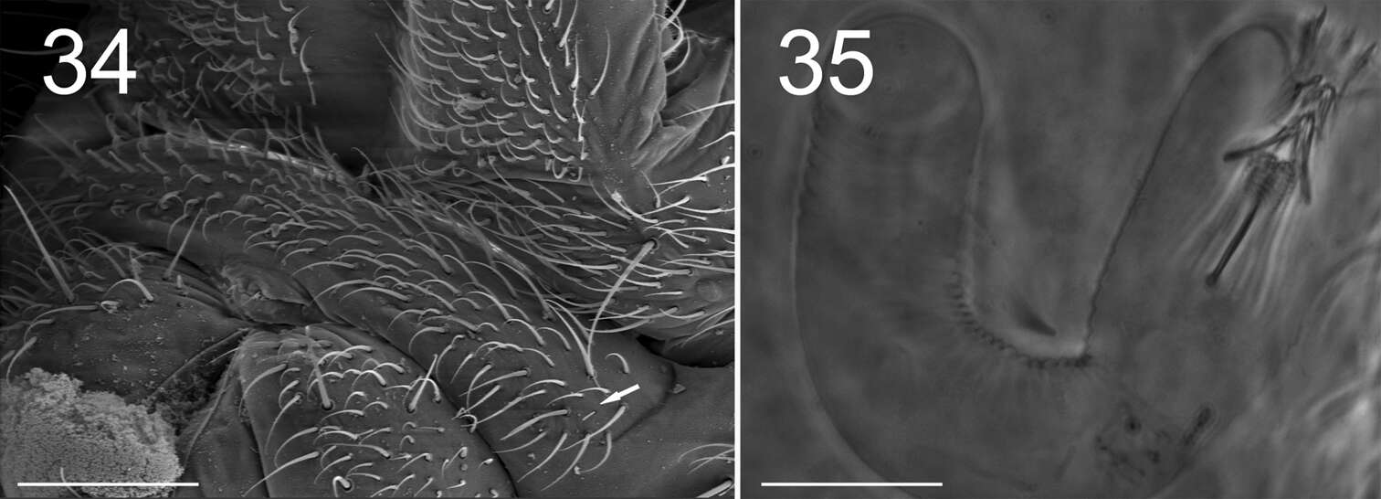
Marko Lukić, Céline Houssin, Louis Deharveng
Zookeys
Figures 34–35. Tritomurus veles sp. n. (34, SEM; 35, optical microscope). 34 Sternite of Abd.V with genital plate (scale 100μm); arrow points to minute lateral S-microchaeta 35 Internal parasitic larva (Nematomorpha) (scale 20 µm).